Navaratnam J1*, Eide N1, Bærland TP1, Rekstad B2, Nilsen T2
1Department of Ophthalmology, Norway
2Department of Medical Physics, Oslo University Hospital, Norway
*Corresponding Author: Jesintha Navaratnam, Department of Ophthalmology, Norway;
Email: [email protected]
Published Date: 25-03-2022
Copyright© 2022 by Navaratnam J, et al. All rights reserved. This is an open access article distributed under the terms of the Creative Commons Attribution License, which permits unrestricted use, distribution, and reproduction in any medium, provided the original author and source are credited.
Abstract
Juxtapapillary retinal capillary haemangiomas are rare benign vascular tumors located adjacent to the optic disc. They may occur sporadically or in association with von Hippel-Lindau disease. Treatment modalities for juxtapapillary retinal capillary haemangiomas vary from observation to aggressive vitreoretinal surgery. A combination of different treatment modalities is often tried with modest success.
We report a patient diagnosed with right-sided juxtapapillary retinal capillary haemangioma at the age of 36 in an amblyopic eye previously treated with patching and surgery. In 2013, at the age of 45 he presented with increasing shadow in his right visual field. The ophthalmological examination revealed a right-sided subtotal serous retinal detachment with 6.2×5.6×3.5 mm juxtapapilllary retinal capillary haemangioma and visual acuity of hand movements. He underwent vitreoretinal surgery, radiation of juxtapapillary retinal capillary haemangioma with intraocular strontium applicator and insertion of silicone oil, all in one session. We describe a novel treatment, the intraocular radiation with strontium. This can be applied in complicated cases where vitrectomy is necessary.
Keywords
Vitreoretinal Surgery; Silicone Oil; Juxtapapillary Retinal Capillary Haemangiomas; Intraocular Radiotherapy
Introduction
Juxtapapillary Retinal Capillary Haemangiomas (JRCH) are well-circumscribed, bright-red tumors that lie adjacent to or on the surface of the optic nerve head. Three distinct growth patterns are described, and these include endophytic, exophytic and sessile types [1,2]. These tumors may occur sporadically or in association with von Hippel-Lindau disease. Von Hippel-Lindau disease is an autosomal dominant genetic disorder associated with tumors arising in multiple organs [3,4]. The JRCH may grow and present with subretinal fluid accumulation, macular edema, hemorrhage from surface neovascularization, exudation, exudative or tractional retinal detachment or epiretinal membrane, resulting in visual deterioration and an amaurotic eye [5]. These tumors are well-known to be difficult to treat due to their locations [6].
Case Presentation
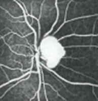
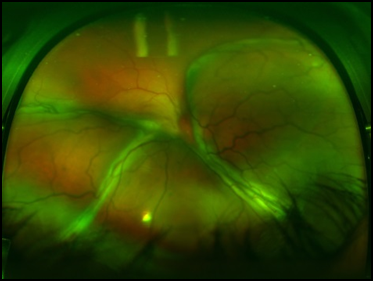
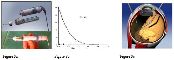
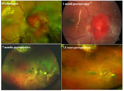
The patient underwent patching and surgery for an anisometropic strabismus in the childhood, but later he developed amblyopia in the right eye. At the age of 36 years he was referred to an ophthalmologist due to floaters in his right eye. During the eye examination, he was diagnosed with right-sided JRCH (Fig. 1). Although the decision of observation of the tumor was made at the time of the diagnosis, he was examined by an ophthalmologist 6 years later and he underwent full work-up including radiological imaging and DNA testing without any evidence of von Hippel-Lindau disease. Three years later, in 2013, he presented with increasing shadow in his right visual field. The ophthalmological examination in September revealed a right-sided subtotal serous retinal detachment with 6.2×5.6×3.5 mm JRCH and visual acuity of hand movements. Ultra-widefield fundus image of right-sided retinal detachment pre-operatively is demonstrated in Fig. 2. In October he underwent vitrectomy, retinotomies, reattachment of the retina, radiation with intraocular strontium, endolaser of suspected retinal capillary hemangioma at 11 O’clock position and cryotherapy of retinotomies and 1000 centistoke silicon oil. The Strontium radiation applicator (NeoVista) consists of a hand piece with a 20- gauge cannula attached. A sclerotomy is made in pars plana, and the applicator is inserted via sclerotomy to assess the tumor. The Strontium-90 source resides in storage position within a radiation sealed canister inside the hand piece in the inactive state. The source is attached to a flexible wire. The position of the source (treatment position in cannula tip- or shielded storing in canister) is controlled by a slide mechanism outside on the hand piece attached to the wire. The Strontium radiation applicator (NeoVista) touched the center of the JRCH in 7 minutes and 45 seconds, which gave a dose of 40 Gy in a depth of 3.5 mm (above the area of the highest thickness of the tumor). Fig. 3 shows the NeoVista strontium applicator and beta-radiation characteristics of Strontium-90. In August 2014, 2 months following removal of the silicone oil, the size of the tumor was reduced and the tumor height went down from 3.5 mm to 1.9 mm. The retina remained reattached after removal of the silicone oil, the patient’s visual field improved and the Best Corrected Visual Acuity (BCVA) was 0.1. Fig. 4 demonstrates perioperative and post-operative images from 1 month to 7.5 years. He underwent cataract surgery of his right eye in January 2017. His visual acuity has been unaltered during more than 8 years follow-up following the surgery in 2013. At the last follow-up in 2021 the BCVA was 0.1 in his right eye and 1.6 in his left eye, JRCH tumor height was 1.4 mm and retina remained attached. There has been no further exudation from the tumor following treatment.

Figure 1: Fluorescein angiography (FA) of juxtapapillary retinal capillary haemangioma in the patient’s right eye in 2004 (image was taken 20 seconds after FA was given).

Figure 2: Ultra-widefield fundus image of right-sided subtotal serous retinal detachment and juxtapapillary retinal capillary haemangioma pre-operatively.


Figure 3: a: Strontium (Sr) applicator (NeoVista) used to treat the juxtapapillary retinal capillary haemangioma. The device consists of a hand piece with a 20- gauge cannula attached. In inactive state the Sr-90 source resides in storage position within a radiation sealed canister inside the hand piece. The source is attached to a flexible wire. The position of the source (treatment position in cannula tip- or shielded storing in canister) is controlled by a slide mechanism outside the hand piece attached to the wire; b: Relative (%) distribution of radiation dose as function of depth, demonstrating beta-radiation characteristics of the St-90 radioactive beta decay radiation (electrons with short depth penetrance, important to avoid radiation hazards);c: Intravitreal setup during treatment, with the cannula and a light inserted (the infusion is not shown in this figure). A mark indicating the mid position of the Sr-90 source (0,52 x 2,5 mm) during treatment is viable near the end of the tip. © NeoVista®, Newark, USA.

Figure 4: Ultra-widefield fundus images of the right eye captured under surgery, 1 month following the surgery with silicon oil insertion, 7 months following the first surgery and 2 months following removal of the silicon oil and 7 ½ years following first surgery. Retina is attached with fibrotic changes around the juxtapapillary retinal capillary tumor and scarring following cryotherapy at 11 o’clock position.
Discussion
The treatment of JRCH remains challenging, and a definitive treatment guideline is not yet established [6]. In asymptomatic patients, observation remains the mainstay of management since the tumor can remain stable for decades. Generally, the treatment of JRCH is not advised before exudation or symptoms develop. Various treatments have been tried, and these include laser photocoagulation, external beam radiation, plaque radiotherapy, transpupillary thermotherapy, photodynamic treatment, intravitreal anti-Vascular Endothelial Growth Factor (anti-VEGF) or steroids, vitreoretinal surgery or combination of treatment modalities [6]. The limitation of all the current therapeutic modalities listed is documented by the high number of options without no clear preference. The efficacy of treating JRCH is evaluated by regression of the tumor and the consequence on subretinal fluid, exudation and visual acuity immediately and long-term. The JRCHs are rare tumor, and are reported as case reports or small series. Therefore, there is sparse information on the results of the various interventions. The series by Sahdeva, et al., reported a modest effect on visual acuity based on three cases of JRCH: one better, one unchanged and one worse [7]. In addition, they stated that visual acuity improved or stabilized in 69% and complications were observed in 50% of cases based on their literature review. However, in their case series with 8 JRCH eyes 38% improved their visual acuity. In another study with average follow-up of 5.4 years the patients’ visual acuity generally worsens with various treatment modalities including laser photocoagulation, external beam radiotherapy, scleral buckling and pars plana vitrectomy [8]. Sahdeva, et al., reported also need for retreatment, and about 50% needed multiple photodynamic treatment [7]. Some patients develop JRCH bilaterally, and in these patients we may consider treating the second eye early. In challenging cases, the intraocular radiation with strontium can be a treatment option. A specific and targeted dose can easily be administered. Strontium-90 is an almost pure beta particle source with short penetrance and high dose rate. It is a by-product of uranium and plutonium fission, acts in-vivo as calcium and has a half-life of 29 years [9]. Due to the characteristics of the beta radiation, with a steep dose fall-off with increasing depth, the most effect will be on the haemangioblastoma and not the other tissues. In addition, the Strontium applicator can be placed directly to the surface to the tumor intravitreally. Exudative age-related macular degeneration have been treated with epimacular brachytherapy [10,11]. The optimal timing and treatment modality of JRCH is yet to be established and is still open for discussion.
Conclusion
Today, commercial intraocular strontium radiation equipment is not available to the best of our knowledge. The development of strontium radiation equipment may be a successful intervention for treatment of tumors located close to the optic disc or macula and due to the long half-life of strontium-90 the equipment will last for decades.
Conflict of Interest
The authors declare no conflict of interest, financial or otherwise.
References
- Gass JD, Braunstein R. Sessile and exophytic capillary angiomas of the juxtapapillary retina and optic nerve head. Arch Ophthalmol. 1980;98:1790-7.
- McCabe CM, Flynn Jr HW, Shields CL, Shields JA, Regillo CD, McDonald HR, et al. Juxtapapillary capillary hemangiomas: Clinical features and visual acuity outcomes. Ophthalmol. 2000;107:2240-8.
- Kaelin WG. Molecular basis of the VHL hereditary cancer syndrome. Nat Rev Cancer. 2002;2:673-82.
- Singh AD. Von Hippel-Lindau disease. Surv Ophthalmol. 2001;46:117-42.
- Magee MA, Kroll AJ, Lou PL, Ryan EA. Retinal capillary hemangiomas and von Hippel-Lindau disease. Semin. Ophthalmol. 2006;21:143-50.
- Saitta A, Nicolai M, Giovannini A, Mariotti C. Juxtapapillary Retinal Capillary Hemangioma: New Therapeutic Strategies. Med Hypothesis Discov Innov Ophthalmol. 2014;3:71-5.
- Sachdeva R, Dadgostar H, Kaiser PK, Sears JE, Singh AD. Verteporfin photodynamic therapy of six eyes with retinal capillary haemangioma. Acta Ophthalmol. 2010;88:e334-40.
- McCabe CM, Flynn Jr HW, Shields CL, Shields JA, Regillo CD, McDonald HR, et al. Juxtapapillary capillary hemangiomas. Clinical features and visual acuity outcomes. Ophthalmol. 2000;107:2240-8.
- https://pubchem.ncbi.nlm.nih.gov/compound/Strontium-90 [Last accessed on: March 18, 2022]
- Dugel PU, Bebchuk JD, Nau J, Reichel E, Singer M, Barak A, et al. Epimacular brachytherapy for neovascular age-related macular degeneration: a randomized, controlled trial (CABERNET). 2013. Ophthalmol. 2013;120:317-27.
- Jackson TL, Soare C, Petrarca C, Simpson A, Neffendorf JE, Petrarca R, et al. Epimacular brachytherapy for previously treated neovascular age-related macular degeneration (MERLOT): A phase 3 randomized controlled trial. Ophthalmol. 2016;123:1287-96.
Article Type
Case Report
Publication History
Received Date: 18-02-2022
Accepted Date: 18-03-2022
Published Date: 25-03-2022
Copyright© 2022 by Navaratnam J, et al. All rights reserved. This is an open access article distributed under the terms of the Creative Commons Attribution License, which permits unrestricted use, distribution, and reproduction in any medium, provided the original author and source are credited.
Citation: Navaratnam J, et al. A Case Report of Intraocular Radiotherapy with Strontium-90 for Juxtapapillary Retinal Capillary Haemangioma. J Ophthalmol Adv Res. 2022;3(1):1-6.

Figure 1: Fluorescein angiography (FA) of juxtapapillary retinal capillary haemangioma in the patient’s right eye in 2004 (image was taken 20 seconds after FA was given).

Figure 2: Ultra-widefield fundus image of right-sided subtotal serous retinal detachment and juxtapapillary retinal capillary haemangioma pre-operatively.


Figure 3: a: Strontium (Sr) applicator (NeoVista) used to treat the juxtapapillary retinal capillary haemangioma. The device consists of a hand piece with a 20- gauge cannula attached. In inactive state the Sr-90 source resides in storage position within a radiation sealed canister inside the hand piece. The source is attached to a flexible wire. The position of the source (treatment position in cannula tip- or shielded storing in canister) is controlled by a slide mechanism outside the hand piece attached to the wire; b: Relative (%) distribution of radiation dose as function of depth, demonstrating beta-radiation characteristics of the St-90 radioactive beta decay radiation (electrons with short depth penetrance, important to avoid radiation hazards);c: Intravitreal setup during treatment, with the cannula and a light inserted (the infusion is not shown in this figure). A mark indicating the mid position of the Sr-90 source (0,52 x 2,5 mm) during treatment is viable near the end of the tip. © NeoVista®, Newark, USA.

Figure 4: Ultra-widefield fundus images of the right eye captured under surgery, 1 month following the surgery with silicon oil insertion, 7 months following the first surgery and 2 months following removal of the silicon oil and 7 ½ years following first surgery. Retina is attached with fibrotic changes around the juxtapapillary retinal capillary tumor and scarring following cryotherapy at 11 o’clock position.


