Comparative Studies of the Tongue of Postnatal (Lactating), Young and Adult Between Two Species of Mammalian Animals of Different Feeding Habit Rattus Rattus and Orcytolagus Cuniculus
Atteyat Selim1*, Ezzar Hafez2, Wafaa Goda3
1Department of Zoology, Faculty of Science, Tanta University, Egypt
2Department of Zoology, Faculty of Science, Tanta University, Egypt
3Department of Zoology, Faculty of Science, Tanta University, Egypt
*Correspondence author: Atteyat Selim, Department of Zoology, Faculty of Science, Tanta University, Egypt; Email: atteyatselim@hotmail.com
Citation: Selim A, et al. Comparative Studies of the Tongue of Postnatal (Lactating), Young and Adult Between Two Species of Mammalian Animals of Different Feeding Habit Rattus Rattus and Orcytolagus Cuniculus. Jour Clin Med Res. 2023;4(1):1-20.
Copyright© 2023 by Selim A, et al. All rights reserved. This is an open access article distributed under the terms of the Creative Commons Attribution License, which permits unrestricted use, distribution, and reproduction in any medium, provided the original author and source are credited.
| Received 03 Apr, 2023 | Accepted 18 Apr, 2023 | Published 25 Apr, 2023 |
Abstract
Background: The study of tongue of mammals is important because the different species of animals with different feeding, so the present study was attended to observe the relation between various feeding and the morphological shape of tongue of present stages of both R. rattus and O. cunniculus using histological, histochemical techniques, ultra structural examination and Statistical analysis.
Results: The Egyptian herbivorous rabbit O. cunniculus and the rodent R. rattus showed that the tongue is elongated with rounded apex. In both species the thicknesses of the tongue increase gradually toward the pharynx. The tongue length of adult rabbit was large than the adult rat.
In the studied animals, the anterior end of the tongue of the herbivorous rabbit O. cunniculus is covered with short horny filiform papillae at the anterior part and giant circumvallate papilla at the posterior part with taste buds, also large enormous numbers of mucus and serous glands but in the rodent Rattus is covered with short pointed horny filiform papillae and others small elongated ones. Few numbers of fungiform papillae and numbers of mucus and serous glands are scattered among the skeletal muscles. The Tongue of adult R. rattus and O. cunniculus have different shapes of filiform papillae, fungiform papillae distributed between the filiform ones, two circumvallate on the surface, number of serrate glands and mucous gland. Give strong reaction with Periodic Acid Schiff (PAS). In young and lactating some undifferentiated and poor developed papillae were observed and serrate and mucous glands less than that in adult stage
Conclusion: The different feeding habit of both rat (rodents and Rabbit (herbivorous) give different shapes of papilla and give different reaction with histochemical stains.
Keywords: Rabbit; Rat; Herbivorous; Tongue; Rodents; Lactating; Young; Adult and Ultra Structure
The Aim of Study
The difference of habits between rodents (rats) and herbivorous (rabbits) were the aim of study, so the research focus on the first part of the alimentary canal, the tongue. The previous researches on the tongue describe it as muscular organ covered with squamous epithelium. It lies between oral cavity and pharynx; it is attached by its muscles to the hyoid bone, mandible, styloid processes, soft plate and the pharyngeal wall describe the specimens of Camelus dromedaries exhibit that the animal has ideal elongated tongue which appear surface [1,2].
Many previous studies had described the foliate papillae which located on the posterior dorsal part of domestic rabbit tongue. In Oryctolagus cuniculus fungiform papillae observed on the apex of tongue body [3]. In detecting protein content of papillae of tongue Egyptian insectivorous bats tongue give more dense reaction with bromophenol blue stain than those Syrian ones [4]. Three shapes of papillae (filiform, fungiform and circumvallate) observed on the surface of Miniopterus schreibersi fuliginosus and Pipistrellus savii tongue [5]. Ultrastructure study on Dasypus hybrid tongue detect taste buds carrying on fungiform and circum allate papillae that both are responsible for taste, while mechanical and protective role carry out by filiform ones [6]. At the first day after birth taste bud were observed on vallate papillae of domestic rabbit tongue [7]. Kulawik, et al., revealed that the filiform papillae are cone or palm shaped with three to seven processes in a study by light and scanning electron microscope of the filiform papillae of the tongue in adult rabbit (Oryctolagus cuniculus) [8].
Differences in the stain ability and distribution of neutral and acid muco polysaccharides in the lingual glands were present in comparative histological studies of three species of Egyptian bats the frugivorous ones, the insectivorous bat Rhinopoma hardwickie and the tomb-inhabiting bat Taphozous perforates [9]. The California sea lion show some morphological adaptations to their aquatic environment however, they possess some structures similar to those of land mammals [10]. Mucous and serous glands were observed especially on posterior ventral surface of insectivore bat (Rhinopoma hardwickii) tongue and single circumvallate papilla were situated in dorsal root of tongue [11]. Some authors observed the three vallate papillae in Marsupials [12]. Others observed the shape of the horse tongue and its papillae, they observed that the papillae are present on the dorsal surface of the tongue while the foliate papillae are found in the horse but not found in the goat [13].
Material and Methods
The animals of Rattus rattus (Sub order Myomorpha). Family: Muridae and Oryctolagus cunniculus (order Lagomorpha). Family Leporidae were brought alive and the specimens were killed by decapitation.
Collected the Animals
The specimens of Oryctolagous cunniculus and Rattus rattus collected alive and classified as follows: Adult group, young group; (after weaning) and Lactating group.
Histological Examination
Organs of adult, prenatal and postnatal Oryctolagous cunniculus and Rattus rattus collected. Parts of tongue were immediately fixed. All preparations were microscopically examined for the histological examination.
Histochemical Examination
Periodic Acid Schiff procedure (PAS): The PAS reaction used to: demonstrate the mucous secretion of the mucosa, and muscles in tongue demonstrate the secretion of mucus and serous glands.
Azan Stain:(Gretchen, 1974). Azan stain used to: examine the activity of the tongue (serous and mucous) and to demonstrate keratinization of epithelia, connective tissue.
Scanning Electron Microscopy
The surface lingual papillae and glands were observed using Scanning Electron Microscope (SEM) [14].
Statistics
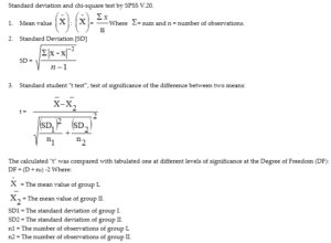
4. Analysis of Variance [ANOVA] tests (f): According to the computer program SPSS for Windows. ANOVA test was used for comparison among different times in the same group in quantitative data [15].
Results
External Morphology
Examination of the tongue of the two studied species, the Egyptian herbivorous rabbit O. cunniculus and the rodent R. rattus showed that the tongue is elongated with rounded apex. In both species the thicknesses of the tongue increase gradually toward the pharynx (Fig. 1).
Morphometric Study
The tongue length of rabbit was in lactating 2.08 ± 0.31 cm, in Young 3.09 ± 0.22 cm and in adult 4.24 ± 0.30 cm. The tongue length of rat was in lactating 1.07 ± 0.20 cm, in Young 2.0 ± 0.21 cm and in adult 2.83 ± 0.21 cm.
Adult Tongue
Light Microscopic Observations: In the studied animals, the apex of the tongue of the herbivorous rabbit O. cunniculus is covered with short horny filiform papillae at the anterior part and giant circumvallate papilla at the posterior part with taste buds, also large enormous numbers of mucus and serous glands (Fig. 2) the rodent Rattus rattus is covered with short pointed horny filiform papillae and others small elongated ones (Fig. 3). Few numbers of fungiform papillae and numbers of mucus and serous glands are scattered among the skeletal muscles (Fig. 3) in the posterior part of the tongue.
Histochemical Investigation
In Oryctolagus cunniculus (Fig. 2) showing numerous mucous glands give strong reaction with (PAS) stain. But the mucus gland gives weak reaction with azan stain. (Fig. 2). In Rattus rattus by using azan stain, mucous glands (Fig. 3) mucous glands give weak reaction while serous gland give moderate reaction with azan and give very moderate reaction with Schiff reagent of (PAS) Fig. 3. The serous glands more developed in Rattus rattus (feed on dry materials) than Oryctolagus cuniculus.
Scanning Electron Microscopic Observations: Oryctolagus cunniculus showed many different shapes of filiform papillae from elongated to short and horny (Fig. 4), while in R. rattus with small, elongate and serrate filiform papillae were observed covering the tongue apex (Fig. 5). In the two bat species studied, the fungiform papillae were few in number and scattered and among the filiform papillae (Fig. 4,5). At the posteriorly of the tongue, the circumvallate papillae and the taste buds were shown in the two studied bats (Fig. 4,5). In addition, the mucous glands were seen in the posteriolyt of the tongue in both species (Fig. 4,5).
Young Tongue
Light Microscopic Observations
The tongue of the young rabbit in the anterior lateral part of O. cunniculus showing very short horny filiform papillae than adult, fungiform papillae and muscle, Fig. 6 while in the posterior part of O. cuniculus tongue showing filiform papillae, mucous glands and serous gland in a few numbers than adult. In Rattus rattus in the anterior part the tongue showing filiform papillae, fungiform papillae, muscles Hand E. in middle line of the anterior part of R. rattus tongue showing short filiform papillae, muscles and lamina propria Hand E. but in the posterior part of R. rattus tongue showing horny filiform papillae fungiform papillae, mucous glands, Serous Gland (SG), Muscles (M) and Lamina Propria (LP) Hand E (Fig. 7).
Histochemical Investigation
Transverse section in the posterior part of O. cunniculus tongue showing Mucous Gland (MG) give negative reaction with azan. G and h- and moderate reaction with PAS (Fig. 7), There is no serous glands developed in the anterior part of R. rattus tongue showing Mucous Glands (MG) give weak reaction while Serous Gland (SG) give moderate reaction with azan. E, f- in the posterior part of R. rattus tongue showing Mucous Glands (MG) and Serous Glands (SG) give moderate reaction with Schiff reagent of (PAS) stain (Fig. 7).
Scanning Electron Microscopic Observations
Examination of the surface of the tongue apex in the Rabbit showed that the median was curved and the tip of the tongue was round with undifferentiated papillae (Fig. 8). At the posterior part of the tongue, the fungiform papillae were shown and some short horny filiform papillae were observed at this stage (Fig. 8), While in Rat at the posterior part of the tongue the is divided by suture and covered with undifferentiated elongated filiform papillae (Fig. 9). In the middle and the posterior part, the circumvallate, less developed fungiform and the irregular filiform papillae began to develop (Fig. 9).
Lactating Tongue
Light Microscopic Observations
In the studied animals the anterior part of the tongue of Rabbit is covered with short horny filiform papillae and the fungiform papillae (Fig. 10) poor developed, but in the middle and the posterior part the circumvallate papillae poor developed (Fig. 10) and the mucus glands were poor than those of adult, but in rat the anterior part of the tongue with poor developed filiform and fungiform papillae the apex is covered with small filiform and fungiform papillae (Fig. 11).
Histochemical Investigation
In Rabbit mucus glands give moderate PAS reaction (Fig. 10), the serous glands were present and react negatively with PAS (Fig. 10) but in rat there are very small amount of mucus glands which scattered around blood vessels of the tongue and give moderate PAS reaction (Fig. 11).
Scanning Electron Microscopic Observations
The median sulcus on the anterior part of the dorsal surface was distinct in the anterior surface of the tongue but in rabbit curved. The filiform papillae undifferentiated (Fig. 12). At the posterior part of the tongue the circumvallate papillae and the taste buds were shown (Fig. 12). In Rat (Fig. 13) the filiform papillae undifferentiated in the middle of the tongue. At the posterior part of the tongue the circumvallate papillae and the taste buds were poor (Fig. 13).
Musculature
An intricate complex of three types of muscle bundle comprises the bulk of the tongue of both species:
- Horizontal muscle bundles attach one lateral surface to the other
- Vertical muscle bundles connect the dorsal and ventral surface
- Longitudinal muscle bundles extend along the tongue
Results Obtained from This Study
Various changes occur in tongue from lactating too young to adult include: Tongue of adult O.cunniculus and R.rattus elongated with rounded apex and the thickness of the tongue increase gradually toward the pharynx its length increase gradually from lactating to adult stage . The apex of the tongue of the O. cunniculus is covered with short small filiform papillae, but in R. rattus, the apex is covered with small horny filiform ones. In both species, few number of fungiform papillae are scattered among the filiform papillae. In the posterior part of the tongue, the circumvallte papillae and the taste buds were observed. Also, two types of glands: mucous and serous were found. In young and lactating O. cunniculus and R. rattus, developed, poor developed and undifferentiated lingual papillae observed found on the dorsal surface of the tongue. Circumvallte papillae and the taste buds were observed. Also, two types of glands: mucous and serous were found. In young and lactating O. cunniculus.
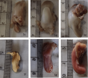
Figure 1: Photographs show external feature of studied animals. (a-b):a- External feature of adult of rabbit O. cuniculus. b- External feature of adult rat R. rattus; c- External feature of adult tongue of O. cuniculus. d- External feature of young tongue of O. cuniculus; e- External feature of lactating tongue of O. cuniculus; f- External feature of adult tongue of R. rattus; g- External feature of young tongue of R. rattus; h- External feature of lactating tongue of R. rattus.
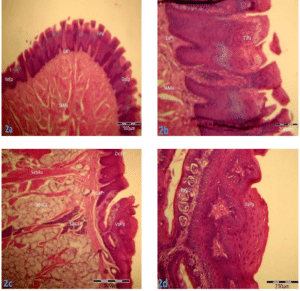
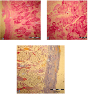
Figure 2: Photomicrographs of adult tongue sections of rabbit O. cuniculus to show different type of papillae. (a-g): a, b – Transverse sections in the anterior lateral part of O. cuniculus tongue showing filiform papillae (Fi Pa), lamina propria (La Pr) and muscles (Mu) Hand E; c- Transverse section in the posterior part of O. cuniculus tongue showing circumvallate papillae (Va Pa), lamina propria (La Pr), mucous gland(Mu Gl) and muscles (Mu) Hand E; d- Transverse section in the posterior part of O. cuniculus tongue showing circumvallate papillae (VaPa), and taste buds (TaBu) Hand E; e, f- Transverse sections in the posterior part of O. cuniculustongue showing numerous Mucous Glands (MuGl) give strong reaction with (PAS) stain; g- Transverse section in the posterior part of O. cuniculus tongue showing the Mucous Gland (MuGl) give weak reaction with azan stain.
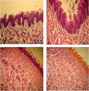
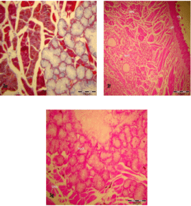
Figure 3: Photomicrographs of adult tongue sections of R. rattus to show different type of papillae. (a-j): a- Transverse section in the anterior part of R. rattus tongue showing filiform papillae (FiPa), muscles (Mu) and lamina propria (La Pr) Hand E; b- Transverse section in the anterior part of R. rattus tongue showing Elongated Horny Filiform Papillae (EHFiPa), Muscles (Mu) and Lamina Propria (LaPr) Hand E; c- Transverse section in the posterior part of R. rattus tongue showing Filiform Papillae (FiPa), Mucous Gland (MuGl), Serous Gland(SeGl), Muscles (Mu) and Lamina Propria (LaPr) Hand E; d, e- Transverse section in the posterior part of R. rattus tongue showing mucous glands (MuGl) give weak reaction while serous gland give moderate reaction with azan stain .fand g- Transverse sections in the posterior part of R. rattus tongue showing Mucous Glands (MuGl) and serous glands (SeGl) give very moderate reaction with Schiff reagent of (PAS) stain.
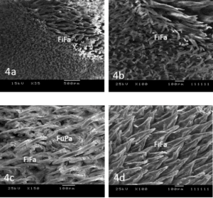
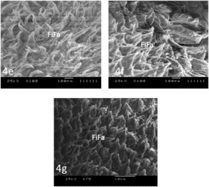
Figure 4: Scanning Electron Micrographs of adult tongue O. cuniculus to show different type of papillae. (a-g): a, b- Scanning electron micrographs of anterior part of O. cuniculus tongue showing Elongated Filiform Papillae (EFiPa) and short filiform papillae (SFiPa); c- Scanning electron micrograph of anterior part of O. cuniculus tongue showing fungiform papillae (FuPa) and filiform papillae (FiPa). d, e- Scanning electron micrograph of middle part of O. cuniculus tongue showing filiform papillae (FiPa); F, g- Scanning electron micrograph of posterior part of R. aegyptiacus tongue showing filiform papillae (FiPa).
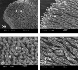
Figure 5: Scanning Electron Micrographs of adult tongue R.rattus to show different type of papillae. (a-d): a, b- Scanning electron micrograph of anterior part of R.rattus tongue showing serrate filiform papillae (FP); c, d- Scanning electron micrograph of middle part of R.rattus tongue showing fungiform (FuP), filiform papillae (FP) .
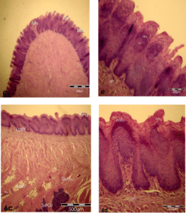
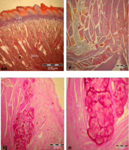
Figure 6: Photomicrographs of young tongue sections of rabbit O. cuniculus to show different type of papillae. (a-h): a – Transverse sections in the anterior lateral part of O. cuniculus tongue showing Filiform Papillae (FiPa), fungiform papillae (FuPa), Lamina Propria (La Pr) and Muscles (SkMu) Hand E; b- Transverse section in the anterior part of the tongue of O. cuniculus showing Filiform Papillae (FiPa), Hand E; c- Transverse section in the posterior part of O. cuniculus tongue showing filiform papillae (FiPa), Lamina Propria (LaPr), Mucous Gland (MuGl), Adipose Tissue (AdTi) and muscles (Mu) Hand E; d- Transverse section in the posterior part of O. cuniculus tongue showing filiform papillae (FiPa) and lamina propria (LaPr) Hand E; e- showing Filiform Papillae (FiPa), Lamina Propria (LaPr), Mucous Gland (MuGl), Adipose Tissue (AdTi) and muscles (Mu) with azan; f- Transverse section in the posterior part of O. cuniculus tongue showing Mucous Gland (MuGl) give negative reaction with azan; g, h- Transverse sections in the posterior part of O. cuniculus tongue showing Mucous Glands (MuGl) give moderate reaction with (PAS) stain.
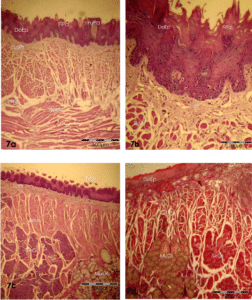
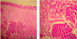
Figure 7: Photomicrographs of young tongue sections of R. rattus to show different type of papillae. (a-f): a- Transverse section in the anterior part of R. rattus tongue showing Filiform Papillae (FiPa), Fungiform Papillae (FuPa), Muscles (Mu) and Lamina Propria (LaPr) Hand E; b- Transverse section in tongue showing filiform propria (LaPr) Hand E. middle line of the anterior part of R. rattus papillae (FiPa), muscles (Mu) and lamina; c- Transverse section in the posterior part of R. rattus tongue showing horny Filiform Papillae (FiPa), Fungiform Papillae (FuPa), Mucous Glands (MuGl), Serous Gland (SeGl), Muscles (Mu) and lamina propria (La Pr) Hand E; d- Transverse section in the posterior part of R. rattus tongue showing Mucous Glands (MuGl) give weak reaction while serous gland (SeGl) give moderate reaction with azan stain; e, f- Transverse sections in the posterior part of R. rattus tongue showing Mucous Glands (MuGl) and Serous Glands (SeGl) give moderate reaction with Schiff reagent of (PAS) stain.
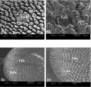
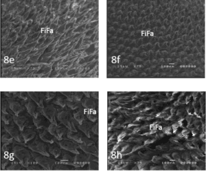
Figure 8: Scanning Electron Micrographs of adult tongue O. cuniculus to show different type of papillae. (a-h): a, b, c, d- Scanning electron micrographs of anterior part of O. cuniculus tongue showing Filiform Papillae (FiPa) and Fungiform Papillae (FuPa; e, f- Scanning electron micrograph of middle part of O. cuniculus tongue showing Filiform Papillae (FiPa); g, h – Scanning electron micrograph of posterior part of R. aegyptiacus tongue showing Filiform Papillae (FiPa).
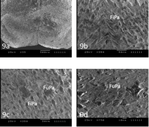
Figure 9: Scanning Electron Micrographs of young tongue R. rattus to show different type of papillae. (a-d): A- Scanning electron micrograph of anterior part of R.rattus tongue showing Filiform Papillae (FiPa), Fungiform Papillae(FiPa); b- Scanning electron micrograph in middle line of anterior part of R.rattus tongue showing Filiform Papillae (FiPa); c- Scanning electron micrograph of middle part of R. rattus tongue showing Filiform Papillae (FiPa) ), Fungiform Papillae(FuPa); d- Scanning electron micrograph of posterior part of R. rattus tongue showing Fungiform (FuPa).
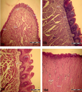
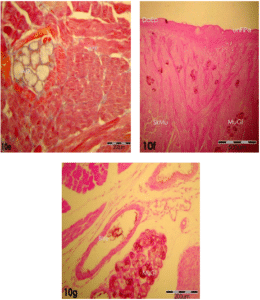
Figure 10: Photomicrographs of lactating tongue sections of rabbit O. cuniculus to show different type of papillae. (a-g): a – Transverse sections in the anterior lateral part of O. cuniculustongue showing Filiform Papillae (FiPa), Lamina Propria (LaPr) and Muscles (Mu) Hand E; b, c- Transverse section in the anterior part of the tongue of O. cuniculus showing filiform papillae (FiPa), Undifferantiated Filiform Papillae (unFiPa),Hand E; d- Transverse section in the posterior part of O. cuniculus tongue showing Undifferantiated Filiform Papillae (unFiPa), Lamina Propria (LaPa), Mucous Gland(MuGl) and muscles (Mu) Hand E; e- Transverse section in the posterior part of O. cuniculus tongue showing Mucous Gland (MuGl) give negative reaction with azan stain; f, g- Transverse sections in the posterior part of O. cuniculus tongue showing Mucous Glands (MuGl) give weak reaction with (PAS) stain.
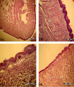
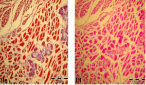
Figure 11: Photomicrographs of lactating tongue sections of R. rattus to show different type of papillae. (a-f): a- Transverse section in the anterior lateral part of R .rattustongue showing Filiform Papillae (FiPa), Fungiform Papillae (FuPa), Muscles (Mu) and Lamina Propria (LaPr) Hand E; b- Transverse section in the anterior part of R. rattus tongue showing Filiform Papillae (FiP), Muscles (Mu) and Lamina Propria (LaPr) Hand E; c, d- Transverse section in the posterior part of R. rattus tongue showing Vallate Papillae (VP), Fungiform Papillae (FuPa), Muscles (Mu) and Lamina Propria (LaPr) Hand E; e- Transverse section in the posterior part of R. rattus tongue showing Mucous Glands (MuGl) give weak reaction with azan; f – Transverse section in the posterior part of R. rattus tongue showing Mucous Glands (MuGl) give very weak reaction with Schiff reagent of (PAS) stain.
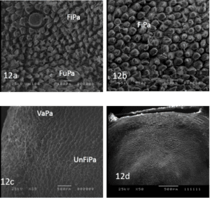
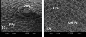
Figure 12: Scanning Electron Micrographs of lactating tongue O. cuniculus to show different type of papillae. (a-f): a- Scanning electron micrographs of anterior part of O .cuniculus tongue showing Filiform Papillae (FiPa) and fungiform papillae (FuPa); b- Scanning electron micrograph of middle part of O. cuniculus tongue showing Filiform Papillae (FiPa); c, d, e- Scanning electron micrograph of posterior part of R. aegyptiacus tongue showing Filiform Papillae (FiPa) and Vallatu Papillae(VaPa); f- Scanning electron micrograph of posterior part of R. aegyptiacus tongue showing filiform papillae (FiPa) and undifferentiated Filiform Papillae (un FiPa).
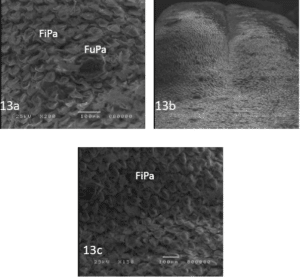
Figure 13: Scanning Electron Micrographs of lactating tongue R.rattus to show different type of papillae. (a-d): a- Scanning electron micrograph of anterior part of R.rattus tongue showing Filiform Papillae (FiPa), Fungiform Papillae(FuFa); b- Scanning electron micrograph in middle line of anterior part of R.rattus tongue showing Filiform Papillae (FiPa); c- Scanning electron micrograph of middle part of R.rattus tongue showing Filiform Papillae (FiPa) ), Fungiform Papillae(FuFa); d- Scanning electron micrograph of posterior part of R.rattus tongue showing Fungiform (FuPa).
Discussion
Adult Tongue
The muscular part of the tongue of Oryctolagus cunniculus and Rattus rattus is complicated as in R. aegyptiacus and T. nudiventris because difference of food habit [15]. Also, in adult Egyptian and Syrian Pipistrillus kuhli [4]. In the current study, the tongue is thick and elongated in Rabbit but moderate in rat with around apex. Iwasaski, et al., revealed that the shape of the tongue affects the feeding habit as it is responsible for the transformation of food to the mouth [16].
In the present study, anteriorly the epithelium is more keratinized than the posterior that make the tongue rigidity to become more efficient for feeding. Keratinization was also described in mammals [17,18]. The filiform papillae observed distributed mainly at the apex on the surface of the tongue of Oryctolagus Cuniculus where the filiform are long and collected together in large groups, also the fungiform papillae are scattered mainly on the dorsal and lateral surface rounded [3]. This observation different from Egyptian bats Roussetus egyptiacus and Taphozous nudiventris in which the fungiform papillae distributed laterally and also different from that found in Egyptian and Syrian P Kuhli [4]. Different from a finding that the tongue is elongated and free anterior part to facilitate the movement of the tongue while swiping the extracts of fruit pulp or licking nectar in fruit bats similar to the case with that of Kunz, et al., but different from the present herbivorous Rabbit and rodent rats [19]. The arrangement of the filiform papillae in the Poznan Poland Egyptian fruit eating bat are different from the studied animals in which there are two subtypes of filiform papillae [20]. The present of fungiform papillae between the filiform in Posteriorly of the tongue may focus to the taste sense n of the food accumulation in this part before swallowing. The presence and the distribution of the circumvallate papillae with their triangle shape observed in the blood drinking bat “Desmodus rotundus” recorded [15]. There were amount of mucus glands give strong reaction with PAS reaction of Rabbit than rat in the present study.
Lactating and Young Tongue
In the development and growth of the tongue at the histological and cytological level must be control by various factors, such as nerves, hormones and many kinds of growth factors. The outline of the tongue in the rat fetus appeared at day 12, only two rows of hemisphere dump shaped eminence which were identified as the rudiment of fungiform papillae were arranged at these stages on either side of the median sulcus on the dorsally of the tongue [21]. At the lactating stage of Rat there were a median sulcus at the posterioly without differentiation of papillae at the anterior part different from the case of R. aegyptiacus about three months, there were a median sulcus without differentiation of papillae at the anterior part of the tongue. But in the tongue of T. nudiventris there is no median sulcus at this lactating stage. Histochemically, at the posterior region of the lactating stage of Rabbit there are few numbers of mucus glands that give strong PAS reaction than those of rat as happen in R. aegyptiacus. In the young the mucus glands were very large in Rabbit than those of rat.
Many studies demonstrate that the dorsal epithelium at the middle or late period of gestation (E15) did not include any rudiments of filiform papillae and no signs of keratinization were observed. By contrast, rudiments of the filiform papillae were clearly recognized in the dorsal epithelium of the tongues of newborn mice just after birth [22]. Thus, morphogenesis of the filiform papillae of mice occurs rapidly during days 2 or 3 just before birth. Significant numbers of keratohyalin granules and keratinized cells are present in new-born mice at P0 as presented by scanning electron microscopy. Paulson, et al., and Iwasaki, et al., showed that filiform papillae at P0 are distributed in the same patterns as those observed in the mature adult, however, their shapes are clearly differed from those of the corresponding papillae in the adult [22,23]. In the fruit and nectar eating bats, the micro structure of the tongue has been investigated in the large flying fox (pteropus vampyrus), short-nosed fruit bat (cynopterus brachyotis), and other species belonging to phllostomidae [24,25]. The findings of these studies showed the presence of two types of mechanical papillae (filiform and conical papillae) for food manipulation and two types of gustatory papillae (fungiform and vallate papllae) for taste precepation. In the dentition fruit –eating bats, the characteristic traits of the tongue include pronounced canines for cutting the surface of fruit and flattened post canines for delete food particles [24].
In the insectivorous R hardwickei and A tridens, the tongue is complex and the epithelium anteriorly of the tongue is keratinized if compared with the posterior which make the tongue with greater rigidity to be help for feeding [26]. The surface of the tongue has four types of lingual papillae: two types of mechanical papillae – filiform and conical papillae and two types of gustatory papillae-fungiform and vallate papillae. Most numerous are the filiform papillae with well-developed keratinized processes. These papillae are represented by four morphological subtypes: small, giant, elongated, and bifid papillae. Iwasaki, et al., studied the morphogenesis of three types of lingual papilla in the mouse by scanning electron microscope after collecting of different stages of development from day 15 of gestation to day 7 after birth [23].
In the fetuses at E15, rudiments of fungiform papillae with a relatively regular, lattice-like pattern were visible on the anterior half of the dorsal surface of the tongue, the rudiments of each of the three different kinds of lingual papilla appeared at different respective stages of development. In the rats, the rudiments of the fungiform and circumvallate papillae, which are related to the sense of taste, were visible earlier than those of the filiform papillae, which are not involved in this property [23]. In the Egyptian and Syrian bats P Kuhli the dorsal surface of the former specimen showed numerous filiform papillae, moderate number of fungiform papillae and small number of circumvallate, while in the latter specimen no circumvallate papillae were observed. The fungiform papillae are scattered mainly on the lateral surface and are mostly rounded in shape and are found in both species [4]. In the Egyptian fruit-eating bat Poznan Poland, the surface of the tongue has four types of lingual papillae, two mechanical and two gustatory [20]. In other mammals, three vallate papillae were distributed in a v-shaped structure each papilla was surrounded by a deep sulcus and an external pad. The filiform papillae exhibited crown or finger like shape structure [27]. In the present work, the filiform papillae are distributed mainly at the apex on the dorsal of the tongue, while the fungiform papillae are mainly scattered on the lateral surface and are rounded in shape. These structures are similar to the case found in the adult Egyptian P. kuhli [4]. In other mammalians such as the rabbit O. Cuniculus the filiform papillae are long and gathered together in large groups. The elongated tongue in the Egyptian fruit bat has a flat surface and free anterior part which facilitate the movement of the tongue while swiping the extracts of fruit pulp or licking nectar [3]. The types and the arrangement of the filiform papillae in the Poznan Poland Egyptian fruit eating bat were different from both the fruit eating bat and the insectivorous bat in which there are only two subtypes of filiform papillae [20]. The occurrence of fungiform papillae among the filiform ones in the posterior part of the tongue may contribute to the taste perception of the food accumulation in this part before swallowing. The distribution of few numbers of circumvallate papillae in the form of triangle observed in the blood drinking bats D. rotundus [28-32].
Summary and Conclusion
Our studies comprised; histological and histochemical observations, ultra structural examination and bio analysis to find the effect of different feeding habit on the structure of tongue of adult, young and lactating of both O. cunniculus and R. rattus.
Conflict of Interest
The authors have no conflict of interest to declare.
Availability of Data and Materials
The data set analyzed during the current study are available from the corresponding author and the main author with facilities in our department.
Contributions
The authors developed the study design, The laboratory analyses of faculty of medicine provide the electron microscope for scanning sections of tongue. The statistical analyses results data was performed by faculty of medicine.
Ethics Approval and Consent to Participate
This research was done in compliance with the ethical standard set by the faculty of science -Tanta University, Egypt.
Acknowledgements
The authors sincerely thank to the Department of Zoology- Faculty of Science, Tanta University, Egypt.
References
- Stevens A, Lowe JS. Human histology (3rd Ed.). Elsevier Limited, USA. 2005;190-4.
- Qayyum MA, Fatani JA, Mohajir AM. Scanning electron microscopic study of the lingual papillae of the one humped camel, Camelus dromedarius. J Anatomy. 1988;160:21.
- Selim AM. Comparative anatomical and histological studies of the tongue between the prenatal, postnatal and adult of rousettus aegyptiacus and comparing the adult one with those of oryctolagus cunniculus and rattus rattus. J Egypt Ger Soc Zool. 2006;52A:130-45.
- Selim A, Nahla NE, Shelfeh M. Comparative anatomical and histological studies of the tongue between the Egyptian bat pipistrillus kuhli and the Syrian bat pipstrillus kuhli. Tishreen University J Res and Scientific Studies-Biological Sciences Series. 2008;30(1):247-55.
- Park JW, Lee JH. Comparative Morphology of the Tongue of Miniopterus schreibersi fuliginosus and Pipistrellus savii. Applied Microscopy. 2009;39(3):267-76.
- Ciuccio M, Estecondo S, Casanave EB. Scanning Electron Microscopy Study of the Dorsal Surface of the Tongue of Dasypus hybridus (Mammalia, Xenarthra, Dasypodidae). Int Morphol. 2010;28(2).
- Kulawik M, Godynicki S, Frąckowiak H. Light microscopic observations of vallate papillae in prenatal and postnatal periods of rabbit (Oryctolagus cuniculus domestica). Elect J Polish Agricultural Universities. 2013;16(3).
- Kulawik M, Godynicki S, Frąckowiak H. Light and scanning electron microscopic study of the filiform papillae of the tongue in adult rabbit (Oryctolagus cuniculus domestica, Linnaeus 1758). Nauka Przyroda Technologie. 2015;9(1):12.
- Taki-El-Deen FM, Sakr SM, Shahin MA. Comparative histological studies on the tongue of three species of Egyptian bats. Life Sci J. 2013;10(2):633-40.
- Yoshimura K, Shindoh J, Kobayashi K. Scanning electron microscopy study of the tongue and lingual papillae of the California sea lion (Zalophus californianus californianus). The Anatomical Record: An Official Publication of the American Association of Anatomists. 2002;267(2):146-53.
- Mohebinia S, Ghassemi F. Histological study of tongue in insectivore bat (Rhinopoma hardwickii). Adv Environmental Biology. 2013:4643-9.
- Kobayashi K, Kumakura M, Yoshimura K, Nonaka K, Murayama T, Henneberg M. Comparative morphological study of the lingual papillae and their connective tissue cores of the koala. Anat Embroyol. 2003;206:247-54.
- Kobayashi K, Jackowak H, Frackowiak H. Comparative morphological studies on the tongue of the selected primates using scanning electron microscope. Ann Anat. 2005;186:525-6.
- Romeis B. Mikroskopische Technik. neubearb. Aufl. München, Wien, Baltimore: Urban u. Schwarzenberg. 1989.
- Greenbaum IF, Phillips CJ. Comparative anatomy and general histology of tongues of long-nosed bats (Leptonycteris sanborni and L. nivalis) with reference to infestation of oral mites. J Mammal. 1974;55(3):489-504.
- Iwasaki SI, Yoshizawa H, Kawahara I. Study by scanning electron microscopy of the morphogenesis of three types of lingual papilla in the rat. The Anatomical Record: An Official Publication of the American Association of Anatomists. 1997;247(4):528-41.
- Ross MH, Kaye GI, Pawlina W. Histology, A text Book and Atlas, 4th Lippincott Williams and Wilkins, Baltimore, Philadelphia. 2003.
- Wu D, Irwin D, Zhang, V. Molecular evolution of the keratin associated protein gene family in mammals, role in evolution of mammalian hair. 2019;9:213-28.
- Kunz TH, Jones DP. Pteropus vampyrus. Mammalian Species. 2000;2000(642):1-6.
- Hanna JA, Joanna TL, Szymon GO. The microstructure of lingual papillae in the Egyptian fruit bat (Rousettus aegyptiacus) as observed by light microscopy and scanning electron microscopy. Arch Histol Cytol. 2009;72(1):13-21.
- Farbman AI, Mbiene JP. Early development and innervation of taste bud‐bearing papillae on the rat tongue. J Comparative Neurol. 1991;304(2):172-86.
- Paulson RB, Hayes TG, Sucheston ME. Scanning electron microscope study of tongue development in the CD-1 mouse fetus. J Craniofacial Genetics Develop Biol. 1985;5(1):59-73.
- Iwasaki SI, Asami T, Chiba A. Ultrastructural study of the keratinization of the dorsal epithelium of the tongue of Middendorff’s bean goose, Anser fabalis middendorffii (Anseres, Antidae). The Anatomical Record: An Official Publication of the American Association of Anatomists. 1997;247(2):149-63.
- Winter Y, von Helversen O. Operational tongue length in phyllostomid nectar-feeding bats. J Mammal. 2003;84(3):886-96.
- Kobayashi K. Comparative morphology of the lingual papillae and their Connective Tissue Cores (CTC) of flying foxes (Pteropus giganteus). Anat Sci Internat. 2004;79:230.
- Madkour GA, Hammouda EM, Ibrahim IG. Histology of the alimentary tract of two common Egyptian bats. Ann Zool. 1982;19:53-74.
- Burity CH, Silva MR, Souza AM, Lancetta CF, Medeiros MF, Pissinatti A. Scanning electron microscopic study of the tongue in golden-headed lion tamarins, Leontopithecus chrysomelas (Callithrichidae: Primates). Zoologia (Curitiba). 2009;26:323-7.
- Gretchen LH. Animal tissue technique. 1974.
- Snedecor GW, Cochran WG. Statistical methods. IOWA. Iowa State University Press. Starkstein, SE, Robinson, RG (1989). Affective disorders and cerebral vascular disease. The British J Psychiatry. 1980;154:170-82.
- Bancroft JD, Gamble M. Theory and practice of histological techniques. Elsevier Health Sciences. 2008.
- Kulawik M, Godynicki S. Vallate papillae in the domestic rabbit (Oryctolagus cuniculus domestica). Polish J Veterinary Sci. 2007;10(1):47-50.
- Kobayashi K, Jackowak H, Frackowiak H. Comparative morphological studies on the tongue of the selected primates using scanning electron microscope. Ann Anat. 2005;186:525-6.
This work is licensed under a Creative Commons Attribution 2.0 International License.


