Comparative Studies of the Tongue of Postnatal (Lactating), Young and Adult Between Two Species of Mammalian Animals of Different Feeding Habit Rattus Rattus and Orcytolagus Cuniculus
Atteyat Selim1*, Ezzar Hafez2, Wafaa Goda3
1Department of Zoology, Faculty of Science, Tanta University, Egypt 2Department of Zoology, Faculty of Science, Tanta University, Egypt 3Department of Zoology, Faculty of Science, Tanta University, Egypt
*Correspondence author: Atteyat Selim, Department of Zoology, Faculty of Science, Tanta University, Egypt; Email: [email protected]
Background: The study of tongue of mammals is important because the different species of animals with different feeding, so the present study was attended to observe the relation between various feeding and the morphological shape of tongue of present stages of both R. rattus and O. cunniculus using histological, histochemical techniques, ultra structural examination and Statistical analysis.
Results: The Egyptian herbivorous rabbit O. cunniculus and the rodent R. rattus showed that the tongue is elongated with rounded apex. In both species the thicknesses of the tongue increase gradually toward the pharynx. The tongue length of adult rabbit was large than the adult rat.
In the studied animals, the anterior end of the tongue of the herbivorous rabbit O. cunniculus is covered with short horny filiform papillae at the anterior part and giant circumvallate papilla at the posterior part with taste buds, also large enormous numbers of mucus and serous glands but in the rodent Rattus is covered with short pointed horny filiform papillae and others small elongated ones. Few numbers of fungiform papillae and numbers of mucus and serous glands are scattered among the skeletal muscles. The Tongue of adult R. rattus and O. cunniculus have different shapes of filiform papillae, fungiform papillae distributed between the filiform ones, two circumvallate on the surface, number of serrate glands and mucous gland. Give strong reaction with Periodic Acid Schiff (PAS). In young and lactating some undifferentiated and poor developed papillae were observed and serrate and mucous glands less than that in adult stage
Conclusion: The different feeding habit of both rat (rodents and Rabbit (herbivorous) give different shapes of papilla and give different reaction with histochemical stains.
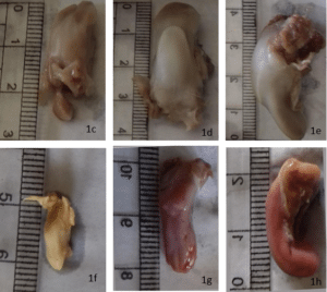
Figure 1: Photographs show external feature of studied animals. (a-b):a- External feature of adult of rabbit O. cuniculus. b- External feature of adult rat R. rattus; c- External feature of adult tongue of O. cuniculus. d- External feature of young tongue of O. cuniculus; e- External feature of lactating tongue of O. cuniculus; f- External feature of adult tongue of R. rattus; g- External feature of young tongue of R. rattus; h- External feature of lactating tongue of R. rattus.
Figure 2: Photomicrographs of adult tongue sections of rabbit O. cuniculus to show different type of papillae. (a-g): a, b – Transverse sections in the anterior lateral part of O. cuniculus tongue showing filiform papillae (Fi Pa), lamina propria (La Pr) and muscles (Mu) Hand E; c- Transverse section in the posterior part of O. cuniculus tongue showing circumvallate papillae (Va Pa), lamina propria (La Pr), mucous gland(Mu Gl) and muscles (Mu) Hand E; d- Transverse section in the posterior part of O. cuniculus tongue showing circumvallate papillae (VaPa), and taste buds (TaBu) Hand E; e, f- Transverse sections in the posterior part of O. cuniculustongue showing numerous Mucous Glands (MuGl) give strong reaction with (PAS) stain; g- Transverse section in the posterior part of O. cuniculus tongue showing the Mucous Gland (MuGl) give weak reaction with azan stain.
Figure 3: Photomicrographs of adult tongue sections of R. rattus to show different type of papillae. (a-j): a- Transverse section in the anterior part of R. rattus tongue showing filiform papillae (FiPa), muscles (Mu) and lamina propria (La Pr) Hand E; b- Transverse section in the anterior part of R. rattus tongue showing Elongated Horny Filiform Papillae (EHFiPa), Muscles (Mu) and Lamina Propria (LaPr) Hand E; c- Transverse section in the posterior part of R. rattus tongue showing Filiform Papillae (FiPa), Mucous Gland (MuGl), Serous Gland(SeGl), Muscles (Mu) and Lamina Propria (LaPr) Hand E; d, e- Transverse section in the posterior part of R. rattus tongue showing mucous glands (MuGl) give weak reaction while serous gland give moderate reaction with azan stain .fand g- Transverse sections in the posterior part of R. rattus tongue showing Mucous Glands (MuGl) and serous glands (SeGl) give very moderate reaction with Schiff reagent of (PAS) stain.
Figure 4: Scanning Electron Micrographs of adult tongue O. cuniculus to show different type of papillae. (a-g): a, b- Scanning electron micrographs of anterior part of O. cuniculus tongue showing Elongated Filiform Papillae (EFiPa) and short filiform papillae (SFiPa); c- Scanning electron micrograph of anterior part of O. cuniculus tongue showing fungiform papillae (FuPa) and filiform papillae (FiPa). d, e- Scanning electron micrograph of middle part of O. cuniculus tongue showing filiform papillae (FiPa); F, g- Scanning electron micrograph of posterior part of R. aegyptiacus tongue showing filiform papillae (FiPa).
Figure 5: Scanning Electron Micrographs of adult tongue R.rattus to show different type of papillae. (a-d): a, b- Scanning electron micrograph of anterior part of R.rattus tongue showing serrate filiform papillae (FP); c, d- Scanning electron micrograph of middle part of R.rattus tongue showing fungiform (FuP), filiform papillae (FP) .
Figure 6: Photomicrographs of young tongue sections of rabbit O. cuniculus to show different type of papillae. (a-h): a – Transverse sections in the anterior lateral part of O. cuniculus tongue showing Filiform Papillae (FiPa), fungiform papillae (FuPa), Lamina Propria (La Pr) and Muscles (SkMu) Hand E; b- Transverse section in the anterior part of the tongue of O. cuniculus showing Filiform Papillae (FiPa), Hand E; c- Transverse section in the posterior part of O. cuniculus tongue showing filiform papillae (FiPa), Lamina Propria (LaPr), Mucous Gland (MuGl), Adipose Tissue (AdTi) and muscles (Mu) Hand E; d- Transverse section in the posterior part of O. cuniculus tongue showing filiform papillae (FiPa) and lamina propria (LaPr) Hand E; e- showing Filiform Papillae (FiPa), Lamina Propria (LaPr), Mucous Gland (MuGl), Adipose Tissue (AdTi) and muscles (Mu) with azan; f- Transverse section in the posterior part of O. cuniculus tongue showing Mucous Gland (MuGl) give negative reaction with azan; g, h- Transverse sections in the posterior part of O. cuniculus tongue showing Mucous Glands (MuGl) give moderate reaction with (PAS) stain.
Figure 7: Photomicrographs of young tongue sections of R. rattus to show different type of papillae. (a-f): a- Transverse section in the anterior part of R. rattus tongue showing Filiform Papillae (FiPa), Fungiform Papillae (FuPa), Muscles (Mu) and Lamina Propria (LaPr) Hand E; b- Transverse section in tongue showing filiform propria (LaPr) Hand E. middle line of the anterior part of R. rattus papillae (FiPa), muscles (Mu) and lamina; c- Transverse section in the posterior part of R. rattus tongue showing horny Filiform Papillae (FiPa), Fungiform Papillae (FuPa), Mucous Glands (MuGl), Serous Gland (SeGl), Muscles (Mu) and lamina propria (La Pr) Hand E; d- Transverse section in the posterior part of R. rattus tongue showing Mucous Glands (MuGl) give weak reaction while serous gland (SeGl) give moderate reaction with azan stain; e, f- Transverse sections in the posterior part of R. rattus tongue showing Mucous Glands (MuGl) and Serous Glands (SeGl) give moderate reaction with Schiff reagent of (PAS) stain.
Figure 8: Scanning Electron Micrographs of adult tongue O. cuniculus to show different type of papillae. (a-h): a, b, c, d- Scanning electron micrographs of anterior part of O. cuniculus tongue showing Filiform Papillae (FiPa) and Fungiform Papillae (FuPa; e, f- Scanning electron micrograph of middle part of O. cuniculus tongue showing Filiform Papillae (FiPa); g, h – Scanning electron micrograph of posterior part of R. aegyptiacus tongue showing Filiform Papillae (FiPa).
Figure 9: Scanning Electron Micrographs of young tongue R. rattus to show different type of papillae. (a-d): A- Scanning electron micrograph of anterior part of R.rattus tongue showing Filiform Papillae (FiPa), Fungiform Papillae(FiPa); b- Scanning electron micrograph in middle line of anterior part of R.rattus tongue showing Filiform Papillae (FiPa); c- Scanning electron micrograph of middle part of R. rattus tongue showing Filiform Papillae (FiPa) ), Fungiform Papillae(FuPa); d- Scanning electron micrograph of posterior part of R. rattus tongue showing Fungiform (FuPa).
Figure 10: Photomicrographs of lactating tongue sections of rabbit O. cuniculus to show different type of papillae. (a-g): a – Transverse sections in the anterior lateral part of O. cuniculustongue showing Filiform Papillae (FiPa), Lamina Propria (LaPr) and Muscles (Mu) Hand E; b, c- Transverse section in the anterior part of the tongue of O. cuniculus showing filiform papillae (FiPa), Undifferantiated Filiform Papillae (unFiPa),Hand E; d- Transverse section in the posterior part of O. cuniculus tongue showing Undifferantiated Filiform Papillae (unFiPa), Lamina Propria (LaPa), Mucous Gland(MuGl) and muscles (Mu) Hand E; e- Transverse section in the posterior part of O. cuniculus tongue showing Mucous Gland (MuGl) give negative reaction with azan stain; f, g- Transverse sections in the posterior part of O. cuniculus tongue showing Mucous Glands (MuGl) give weak reaction with (PAS) stain.
Figure 11: Photomicrographs of lactating tongue sections of R. rattus to show different type of papillae. (a-f): a- Transverse section in the anterior lateral part of R .rattustongue showing Filiform Papillae (FiPa), Fungiform Papillae (FuPa), Muscles (Mu) and Lamina Propria (LaPr) Hand E; b- Transverse section in the anterior part of R. rattus tongue showing Filiform Papillae (FiP), Muscles (Mu) and Lamina Propria (LaPr) Hand E; c, d- Transverse section in the posterior part of R. rattus tongue showing Vallate Papillae (VP), Fungiform Papillae (FuPa), Muscles (Mu) and Lamina Propria (LaPr) Hand E; e- Transverse section in the posterior part of R. rattus tongue showing Mucous Glands (MuGl) give weak reaction with azan; f – Transverse section in the posterior part of R. rattus tongue showing Mucous Glands (MuGl) give very weak reaction with Schiff reagent of (PAS) stain.
Figure 12: Scanning Electron Micrographs of lactating tongue O. cuniculus to show different type of papillae. (a-f): a- Scanning electron micrographs of anterior part of O .cuniculus tongue showing Filiform Papillae (FiPa) and fungiform papillae (FuPa); b- Scanning electron micrograph of middle part of O. cuniculus tongue showing Filiform Papillae (FiPa); c, d, e- Scanning electron micrograph of posterior part of R. aegyptiacus tongue showing Filiform Papillae (FiPa) and Vallatu Papillae(VaPa); f- Scanning electron micrograph of posterior part of R. aegyptiacus tongue showing filiform papillae (FiPa) and undifferentiated Filiform Papillae (un FiPa).
Figure 13: Scanning Electron Micrographs of lactating tongue R.rattus to show different type of papillae. (a-d): a- Scanning electron micrograph of anterior part of R.rattus tongue showing Filiform Papillae (FiPa), Fungiform Papillae(FuFa); b- Scanning electron micrograph in middle line of anterior part of R.rattus tongue showing Filiform Papillae (FiPa); c- Scanning electron micrograph of middle part of R.rattus tongue showing Filiform Papillae (FiPa) ), Fungiform Papillae(FuFa); d- Scanning electron micrograph of posterior part of R.rattus tongue showing Fungiform (FuPa).
How to Cite this paper?
Citation: Selim A, et al. Comparative Studies of the Tongue of Postnatal (Lactating), Young and Adult Between Two Species of Mammalian Animals of Different Feeding Habit Rattus Rattus and Orcytolagus Cuniculus. Jour Clin Med Res. 2023;4(1):1-20.