Hassane Amadou Bouba Traore1*, Moctar Issiaka2, Nouhou Hama Agali3, Abba Kaka Yakoura4, Abdou Amza5
1Faculty of Health Sciences, Dan Dicko Dan Koulodo University of Maradi, Department of Ophthalmology, Maradi Regional Hospital, Niger
2Makkah Maradi Ophthalmological Hospital, Niger
3Faculty of Health Sciences, Dan Dicko Dan Koulodo University of Maradi, Histo-embryology and cytogenetics department, Maradi Referral Hospital, Niger
4Faculty of Health Sciences, Abdou Moumouni University of Niamey, Department of Ophthalmology, Niamey National Hospital, Niger
5Faculty of Health Sciences, Université Abdou Moumouni de Niamey, Department of Ophthalmology, Amirou Boubacar Diallo Hospital of Niamey, Niger
*Correspondence author: Hassane Amadou Bouba Traore, Faculty of Health Sciences, Dan Dicko Dan Koulodo University of Maradi, Department of Ophthalmology, Maradi Regional Hospital, Niger; Email: [email protected]
Published Date: 14-10-2023
Copyright© 2023 by Traore HAB, et al. All rights reserved. This is an open access article distributed under the terms of the Creative Commons Attribution License, which permits unrestricted use, distribution, and reproduction in any medium, provided the original author and source are credited.
Abstract
Congenital eversion of the eyelids is a rare entity, usually presenting at birth and most frequently involving the upper eyelid. Most often, congenital eversion presents bilaterally, although unilateral cases also exist. We report four (4) cases in infants treated at our centers. We noted: 3 cases with bilateral total eversion of both upper eyelids and one case of unilateral total eversion of the left upper eyelid. We observed two cases in newborns with a clinical profile of collodion baby, which is a very rare and typical entity and two other cases in newborns with no other associated organic anomaly.
The management of the four cases in our series consisted of subtarsal injections of dexamethasone, use of antibiotic-corticoid eye drops and ointments and local care, which represents a revolution in the conservative method, resulting in a total and spectacular remission after one week of treatment, with normalization of palpebral statics and spontaneous palpebral movements. In addition to conservative treatment other than our own, other authors have proposed surgical treatment using a variety of invasive procedures.
Keywords: Congenital Eversion; Eyelid; Corticosteroids; Antibiotics; Niger
Introduction
Congenital eversion of the eyelid corresponds to the externalization of the palpebral conjunctiva associated with chemosis; it may be uni or bilateral. It is a benign condition, usually treated conservatively [1,2]. This rare condition was first described by Adams in 1896. Most of the cases reported concerned black newborns and collodion babies [2,3]. Several therapeutic approaches have been proposed, but the conservative approach seems to bring better results with fewer invasive explorations [4]. We report four (4) cases of newborn observations in this case series. We noted: 3 cases with bilateral total eversion of both upper eyelids and one case of unilateral total eversion of the left upper eyelid. We observed two cases in newborns with a clinical profile of collodion baby, which is a very rare and typical entity and two cases in newborns with no other associated organic anomalies. All four cases were adequately and perfectly managed by a conservative method according to a protocol we have set up with encouraging results to be shared.
Case Reports
Management Protocol
To manage the various cases of congenital eversion of the upper eyelid, we set up a personalized protocol consisting of:
- Careful examination of the eyeball using a blepharostat, followed by debridement with cotton swabs and eyewash (Fig. 1). A secretion sample and chemosis puncture can be taken at the same time for biological analysis
- A local injection under the tarsus of 0.3 cc of corticosteroid (Dexamethasone: 4 mg/ml) using a 25G needle. It’s best to inject with the blepharostat in place, to ensure that the globe is not perforated. The injection is made at the upper edge of the tarsus, tangentially and towards the free edge (Fig. 2)
- local antibiotic-corticotherapy in eye drops (dexamethasone phosphate 100 mg/100ml, neomycin sulfate 350,000 IU/100ml) 4 times a day, after eyewash with isotonic serum
- A local application of tetracycline ointment 1%, twice a day, followed by an occlusive dressing reinforced with adhesive tape without compression, renewed daily (Fig. 3,4)
- A single sub-tarsal injection of Corticosteroids was required in three cases and only one required a second injection on day 5
Clinical Observation 1
This is a 52-day-old infant from a well-monitored, full-term pregnancy, delivered vaginally. There was no evidence of consanguinity and she was the youngest of 3 siblings. She was referred to the pediatric emergency department for a 6-day history of crying, incessant crying and refusal to feed. An ophthalmological examination revealed congenital eversion of both upper eyelids, highly inflammatory with superinfection of yellowish secretions and impossible spontaneous opening (Fig. 5). Examination with a blepharostat revealed an intact eyeball with preserved motility and pupillary reactions were vivid (Fig. 2). A thorough general examination by the paediatrician revealed no malformations or other abnormalities. Local swabbing was sterile. Progress was very favourable on day 3, encouraging continuation of the protocol. After a week of treatment, palpebral statics were normalized, with spontaneous opening and closing of the globe (Fig. 6,7).
Clinical Observation 2
This was a newborn male, two days old, from a well-monitored pregnancy with spontaneous eutoctal delivery; labor was not prolonged. He was the 4th sibling; his other brothers are all alive and well, with no abnormalities reported to date. There was no evidence of consanguinity. Ophthalmological examination revealed total eversion of the upper left eyelid and significant chemosis with positive transillumination (Fig. 8). Blepharostat examination revealed an intact eyeball with preserved motility and pupillary reactions were vivid.
The presence of dermatological manifestations prompted a referral to a pediatric dermatologist. The somatic anthropometric examination revealed a newborn weighing 3200 g, a height of 52 cm and a cranial perimeter of 33 cm. Dermatological examination revealed congenital ichthyosis sicca, consisting of dry scaly lesions with intervals of healthy skin, periumbilical ulceration and fissures in the flexural folds of the limbs against a background of generalized xerosis (Fig. 9). In view of these signs, the diagnosis of collodion baby was made and the patient was put on symptomatic treatment with local dermatological care and a consultation with the cytogeneticist. A skin biopsy confirmed the orthokeratotic hyperkeratosis. Genetic analysis revealed no mutation. Genetic counseling was offered to the family. Ophthalmological management consisted of the same treatment regimen as our therapeutic protocol. At D2, good progress was noted under treatment, with the possibility of palpebral opening and closing (Fig. 10). The evolution was marked by a total disappearance of signs of infection and complete remission, with the left eyelid in a normal position; but as a matter of custom and tradition, the parents returned to the village with the baby, whom we unfortunately lost sight of.
Clinical Observation 3
This is also a newborn male, two days old, from a well-followed vaginal delivery with no complications. He is the 2nd of the siblings. We noted a notion of consanguinity. Ophthalmological examination revealed a bilateral congenital palpebral eversion, with inflammatory signs and early superinfection, with no other ocular abnormalities (Fig. 11). Given the appearance of the skin and scalp, a dermatological consultation was requested from the dermopediatrician. The clinical dermatological examination confirmed the diagnosis of collodion baby and he was put on symptomatic treatment with local dermatological care and consultation with the cytogeneticist. The somatic anthropometric examination revealed a newborn weighing 2600 g, with a height of 48 cm and a cranial perimeter of 32 cm. A skin biopsy confirmed orthokeratotic hyperkeratosis. This was the only case found in the family and genetic counseling was proposed. Management of the palpebral eversion was exactly the same as in our previously described cases, using the same protocol, with a spectacular result after a one-week return visit (Fig. 12).
Clinical Observation 4
Newborn male, 24 hours old, from a well-monitored pregnancy, three prenatal consultations. Delivery by vaginal delivery with no complications. He is the 6th of the siblings. No evidence of consanguinity. No family anomalies to report. On ophthalmological examination, he presented congenital eversion of both eyelids with chemosis and a few yellowish secretions (Fig. 13). Anthropometric somatic examination revealed a newborn weighing 3200 g, a height of 50 cm and a cranial perimeter of 33 cm. The newborn was in good general condition, with no other organic abnormalities. Secretions were swabbed and returned sterile.
The management of this case responds positively to the same therapeutic scheme we have initiated, which offers complete resolution of the cases observed (Fig. 14).
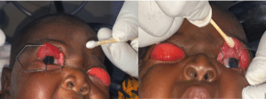
Figure 1: Blepharostat examination finds both globes intact with debridement.
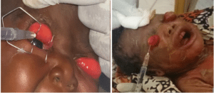
Figure 2: Subtarsal injection of dexamethasone.
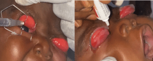
Figure 3: Applying ointment.
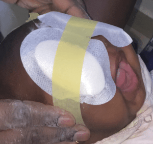
Figure 4: Closure with tape-reinforced eye washers on both eyes.
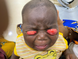
Figure 5: Infant 52 days old born with bilateral total eversion of the upper eyelids with chemosis and thick yellowish secretions.
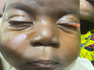
Figure 6: Appearance on day 3 of treatment: disappearance of chemosis with spontaneous opening and closing of the globe, but persistence of slight eversion.
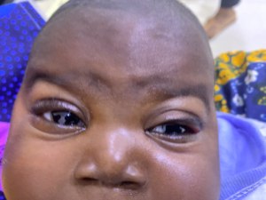
Figure 7: Appearance after one week of treatment: complete remission.
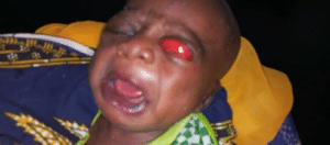
Figure 8: Total eversion of the upper left eyelid with chemosis.
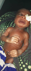
Figure 9: Collodion baby with large adherent scales and healthy skin interval showing total eversion of the left upper eyelid.
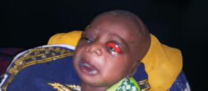
Figure 10: Spontaneous opening of the upper eyelid with good corneal transillumination on day 2 of treatment.
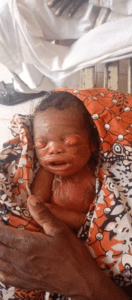
Figure 11: Newborn at 2 days of age on admission to our unit (note the appearance of the collodion baby).

Figure 12: Total resolution with spontaneous opening of both eyes after 8 days.
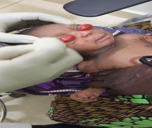
Figure 13: 24-hour-old newborn with bilateral congenital eversion of the eyelids.
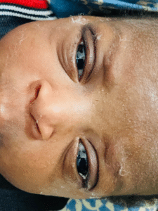
Figure 14: Complete resolution and spontaneous opening of both eyes at 8th day.
Discussion
Congenital eversion of the eyelids is a rare entity that usually presents at birth and most frequently involves the upper eyelid. It can be described as a pathology in which the eyelid is completely inverted and is usually associated with swelling, conjunctival prolapse and chemosis [4]. Although both upper eyelids are usually affected symmetrically, unilateral forms do occur [6]. The incidence is higher in black babies, infants with Down’s syndrome and collodion babies [1,2,6]. Our article presents four cases of congenital eversion of the upper eyelids, including 3 cases with bilateral total eversion of both upper eyelids and one case of unilateral total eversion of the left upper eyelid.
At present, no cases of congenital eversion of the eyelid in Niger have been reported in the literature and we believe we are the first to do so. In addition, many publications in the literature have reported cases of this condition, particularly in countries south of the Sahara; among other countries, we can cite cases in Guinea Conakry, Ghana, Cameroon and Nigeria [2,3,6,7]. Its worldwide incidence is unknown. A few cases have been reported in Europe, Asia and Africa.
Among the pathophysiological mechanisms and possible factors for the occurrence of congenital eversion of the upper eyelids we can cite: Abnormalities such as orbicular hypotonia, birth trauma, vertical shortening of the anterior lamella or vertical lengthening of the posterior lamella of the eyelid and lack of fusion of the orbital septum with the levator aponeurosis, absence of an effective lateral canthal ligament and lateral lengthening of the eyelid [6]. Multiparity, duration of labour and mode of delivery have not been shown to be determining factors in the development of this pathology [7]. The paediatric examination of a newborn with congenital eversion of the eyelids is essential in order to detect any associated anomaly as quickly as possible and to manage it rapidly, in order to prevent and avoid any risk that could be detrimental to the sight and life of the newborn. In our series, two newborns were collodion babies. The collodion baby is a severe form of congenital ichthyosis with neonatal onset. The clinical picture is often characteristic. When not fatal, the disease usually progresses to ichthyosis sicca [8]. Collodion baby syndrome must be differentiated from simple collodion hyperkeratoses of post-maturity, from Christ-Siemens-Touraine syndrome or X-linked hypohidrotic ectodermal dysplasia and from malignant keratoma (harlequin fetus): which is the most serious form of ichthyosis known, in which the fetus appears covered by a rigid, fissured “shell” and whose evolution is most often fatal in the first few days of life [9]. The positive diagnosis of collodion baby, as well as differential diagnoses, is primarily clinical. In case of doubt, a skin biopsy will confirm orthokeratotic hyperkeratosis.
The two collodion babies we followed were quickly managed by the pediatric dermatologist with appropriate treatment. Their skin biopsies confirmed orthokeratotic hyperkeratosis, even though the genetic test carried out on one baby found no chromosomal abnormality. This palpebral anomaly poses a high risk of amblyopia and corneal perforation if management is delayed [2]. Management includes surgical treatment with temporary tarsorrhaphy, fornix sutures and full-thickness skin grafting to the upper eyelid, as well as subconjunctival injection of hyaluronic acid [5-7,11]. However, several authors have reported success with the use of a non-invasive conservative method to achieve complete resolution of this condition [6,10,12-14]. Another type of conservative treatment is the gauze dressing soaked in 5% hypertonic saline solution, which has worked well and this is explained by the hypertonicity having enabled the movement of fluid from the edematous tissues through the semi-permeable conjunctival membrane by the process of osmosis, thus leading to resolution of the edema and subsequent reversion of the eyelid [5].
Our treatment protocol, accessible to all practitioners, especially pediatricians and general practitioners, has proved highly effective, avoiding the need for invasive surgery even in complicated cases. Sub-tarsal injections of corticosteroids act very rapidly against major or intense inflammation, thus combating chemosis and enabling the eyelid to quickly regain its statics and mobility. One or two injections are all that’s needed, followed by local administration. Indeed, in 3 of our patients, a single injection was sufficient, although the 4th patient required a second injection after 5 days without remission. And it is thought to have accelerated healing, which was finally obtained after 8 days: this was the case for the 4th patient reported in our series. Antibiotic therapy will prevent any major infectious complications of the globe. Based on our observations, 8 days of treatment is sufficient for complete remission. Our results are very encouraging, with rapid effects and complete resolution of all cases. It is worth mentioning that larger series need to be evaluated to see the efficacy of this treatment compared with current conservative treatment and it might be interesting to see in bilateral cases what would happen if only one eyelid were treated with corticosteroids, leaving the other eyelid as a control.
Conclusion
Congenital eversion of the eyelid may be a rare malformation, but it is curable by simple and effective means. Raising awareness among the general public and healthcare professionals of its benign nature and of the existence of a very simple and accessible therapy, is necessary in more ways than one. We are adding another revolutionary treatment to the therapeutic arsenal.
Consent of Patients
Written informed consent was obtained from the parents for publication of these case reports and accompanying images. A copy of the written consent is available for review by the Editor-in-Chief of this journal on request.
Conflict of Interest
The authors have no conflict of interest to declare.
References
- Blanc J, Virlouvet AL, Bui-Quoc E, Bourrat E. Neither tumor nor ectropion: congenital eyelid eversion, a diagnostic trap for the dermatopediatrician. InAnnales de Dermatologie et de Venereologie. 2018;145(12):S191-2.
- Farhadi R, Farrokhfar A. Unilateral congenital upper eyelid eversion in a newborn: Conservative management and outcome. Clin Case Rep. 2022;10(1):e05295.
- Krishnappa N, Poddar C, Deb A. Congenital total eversion of upper eyelids in a newborn with Down′s syndrome. Oman J Ophthalmol. 2014;7(2):98.
- Maheshwari R, Maheshwari S. Congenital eversion of upper eyelids: Case report and management. Indian J Ophthalmol. 2006;54(3):203.
- Adeoti CO, Ashaye AO, Isawumi MA, Raji RA. Non-surgical management of congenital eversion of the eyelids. J Ophthalmic Vis Res. 2010;5(3):188-92.
- Monebenimp F, Kagmeni G, Chelo D, Bilong Y, Moukouri E. Eversion congénitale bilatérale des paupières: prise en charge d’un cas selon l’approche conservatrice au Centre Hospitalier Universitaire de Yaoundé, Cameroun. Pan Afr Med J. 2012;11:34.
- Isawumi MA, Adeoti CO, Umar IO, Oluwatimilehin IO, Raji RA. Congenital bilateral eversion of the eyelids. J Pediatr Ophthalmol Strabismus. 2008;45(6):371‑3.
- Fatnassi R, Marouen N, Ragmoun H, Marzougui L, Hammami S. Le bébé collodion: aspects cliniques et intérêt du diagnostic anténatal. The Pan African Medical J. 2017;26.
- Happle R. Carte chromosomique et biologie moléculaire des génodermatoses. Dermatol Infect Sex Transm. 2004:478-85.
- Dohvoma VA, Nchifor A, Ngwanou AN, Attha E, Ngounou F, Bella AL, et al. Conservative management in congenital bilateral upper eyelid eversion. Case Reports in Ophthalmological Med. 2015;2015:1‑3.
- Ruban JM, Baggio E. Chirurgie des malpositions palpébrales congénitales de l’enfant. J Français d’Ophtalmologie. 2004;27(3):304‑26.
- Bentsi-Enchill KO. Congenital total eversion of the upper eyelids. British J Ophthalmol. 1981;65(3):209‑13.
- Othmane B. Monitoring and management of congenital entropion: about 6 cases. East African Scholars J Med Sci. 2022;5(12):317‑9.
- Raab EL, Saphir RL. Congenital eyelid eversion with orbicularis spasm. J Pediatric Ophthalmol Strabismus. 1985;22(4):125‑8.
Article Type
Case Report
Publication History
Received Date: 04-09-2023
Accepted Date: 08-10-2023
Published Date: 14-10-2023
Copyright© 2023 by Traore HAB, et al. All rights reserved. This is an open access article distributed under the terms of the Creative Commons Attribution License, which permits unrestricted use, distribution, and reproduction in any medium, provided the original author and source are credited.
Citation: Traore HAB, et al. Congenital Palpebral Eversion: A New Conservative Management Method (About Four Cases). J Ophthalmol Adv Res. 2023;4(3):1-10.

Figure 1: Blepharostat examination finds both globes intact with debridement.

Figure 2: Subtarsal injection of dexamethasone.

Figure 3: Applying ointment.

Figure 4: Closure with tape-reinforced eye washers on both eyes.

Figure 5: Infant 52 days old born with bilateral total eversion of the upper eyelids with chemosis and thick yellowish secretions.

Figure 6: Appearance on day 3 of treatment: disappearance of chemosis with spontaneous opening and closing of the globe, but persistence of slight eversion.

Figure 7: Appearance after one week of treatment: complete remission.

Figure 8: Total eversion of the upper left eyelid with chemosis.

Figure 9: Collodion baby with large adherent scales and healthy skin interval showing total eversion of the left upper eyelid.

Figure 10: Spontaneous opening of the upper eyelid with good corneal transillumination on day 2 of treatment.

Figure 11: Newborn at 2 days of age on admission to our unit (note the appearance of the collodion baby).

Figure 12: Total resolution with spontaneous opening of both eyes after 8 days.

Figure 13: 24-hour-old newborn with bilateral congenital eversion of the eyelids.

Figure 14: Complete resolution and spontaneous opening of both eyes at 8th day.


