Manu Saini1*, Arun K Jain1, Shubham Manchanda1, Kulbhushan Saini2, Pankaj Gupta1
1Advanced Eye Centre, Post Graduate Institute of Medical Education and Research, Chandigarh-160012, India
2Department of Anaesthesia and intensive care, Post Graduate Institute of Medical Education and Research, Chandigarh-160012, India
*Corresponding Author: Manu Saini, Assistant Professor, Advanced Eye Centre, Post Graduate Institute of Medical Education and Research, Chandigarh-160012, India; Email: [email protected]
Published Date: 15-07-2022
Copyright© 2022 by Saini M, et al. All rights reserved. This is an open access article distributed under the terms of the Creative Commons Attribution License, which permits unrestricted use, distribution and reproduction in any medium, provided the original author and source are credited.
Abstract
A 13-year-old female reported a one-year history of hemolacria in her left eye following an episode of a migraine attack and anger outburst. Ophthalmic examination revealed inferior fornix follicles with petechia and absent inflammatory reaction. The patient was treated with topical antibiotics, steroids followed by topical 1% cyclosporine and vitamin C. Bloody tear symptoms resolved and no bloody tear was observed for the three months insinuating that conjunctival folliculosis should be considered in the differential diagnosis of hemolacria and entity needs to be differentiated from follicular conjunctivitis. Through this case report we would like to emphasize the importance of the conjunctival folliculosis identification and possibility of shedding a bloody tear in presence of vascular dysfunction. Therefore, necessitate treatment of the benign entity similar to reactive lymphoid hyperplasia.
Keywords
Hemolacria; Folliculosis; Follicular Conjunctivitis; Cyclosporine
Introduction
Hemolacria has continued to be inscrutable and periodically reported in the literature with varying etiologies. However, broadly, hemolacria is attributed to either lacrimal system pathology or conjunctival lesions. Due to the non-specificity of signs and symptoms a thorough medical, ophthalmic history with meticulous clinical examination should be done, especially in recurrent hemolacria. Herein the authors describe the first reported case of conjunctival folliculosis with hemolacria during migraine and sympathetic overactivation that got resolved with topical 1% cyclosporine and vitamin C administration.
Case Report
A 13-year-old female presented a one-year history of unilateral blood-stained tear in the left eye. There was no history of trauma, bleeding diathesis, drug allergy, or association with the menstrual cycle. She complained of around five to six episodes of bloody tear in a day, repeated almost every third or fourth day, that lasted a few minutes. Therefore, she suffered around 8-10 episodes in a month. Her medical history included the onset of migraine headache and anger outbursts followed by episodes of blood-stained tear during the initial six-seven months. Subsequently bloody tear were observed without preceding events of the migraine attacks as well. A contrast-enhanced computed tomography scan head and orbit and EEG were normal. She had received systemic flunarizine (nonsteroidal anti-inflammatory drug) for a period of three months, prescribed outside, one year ago for the migraine. Family history was negative for bleeding diathesis or similar condition.
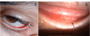
There was no visible mass dilated tortuous vessels or palpable lymphadenopathy or tenderness appreciated. Ophthalmic examination revealed bilateral Snellen visual acuities of 20/20 with normal pupillary reactions, normal intraocular pressure and full extraocular movements. Her lacrimal puncta seemed patent, with no tenderness and no discharge noted on digital pressure along with the lacrimal apparatus. A slit-lamp examination of the left eye revealed the follicles at the inferior fornix with petechia and absent inflammatory signs (Fig. 1). The rest of the ocular examination and fellow eye were unremarkable. Following ocular examination, an episode of bloody tear was witnessed by the health care assistant in the waiting area and a clinical photograph was taken (Fig. 1). A cotton-tipped applicator was rolled on, from medial to lateral direction, over the inferior palpebral conjunctiva and fornix, which confirmed the bleeding site, lateral to the puncta and from the inferior fornix conjunctival surface.
The patient underwent systemic investigations that included- complete blood count, bleeding time, thyroid profile, coagulation profile comprised prothrombin time and activated partial thromboplastin time. Laboratory tests revealed no abnormality. There was no microbial growth or micro-organism detected from the rolled over cotton tipped applicator conjunctival swab. A provisional diagnosis of conjunctival folliculosis manifesting as hemolacria was made. The patient was started on topical medications, tobramycin sulphate plus fluorometholone ophthalmic suspension in tapering dose for four weeks, topical azithromycin ointment in the twice-daily dose for two weeks and tear substitute four times a day. After three weeks of the treatment, the frequency of bloody tears decreased from five-six times to two times a day, nevertheless bloody tears did not subside completely. During a follow-up visit, inferior fornix follicles were regressed but still appreciated, therefore topical 1% cyclosporine two times a day and oral vitamin-C 500 mg twice daily was added. After four weeks of the treatment, the patient was symptom-free and no bloody tear was observed for the three months. Consent to publish the case and clinical photographs in a scientific journal was obtained from the patient’s father. This case report conforms to the principles of the Helsinki Declaration and has received approval from the Institute Ethics Committee- Postgraduate Institute of Medical Education and Research, Chandigarh, India with Ref no- NK/7030/Study/047.

Figure 1: a) Clinical photograph of bloody tear witnessed by the health assistant after ocular examination, b) Slit lamp microscopic examination showing follicles at the palpebral conjunctiva and inferior limbus (arrow) with petechial.
Author (s) | Number of cases | Age (years)/Sex | Migraine/ headache | Aetiology of hemolacria | Treatment | Outcome of treatment |
Ho VH, et al., [2] | 2/4 | Both patients 12/ Female | Present | Unknown Cause
| Psychiatric evaluation and steady compression | Symptom free |
Li E, et al., [10] | 1/1 | 22/Male | Present | Nasolacrimal lymphangioma | Confirmed with frozen specimens of nasolacrimal duct mucosa and bicanalicular stent placement | Symptom free- No episode of hemolacria |
Table 1: Studies with documented history of Migraine/headache in hemolacria presentation.
Discussion
Hemolacria has been related to the lacrimal gland, lacrimal sac tumours and conjunctival lesions including haemangiomas, fibromas, inflammatory granulomas, malignant melanomas and hereditary hemorrhagic telangiectasia [1]. Idiopathic haemolacria is an extremely rare diagnosis of exclusion entity [2]. Previously hemophilia and coagulopathies were considered as the most common aetiologies, however, with the accessibility of replacement therapy for these conditions, disorders of the conjunctiva appear to be the leading cause of hemolacria [3].
The authors report a case of hemolacria resulting from lower lid conjunctival folliculosis which represents the first case of bleeding folliculosis in the literature to the best of their knowledge. Bleeding in follicular conjunctivitis has been reported. Therefore, folliculosis needs to be differentiated from follicular conjunctivitis, as in the former condition no signs of active inflammation are seen. When no sign of inflammation is present, then the follicles show no change in colour from the uninvolved conjunctiva. Histologically, folliculosis is referred to as a sympathetic proliferation of lymphoid tissue of the conjunctiva, secondary to hypertrophy of this tissue elsewhere in the body and generally they do not have a propensity to bleed [4]. In the literature, only a few bleeding lymphoid hyperplasias are enumerated. Bloody epiphora from lymphoid hyperplasia of the nasolacrimal duct was reported by Wolf EJ, et al., [5]. Nevertheless, our patient had conjunctival folliculosis with petechiae that shed blood following an episode of migraine and anger outburst. In the literature, little is known about the association of sympathetic overactivation and bloody tears. Wang, et al., proposed that sympathetic activation played a role in hematidrosis (bloody sweat) and beta-adrenoceptor antagonists can serve as an effective agent to ameliorate symptoms [6]. Their observation bound us to ponder upon the role of sympathetic overactivation during anger outbursts in our case, as the contributory factor for hemolacria like hematidrosis.
During migraine attack constriction preceding and dilatation of both large and minute conjunctival vessels have been observed [7]. It is interesting to acknowledge that the history of migraine was also reported in the documented hemolacria cases (Table 1). Therefore, we speculate a causal link between folliculosis and bloody tear during migraine attack and sympathetic over activation, which is supported by slit-lamp bio microscopic examination of follicles in the only affected eye, no signs of the cicatricial formation, any discharge in a cul-de-sac and hemolacria resolution after cyclosporine and vitamin C therapy. The role of vitamin C in regulating vascular permeability was interestingly reported by Karslıoglu, et al., in recurrent bloody tears following a cefazolin-related erythema multiform-like reaction [8]. Conjunctival folliculosis in our case was treated using cyclosporine 1% twice daily like benign reactive lymphoid hyperplasia of the conjunctiva [9].
Usually, folliculosis do not have tendency to bleed and do not require treatment, hence, possess a diagnostic dilemma for contributing a bloody tear in children. Through this case report we would like to emphasize the importance of the conjunctival folliculosis identification and possibility of shedding a bloody tear in presence of vascular dysfunction. Therefore, necessitate treatment of the benign entity similar to reactive lymphoid hyperplasia. It is believed that the conjunctival vessels became fragile probably because of sympathetic overactivation and repeated migraine attack, therefore, when the lid was put on stretch the minute vessels ruptured, the most likely annotation for recurrent hemolacria.
Conflict of Interest
The authors declare that there is no conflict of interest.
References
- Roy FH. Ocular differential diagnosis. 4th Ed. Philadelphia: Lea and Febiger. 1989:120-1.
- Ho VH, Wilson MW, Linder JS. Bloody tears of unknown cause: case series and review of the literature. Ophthal Plast Reconstr Surg. 2004;20:442-7.
- Jordan DR, McCunn PD. Spurious sanguineous lacrimation. Can J Ophthalmol. 1984;19:315-6.
- Milton V Veldee. An epidemiological study of folliculosis of the conjunctiva. Public Health Reports. 1923;38:2877-87.
- Wolf EJ, Wessel MM, Hirsch MD. Reactive lymphoid hyperplasia of the nasolacrimal duct presenting as bloody epiphora. Ophthalmic Plast Reconstr Surg. 2007;23:242-3.
- Wang Z, Yu Z, Su J. A case of hematidrosis successfully treated with propranolol. Am J Clin Dermatol 2010;11:440-3.
- Ostfeld AM, Wolff HG. Studies on headache: participation of ocular structures in the migraine syndrome. Bibl Ophthalmol. 1957;12:634-47.
- Karslıoglu S, Simsek IB, Akbaba M. A case of recurrent bloody tears. Clin Ophthalmol. 2011;5:1067-9.
- Parikh RN, Sandhu HS, Massaro-Giordano. Benign reactive lymphoid hyperplasia of the conjunctiva treated with cyclosporine. Cornea. 2018;37:255-7.
Article Type
Case Report
Publication History
Received Date: 15-06-2022
Accepted Date: 08-07-2022
Published Date: 15-07-2022
Copyright© 2022 by Saini M, et al. All rights reserved. This is an open access article distributed under the terms of the Creative Commons Attribution License, which permits unrestricted use, distribution, and reproduction in any medium, provided the original author and source are credited.
Citation: Saini M, et al. Conjunctival Folliculosis Presenting as Hemolacria. J Ophthalmol Adv Res. 2022;3(2):1-5.

Figure 1: a) Clinical photograph of bloody tear witnessed by the health assistant after ocular examination, b) Slit lamp microscopic examination showing follicles at the palpebral conjunctiva and inferior limbus (arrow) with petechial.
Author (s) | Number of cases | Age (years)/Sex | Migraine/ headache | Aetiology of hemolacria | Treatment | Outcome of treatment |
Ho VH, et al., [2] | 2/4 | Both patients 12/ Female | Present | Unknown Cause
| Psychiatric evaluation and steady compression | Symptom free |
Li E, et al., [10] | 1/1 | 22/Male | Present | Nasolacrimal lymphangioma | Confirmed with frozen specimens of nasolacrimal duct mucosa and bicanalicular stent placement | Symptom free- No episode of hemolacria |
Table 1: Studies with documented history of Migraine/headache in hemolacria presentation.


