Shreya Thatte1*, Haritima Sharma2, Shruti Patidar3
1Professor and Head of Department of Ophthalmology, Sri Aurobindo Medical College and PG Institute, Indore, Madhya Pradesh, India
2Junior Resident (Third year) Department of Ophthalmology, Sri Aurobindo Medical College and PG Institute, Indore, Madhya Pradesh, India
3Junior Resident (Second year Department of Ophthalmology, Sri Aurobindo Medical College and PG Institute, Indore, Madhya Pradesh, India
*Correspondence author: Shreya Thatte, Professor and Head of Department of Ophthalmology, Sri Aurobindo Medical College and PG Institute, Indore, Madhya Pradesh, India; Email: [email protected]
Published Date: 25-03-2023
Copyright© 2023 by Thatte S, et al. All rights reserved. This is an open access article distributed under the terms of the Creative Commons Attribution License, which permits unrestricted use, distribution, and reproduction in any medium, provided the original author and source are credited.
Abstract
Background: Contrast sensitivity, is defined as the ability to detect the lowest lumination difference between an object and the background. It is one of the main requisites for good quality vision as the eye works by perceiving an object by comparing the difference between the target and the background contrast difference.
Aims and Objectives: To assess patterns of contrast sensitivity functions in patients in different types of refractive errors using Pelli Robson chart and also to measure the severity of refractive error within each group and compared the contrast sensitivity function with severity in different types of refractive errors.
Material and Method: This cross-sectional study was conducted on 500 patients (Age range from 10 to 80 years) who presented with chief complaint of refractive errors mainly myopia and hypermetropia. Patients who had previous history of ocular surgery and other ocular co- morbidity were excluded from study. Selected patients were enrolled for the study after taking a written informed consent. The patterns of contrast sensitivity function in different types of refractive errors were recorded on a prescribed proforma with respect to: Visual acuity, type of refractive error, severity of refractive error, duration of refractive error, age of the patient, fundus examination and Contrast sensitivity. Statistical analysis was undertaken with P<0.05 significant.
Results: A mild decrease in contrast sensitivity was recorded in majority of patients i.e 250 patients (50%). Maximum decrease in contrast sensitivity was seen in 31-60 years age group. A statistically significant direct correlation (P<0.05) was observed between duration and variation in contrast sensitivity. Both the refractive error showed definite decrease in contrast sensitivity and compound astigmatism showed severe decrease in contrast sensitivity.
Conclusion: Despite having BCVA of 6/6, patients showed reduced contrast sensitivity, even without any retinal pathology, making it an essential part of a routine ophthalmic examination.
Keywords: Contrast Sensitivity; Refractive Errors; Myopia; Hypermetropia; Pelli-Robson Chart
Introduction
Contrast sensitivity is the ability to perceive slight changes in luminance between regions which are not separated by definite borders and is just as important as the ability to perceive sharp outlines of relatively small objects [1].
Since refractive errors diminish contrast sensitivity, therefore sometimes a person with visual acuity of 6/6, may not be satisfied with the quality of their vision and still complain of decreased vision because of low contrast sensitivity [1,2].
Contrast Sensitivity can be measured by different methods, most commonly used is Pelli Robson chart that measures contrast sensitivity thresholds ranging from 100% to 0.56% and is a simple, easy-to-use, low-cost test with strong test-retest repeatability.
In advent of same, we undertook the present study to assess the effect of different types of refractive errors (Myopia and hypermetropia) on patterns of contrast sensitivity functions using Pelli Robson chart. Further, we also assessed correlation of the severity of refractive error within each group and the contrast sensitivity function.
Materials and Methods
This cross-sectional study was conducted on 500 patients (Age range from 10 to 80 years) who presented with chief complaint of refractive errors mainly myopia and hypermetropia to OPD of Department of Ophthalmology after approval by institutional ethical committee and Research Committee. Patients who qualified the inclusion criteria were enrolled for the study after taking a written informed consent.
Inclusion criteria:
- OPD patients presenting with refractive errors in all age groups
- Patients with BCVA of 6/6 with glasses
- Patients without any retinal pathology
Exclusion Criteria:
- Patients who did not gave consent for the study
- Patients with history of any ocular surgery/ or ocular pathology
- Uncompliant patients
Methodology
After taking written informed consent, the patient was evaluated and the patterns of contrast sensitivity function in different types of refractive errors were recorded on a prescribed proforma with respect to: Visual acuity, type of refractive error, severity of refractive error, duration of refractive error, age of the patient, fundus examination and Contrast sensitivity.
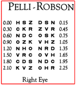
Visual acuity was assessed by the Snellen chart. The refractive error was recorded on the basis of refraction and Post Mydriatic Test (PMT) Contrast sensitivity was recorded by the Pelli-Robson chart (Fig. 1). The data included degree, duration of myopia and hypermetropia, and the degree of astigmatism and were collected in the form of excel sheet.

Figure 1: Pelli Robson chart.
The chart is wall mounted at one meter from the person to be examined and the letter size is 4.9 × 4.9 cm and consists of eight rows of letters. Contrast sensitivity grouping is done on basis of log-MAR values as shown in Table 1.
|
Log MAR Values |
Grade |
|
From 1.5 To Less than 2 |
Mild |
|
From 1 To Less than 1.5 |
Moderate |
|
Less than 1 |
Severe |
Table 1: Grouping of contrast sensitivity.
Contrast sensitivity is determined by the last triplet letter of which the patient should be able to read at least two. The degree of myopia is shown in Table 2 and the degree of hypermetropia is shown in Table 3.
|
Myopia in Dioptres |
Degree of Myopia |
|
<3D |
Low Myopia |
|
>3D to <6D |
Moderate |
|
>=6D |
High |
Table 2: Grading of myopia.
|
Hypermetropia in Dioptres |
Degree of Hypermetropia |
|
<+2.25D |
Low Hypermetropia |
|
>2.25D to +5.00D |
Moderate |
|
>=+5.00D |
High |
Table 3: Grading of hypermetropia.
Statistical Analysis
The data was collected and entered in Microsoft Excel 2010 (Microsoft corp.) and analyzed using the SPSS version 20.0 operating on Windows 10. All the descriptive data were presented as mean, standard deviation, frequency and percentages represented as the pie charts and bar diagrams. Qualitative and Quantitative data pertaining to the research was collected and an appropriate test was applied. Chi square test was used for associated categorical parameters. If p-value is less than 0.5 then it would be considered statistically significant.
Results
A decline in Contrast sensitivity is most commonly caused by refractive errors in patients. In the present study we addressed factors such as age, degree and duration refractive error and the degree of astigmatism as for their role in causing decline in contrast sensitivity of myopic and hypermetropic patients.
Patients were studied in three age groups i.e., under 30,31-60 and more than 61 years. 160 patients (32%) were under 30 years while 231 (46.2%) were 31-60 years and remaining 109 (21.8%) were over 61 years.
|
Age Group |
Contrast Sensitivity |
Total |
Chi Square |
||
|
Mild |
Moderate |
Severe |
|||
|
<=30 Years |
71 |
35 |
54 |
160 |
Х² = 48.912 |
|
28.40% |
21.70% |
60.70% |
32.00% |
||
|
31-60 Years |
127 |
76 |
28 |
231 |
|
|
50.80% |
47.20% |
31.50% |
46.20% |
||
|
>=61 Years |
52 |
50 |
7 |
109 |
p-value = 0.000 |
|
20.80% |
31.10% |
7.90% |
21.80% |
||
|
Total |
250 |
161 |
89 |
500 |
Significant |
|
100.00% |
100.00% |
100.00% |
100.00% |
||
|
100.00% |
100.00% |
100.00% |
100.00% |
||
|
100.00% |
100.00% |
100.00% |
100.00% |
||
Table 4: Variation in contrast sensitivity in different age group.
A statistically significant correlation (P<0.05) was observed between age group and variation in contrast sensitivity. A decline in CS was seen in all age groups. Mild decrease in contrast sensitivity was recorded in majority of patients i.e.,250 patients (50%) i.e., 71 (28.4%), 127 (50.8%), 52 (20.8%) for <=30 years, 31-60 years and >=61 years respectively. Max mild decrease was seen in (31-60) years group and max severe decrease was seen in <=30 years age group.
|
Duration |
Mild |
Moderate |
Severe |
Total |
Chi Square |
|
Upto 6 Months |
105 |
87 |
18 |
210 |
Х² = 85.487 |
|
42.00% |
54.00% |
20.20% |
42.00% |
||
|
7-12 Months |
45 |
61 |
49 |
155 |
|
|
18.00% |
37.90% |
55.10% |
31.00% |
||
|
> =13 Months |
100 |
13 |
22 |
135 |
P-value = 0.000 |
|
40.00% |
8.10% |
24.70% |
27.00% |
||
|
250 |
161 |
89 |
500 |
Significant |
|
|
100.00% |
100.00% |
100.00% |
100.00% |
Table 5: Variation in contrast sensitivity according to duration.
According to duration of refractive error, patients were studied in 3 groups i.e., upto 6 months, 7-12 months and more than 1 year. A statistically significant correlation (P<0.05) was observed between duration of refractive error and variation in contrast sensitivity. Mild decrease was recorded in 105 patients (42%) in upto 6 months duration group followed by 45 patients (18%) in 7-12 months and 100 patients (40%) in >1 year group. Mild decrease in C.S is seen upto 6 months and severe decrease in CS seen in 7-12 months duration.
|
Refractive Errors |
Mild |
Moderate |
Severe |
Total |
Chi Square |
|
Compound Myopic Astigmatism |
48 |
42 |
82 |
172 |
Х² = 255.838 |
|
19.20% |
26.10% |
92.10% |
34.40% |
||
|
Simple Myopic Astigmatism |
56 |
68 |
0 |
124 |
|
|
22.40% |
42.20% |
0.00% |
24.80% |
||
|
Simple Myopia |
26 |
1 |
5 |
32 |
P-value = 0.000 |
|
10.40% |
0.60% |
5.60% |
6.40% |
||
|
Compound Hypermetropic Astigmatism |
17 |
34 |
0 |
51 |
|
|
6.80% |
21.10% |
0.00% |
10.20% |
||
|
Simple Hypermetropic Astigmatism |
77 |
10 |
2 |
89 |
Significant |
|
30.80% |
6.20% |
2.20% |
17.80% |
||
|
Simple Hypermetropia |
26 |
6 |
0 |
32 |
|
|
10.40% |
3.70% |
0.00% |
6.40% |
||
|
Total |
250 |
161 |
89 |
500 |
|
|
100.00% |
100.00% |
100.00% |
100.00% |
Table 6: Variation in contrast sensitivity in refractive error.
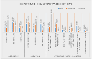
A statistically significant correlation (P<0.05) was observed between refractive errors and variation in contrast sensitivity. Out of 500 patient’s majority of patients i.e.,172 (34.4%) had Compound Myopic Astigmatism with 48 having mild, 42 having moderate and 82 having severe variation in CS. 124 (24.8%) had Simple Myopic Astigmatism, 89 (17.8%) had Simple Hypermetropic Astigmatism, 51 (10.2%) had Compound Hypermetropic Astigmatism and only 32(6.4%) each had Simple Myopia and Simple Hypermetropia. A mild decrease in CS was observed in all refractive errors with maximum 77 patients with Simple Hypermetropic Astigmatism followed by 56 patients with Simple Myopic Astigmatism and least 17 (6.8%) for Compound Hypermetropic Astigmatism. Severe decline in CS seen in Compound Myopic Astigmatism (Fig. 2).

Figure 2: Variation in contrast sensitivity.
Discussion
Contrast sensitivity is one of the most important requirements for healthy vision and can be influenced by many variables unlike visual acuity. Especially with newly developed multifocal intraocular lenses and other refractive procedures, the success of the procedure depends on the contrast sensitivity test results, even if the visual acuity is accurate [5-8]. Therefore, contrast sensitivity testing gained popularity in recent era. Refractive errors are a known cause of decline in contrast sensitivity in patients. To improve the quality of life of a patient with refractive error, sharpness vision plays an important role [9-13].
In the present study, factors such as age, degree and duration of myopia and hypermetropia, and the degree of astigmatism as for their role in causing decline in contrast sensitivity of myopic and hypermetropic patients was evaluated, compared and studied. Patients in age groups of 10-80 were included in our study which were similar to study done by Zhouyue Li, Yin Hu, et al., and Shreya T, et al., which had patients in the age group 20-70 years [2,10,15]. A statistically significant inversely proportionate correlation (P<0.05) was observed between CS and age. In our study a decline in contrast sensitivity was seen in all the age groups. Severe decline in CS was recorded maximum in <=30 years age group. With increasing age severity of C.S function decreased but no definite pattern was observed in our study which was similar to studies done Packer M, et al., Li J. et al. However, studies done Hinds A, et al., Shah P, et al., Bart VA, et al., indicated declines in both photopic and scotopic contrast sensitivity with aging that contrast sensitivity functions were directly proportional to increasing age and are more severely affected in the old age group of >69 years [11,13,16,21]. The possible reason being there is a much larger decrease in the number of rods compared to that of cones [17,18]. These changes explain the decreases in light sensitivity, contrast sensitivity, and visual acuity as well as prolonged dark adaptation that affect individuals over the age of 50 [16]. This contrast with other studies can be attributed to smaller sample size (92 patients) in these studies as compared to ours (500 patients) [22]. Majority of patients i.e.,78.2% belonged to age group <60 years in contrast to older patients in data collected by other studies.
We have observed that duration of myopia was a significant factor which was in concurrence with the results of the study conducted by Bistra Stoimenova, et al., which suggested that contrast sensitivity is negatively related to the degree and duration of myopia [23]. He studied 60 myopes and showed that 89% subjects with myopia of more than 10 years had severe decline in contrast functions [22].
On comparing contrast sensitivity between astigmatisms in myopic patients i.e., Compound myopic astigmatism, simple myopic astigmatism and Simple myopia, mild decrease observed maximally in Simple myopic astigmatism. Severe decline in contrast sensitivity was seen in compound myopic astigmatism, showing a direct relationship between astigmatism and contrast sensitivity. This was similar to the study conducted by Yumi Hasegawa, et al., on 12 emmetropic volunteers, which also suggested that astigmatism deteriorates contrast sensitivity depending on the amount of astigmatic power [24]. The only difference between the studies was that we did not have any comparison group of emmetropic patients.
In present study among hypermetropic patients, simple hypermetropia has the majority of patients with mild decrease in CS which are in concurrence with study done by Karatepe, et al., and severe decline seen in simple hypermetropic astigmatism [25].
On comparing different refractive error, maximum severe decline in CS was seen in compound myopic astigmatism.
Conclusion
It is crucial to create databases of contrast sensitivity values standardized according age, refraction. In our study no definite relationship seen between age and CS. Direct relationship seen between duration of refractive error and contrast sensitivity. Among the astigmatic patients compound myopic astigmatism showed severe decline in contrast sensitivity. Despite having fully corrected refractive error with a visual acuity of 6/6, myopic and hypemetropic patients showed reduced contrast sensitivity, even without any retinal pathology, making it an essential part of a routine ophthalmic examination.
Limitations
Demographic factors like sex, occupation needs to be addressed. CS in patients with refractive errors was not compared with emmetropic patients.
Conflict of Interest
The authors have no conflict of interest to declare.
References
- Daiber HF, Gnugnoli DM. Visual Acuity. In: StatPearls. StatPearls Publishing, Treasure Island(FL). 2021.
- Thatte S, Monga A, Sharma H. A clinical study to assess pattern of contrast sensitivity functions in patients with myopia. BOHR Int J Curr Res Optometry and Ophthalmol. 2022;1(1):67-73.
- Bilal A, Iqbal S, Mateen M, Azam A. Comparison of contrast sensitivity in Myopes and Hyperopes. 2020.
- Amesbury EC, Shallhorn SC. Contrast sensitivity and limits vision. Int Ophthalmol Clin. 2003;43:31-42.
- Tang W, Zhuang S, Liu G. Comparison of visual function after multifocal and accommodative IOL implantation. Eye Sci. 2014;29:95-9.
- Vilupuru S, Lin L, Pepose JS. Comparison of contrast sensitivity a through focus in small aperture inlay, accommodating intraocular lens, or multifocal intraocular lens subjects. Am J Ophthalmol. 2015;160:150-62.
- Çetin B, Arici MK, Özeç AV, Toker Mİ, Erdoğan H, Topalkara A. Zaraccom Ultraflex veya F260 Göz İçi Lens Takilan Hastalarda Kontrast Duyarliliğin Değerlendirilmesi ve Karşilaştirilmasi. Turkish J Ophthalmol. 2011;41(4):230-5.
- Taşkın O, Özbek Z. Miyop ve astigmatizma nedeniyle LASIK ve LASEK uygulanan hastalarda kontrast duyarlık sonuçlarının değerlendirilmesi. Turk J Ophthalmol. 2014;44:436-9.
- Atowa UC, Wajuihian SO, Hansraj R. A review of paediatric vision screening protocols and guidelines. Int J Ophthalmol. 2019;12(7):1194.
- Dudovitz RN, Izadpanah N, Chung PJ, Slusser W. Parent, teacher and student perspectives on how corrective lenses improve child wellbeing and school function. Maternal and Child Health J. 2016;20(5):974-83.
- Hinds A, Sinclair A, Park J, Suttie A, Paterson H, Macdonald M. Impact of an interdisciplinary low vision service on the quality of life of low vision patients. British J Ophthalmol. 2003;87(11):1391-6.
- Lundy C, Hill N, Wolsley C, Shannon M, McClelland J, Saunders K, et al. Multidisciplinary assessment of vision in children with neurological disability. The Ulster Medical J. 2011;80(1):21.
- Shah P, Schwartz SG, Gartner S, Scott IU, Flynn Jr HW. Low vision services: a practical guide for the clinician. Therapeutic Adv Ophthalmol. 2018;10:2515841418776264.
- Holden BA, Fricke TR, Wilson DA, Jong M, Naidoo KS, Sankaridurg P, et al. Global prevalence of myopia and high myopia and temoral trends from 2000 through 2050. Ophthalmol. 2016;123:1036-42.
- Li Z, Hu Y, Yu H, Li J, Yang X. Effect of age and refractive error on quick contrast sensitivity function in Chinese adults: a pilot study. Eye. 2021;35(3):966-72.
- Bart van Alphen B, Winkelman BH, Frens MA. Age-and sex-related differences in contrast sensitivity in C57BL/6 mice. Invest Ophthalmol Visual Sci. 2009;50(5):2451-8.
- Curcio CA. Photoreceptor topography in ageing and age-related maculopathy. Eye (Lond). 2001;15:376-83.
- Curcio CA, Medeiros NE, Millican CL. Photoreceptor loss in age-related macular degeneration. Invest Ophthalmol Vis Sci. 1996;37:1236-49.
- Packer M, Ginsburg AP. Contrast sensitivity and aging. Ophthalmol. 2007;114:1589-90.
- Li J, Zhao JL. Contrast visual acuity in adults with normal visual acuity. Zhonghua Yan Ke Za Zhi. 2012;48:403-8.
- Weale RA. Aging and vision. Vision Res. 1986;26:1507-12.
- Liou SW, Chiu CJ. Myopia and contrast sensitivity function. Curr Eye Res. 2001;22(2):81-4.
- Stoimenova BD. The effect of myopia on contrast thresholds. Invest Ophthalmol Vis Sci. 2007;48(5):2371-4.
- Hasegawa Y, Hiraoka T, Nakano S, Oshika T. Effects of astigmatic defocus on contrast sensitivity. Invest Ophthalmol Vis Sci. 2012;53(14):4805.
- Contrast sensitivity in healthy individuals. Turk J Ophthalmol. 2017;47:80-4.
Article Type
Research Article
Publication History
Received Date: 24-02-2023
Accepted Date: 19-03-2023
Published Date: 25-03-2023
Copyright© 2023 by Thatte S, et al. All rights reserved. This is an open access article distributed under the terms of the Creative Commons Attribution License, which permits unrestricted use, distribution, and reproduction in any medium, provided the original author and source are credited.
Citation: Thatte S, et al. Correlation Between Contrast Variability and Refractive Error. J Ophthalmol Adv Res. 2023;4(1):1-7.

Figure 1: Pelli Robson chart.

Figure 2: Variation in contrast sensitivity.
Log MAR Values | Grade |
From 1.5 To Less than 2 | Mild |
From 1 To Less than 1.5 | Moderate |
Less than 1 | Severe |
Table 1: Grouping of contrast sensitivity.
Myopia in Dioptres | Degree of Myopia |
<3D | Low Myopia |
>3D to <6D | Moderate |
>=6D | High |
Table 2: Grading of myopia.
Hypermetropia in Dioptres | Degree of Hypermetropia |
<+2.25D | Low Hypermetropia |
>2.25D to +5.00D | Moderate |
>=+5.00D | High |
Table 3: Grading of hypermetropia.
Age Group | Contrast Sensitivity | Total | Chi Square | ||
Mild | Moderate | Severe | |||
<=30 Years | 71 | 35 | 54 | 160 | Х² = 48.912 |
28.40% | 21.70% | 60.70% | 32.00% | ||
31-60 Years | 127 | 76 | 28 | 231 | |
50.80% | 47.20% | 31.50% | 46.20% | ||
>=61 Years | 52 | 50 | 7 | 109 | p-value = 0.000 |
20.80% | 31.10% | 7.90% | 21.80% | ||
Total | 250 | 161 | 89 | 500 | Significant |
100.00% | 100.00% | 100.00% | 100.00% | ||
100.00% | 100.00% | 100.00% | 100.00% | ||
100.00% | 100.00% | 100.00% | 100.00% | ||
Table 4: Variation in contrast sensitivity in different age group.
Duration | Mild | Moderate | Severe | Total | Chi Square |
Upto 6 Months | 105 | 87 | 18 | 210 | Х² = 85.487
|
42.00% | 54.00% | 20.20% | 42.00% | ||
7-12 Months | 45 | 61 | 49 | 155 | |
18.00% | 37.90% | 55.10% | 31.00% | ||
> =13 Months | 100 | 13 | 22 | 135 | P-value = 0.000 |
| 40.00% | 8.10% | 24.70% | 27.00% | |
250 | 161 | 89 | 500 | Significant | |
100.00% | 100.00% | 100.00% | 100.00% |
Table 5: Variation in contrast sensitivity according to duration.
Refractive Errors | Mild | Moderate | Severe | Total | Chi Square |
Compound Myopic Astigmatism
| 48 | 42 | 82 | 172 | Х² = 255.838
|
19.20% | 26.10% | 92.10% | 34.40% | ||
Simple Myopic Astigmatism
| 56 | 68 | 0 | 124 | |
22.40% | 42.20% | 0.00% | 24.80% | ||
Simple Myopia
| 26 | 1 | 5 | 32 | P-value = 0.000
|
10.40% | 0.60% | 5.60% | 6.40% | ||
Compound Hypermetropic Astigmatism
| 17 | 34 | 0 | 51 | |
6.80% | 21.10% | 0.00% | 10.20% | ||
Simple Hypermetropic Astigmatism
| 77 | 10 | 2 | 89 | Significant
|
30.80% | 6.20% | 2.20% | 17.80% | ||
Simple Hypermetropia
| 26 | 6 | 0 | 32 | |
10.40% | 3.70% | 0.00% | 6.40% | ||
Total
| 250 | 161 | 89 | 500 | |
100.00% | 100.00% | 100.00% | 100.00% |
Table 6: Variation in contrast sensitivity in refractive error.


