Hassane Amadou Bouba Traore1*, Moctar Issiaka2, Laminou Laouali3, Abba Kaka Yakoura4, Abdou Amza5
1Faculty of Health Sciences, Dan Dicko Dan Koulodo University of Maradi, Department of Ophthalmology, Maradi Regional Hospital, Niger
2Makkah Maradi Ophthalmological Hospital, Niger
3Faculty of Health Sciences, Université André Saliffou de Zinder, Department of Ophthalmology, Zinder National Hospital, Niger
4Faculty of Health Sciences, Abdou Moumouni University of Niamey, Department of Ophthalmology, Niamey National Hospital, Niger
5Faculty of Health Sciences, Université Abdou Moumouni de Niamey, Department of Ophthalmology, Amirou Boubacar Diallo Hospital of Niamey, Niger
*Correspondence author: Hassane Amadou Bouba Traore, Faculty of Health Sciences, Dan Dicko Dan Koulodo University of Maradi, Department of Ophthalmology, Maradi Regional Hospital, Niger; Email: [email protected]
Published Date: 30-08-2023
Copyright© 2023 by Traore HAB, et al. All rights reserved. This is an open access article distributed under the terms of the Creative Commons Attribution License, which permits unrestricted use, distribution, and reproduction in any medium, provided the original author and source are credited.
Abstract
Oculomotor paralysis is a rare complication of diabetes. It is a rare form of diabetic neuropathy, with a prevalence of 1 to 14% in diabetics. This is a 52-year-old patient with no known personal history, who presented himself in ophthalmological consultation for left ocular pain and fall of the upper left eyelid, occurred 6 days ago and rapidly progressive in an atraumatic context. Objective ophthalmological examination a moderate left ptosis, with incomplete ophthalmoplegia sparing abduction and twists, without vicious attitude. Visual acuity at a distance without correction was 6/10 in both eyes, P3. At bio microscopy, RPM kept bilaterally, without surface disturbance with normal anterior and posterior segments. This was an incomplete paralysis of the extrinsic component of the III. Emergency radiological checkup ruled out a central vascular and neurological cause. The etiological biological assessment finds fasting blood glucose high at 1.92 g/l. It is sent to the diabetologist for investigations. The degenerative assessment is normal but with a glycated hemoglobin at 10.5%, testifying to an unknown diabetic. It is retained the diagnosis of a paralysis of III incomplete inaugural type 2 diabetes. The evolution after 2 months was marked by the progressive regression of its paralysis after glycemic balance and correction of risk factors. Oculomotor paralysis is not uncommon during diabetes, it is necessary to think about it during etiological assessment for early and adapted management.
Keywords: Mononeuritis; Oculomotor Paralysis; Diabetes
Introduction
Mononeuritis with oculomotor nerve involvement is a rare form of diabetic neuropathy; with a prevalence of 1-14% in diabetics [1]. It is often a complication of a fairly advanced stage or an unbalanced diabetes but it can be indicative of diabetes in 2.2 to 3.6% [2]. It is a form of nerve degeneration, which is usually reversible without sequelae. All oculomotor nerves are affected but the most affected are the external oculomotor nerves VI and common III [3]. It is manifested by a table of oculomotor paralysis of interest to the affected nerve or nerves. The etiological paraclinical assessment allows to rule out another cause and to establish the origin of diabetes, thus allowing the proper management.
We present through this observation a case of extrinsic paralysis of nerve III, rare, revealing type 2 diabetes and discuss the epidemiological and clinical aspects, the diagnostic approach as well as the management and evolution.
Clinical Observation
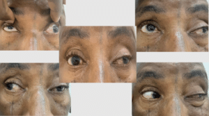
This is a 52-year-old patient, with no known personal history, who presented for ophthalmological consultation for moderate left eye pain and left upper eyelid fall, which occurred 6 days ago and rapidly progressing in an atraumatic context. The objective ophthalmological examination a left moderate ptosis, with incomplete ophthalmoplegia of the III sparing abduction and twists (moderate ptosis, paralysis of the adduction, elevation as well as a limitation of the lowering of the look), without vicious attitude of the head. There is no exophthalmos, no inflammatory signs, no vascular breath, no thrill and no vessels at the head of the jellyfish. There was no palpable mass (Fig. 1). There were no sensitivity abnormalities in the trigeminal territory. Visual acuity at a distance without correction was 6/10 in both eyes, P3. At biomicroscopy, the photomotor reflex is kept bilaterally, without surface disturbance with at the anterior segment a cataract subcapsular beginner and a posterior segment strictly normal. The complete neurological examination found no other involvement of the cranial pairs. This was an incomplete paralysis of the extrinsic component of the III.
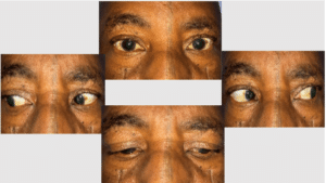
The emergency radiological check-up, namely the injected orbito-cerebral scanner and Angio-MRI, eliminated a central vascular and/or neurological cause. The inflammatory balance is negative but we find in the rest of the etiological biological balance a fasting glucose high at 2.92 g/l with a glycated hemoglobin at 10,5%. The diagnosis of incomplete oculomotor paralysis of type 2 diabetes was retained. It is addressed to the diabetologist for further investigation. The degenerative balance is normal apart from an overweight estimated by BMI at 24 kg/m2 and high cholesterol. No other neuropathy or microangiopathy was found. He is put on oral and statin antidiabetics with a low dose of corticosteroids at 0.75 mg/kg for 3 weeks. The evolution after 2 months was marked by the progressive regression of his paralysis after glycemic balance and correction of risk factors. He fully recovers his oculo-motor after 6 months (Fig. 2). There is no recurrence or complication after a one-year setback.

Figure 1: Oculomotricity examination shows extrinsic paralysis of the III of the left eye.

Figure 2: Complete regression of paralysis and suppression of left ptosis.
Discussion
Ophthalmopathy is a common micro-angiopathic complication in diabetic patients [4]. The pathophysiology of degenerative complications of diabetes mellitus involves vascular and genetic metabolic factors that interact with each other. Chronic hyperglycemia is responsible for metabolic and vascular abnormalities [2]. It is true that retinopathy is still the most obvious ocular disease in diabetes, but all ocular structures can be affected, including nerve structures whose mechanism is said to be ischemic [4-6]. The incidence of ophthalmoplegia in diabetics is estimated to range from 0.4% to 5.0% in series [7,8]. In his review, Said, et al., reports that it is most often diabetic polyneuritis and that mononeuropathy in general in diabetes is rare [7,9]. All nerves can be affected both the cranial pairs or other. Concerning the oculo-motor nerves, it is often a picture of mononeuritis with isolated involvement of VI, III, more rarely IV, sudden installation or over a few days, on a diabetic ground often light, rarely unknown as it is the case of our patient. A recent study shows that 26% of III paralysis manifests as complete extrinsic involvement, with an identical proportion regardless of the etiology [10]. This sufficiently demonstrates the rarity of this disease and this is the case for our patient. In adults, the main cause of paralysis of III corresponds to microvascular involvement (35 to 40%), classically described in the books as brutal and maximum right away, completely affecting the extrinsic musculature, sparing the pupil. The average age of patients with microvascular involvement is 68 years [10]. Pain makes oculomotor paralysis a diagnostic emergency, even if our patient has not reported orbital or periorbital pain or headache. The most feared cause is aneurysm involvement (6 to 16%), to be mentioned systematically in case of mydriasis, focal involvement or paresis. Progressive involvement may be related to compression (neoplasia 6% to 11%, aneurysm, etc.). Traumatic cause (12-19%) is evoked in the context of severe head trauma. In practice, only neurologically isolated and extrinsic full III with total pupil saving, in a patient over 50 years without risk factors other than cardiovascular history, can actually evoke first a microvascular involvement [11]. However, even in this situation, the systematic conduct of cerebral and orbital magnetic resonance imaging (MRI) with angio-MRI finds another etiology in 9% of cases [12]. In all other cases, urgent imaging is required to eliminate a pituitary aneurysm and apoplexy and to do a biological assessment to look for Horton’s disease. In case of diabetes, very often, during the etiological diagnosis, the assessment is very telling. Thus, all the etiological assessment in our patient reveals a type 2 diabetes unknown. The probability of spontaneous recovery after acquisition of the third cranial pair, depending on the etiological mechanism, is possible [11]. Management is etiological in the case of diabetes, including oral anti-diabetics and correction of risk factors [1,3,9]. Some authors advocate low-dose corticosteroids of 0.75 mg/kg/day or acetylsalicylic acid and benfotiamine with good clinical progression and recovery often without sequelae [4,12].
Conclusion
Oculomotor paralysis almost always occurs after 50 years. In case of diabetes, they can occur as a result of a chronic glycemic imbalance or decompensation but can be indicative of type 2 diabetes. The picture is very often that of a complete extrinsic disease. It is necessary to think about it during the etiological assessment of a paralysis of oculomotor nerves, especially after having eliminated an emergency, for an early and adapted management.
Consent of Patients
Written informed consent was obtained from the patients for publication of these case reports and accompanying images.
Conflict of Interest
The authors have no conflict of interest to declare.
References
- Touiti A, El Mghari G, El Ansari N. P2126 Ptosis révélant le diabète : À propos de 2 Observations. Diabetes Metab. 2013;39:A97‑8.
- Raccah D. Epidémiologie et physiopathologie des complications dégénératives du diabète sucré. EMC-Endocrinologie. 2004;1(1):29-42.
- Zbadi R, Doubi S, Rchachi M, El Ouahabi H, Ajdi F. Ptosis révélateur d’un diabète de type 2 méconnu : à propos de deux cas. Ann Endocrinol. 2015;76(4):551
- Laaribi N, Khlifi A, Ajhoun Y, Messaoudi R, Reda K, Oubaaz A. Extrinsic paralysis of the common ocular motor nerve, revealing type 2 diabetes. La Presse Médicale. 2017;46(6):630-3.
- Dreyfus PM, Hakim S, Adams RD. Diabetic ophthalmoplegia. Arch Neurol Psychiat. 1957;77:337-49.
- Annabi A, Lasjaunias P, Lapresle J. Paralysies de la IIIe paire au cours du diabète et vascularisation du moteur oculaire commun. J Neurol Sci. 1979;41(3):359-67.
- Said G. Diabetic neuropathy – a review. Nat Clin Pract Neurol. 2007;3:331-40.
- Watanabe K, Hagura R, Akanuma Y, Takasu T, Kajinuma H, Kuzuya N, et al. Characteristics of cranial nerve palsies in diabetic patients. Diabetes Res Clin Pract. 1990;10:19-27.
- Obbiba A, El Aziz S, El Chadli A, El Ghomari H, Farouqi A. La mononeuropathie phrénique secondaire au diabète: une complication rare. Med Mal Metab. 2013;7(3):247-9
- Fang C, Leavitt JA, Hodge DO, Holmes JM, Mohney BG, Chen JJ. Incidence and etiologies of acquired third nerve palsy using a population-based method. JAMA Ophthalmol. 2017;135(1):23-8.
- Chou KL, Galetta SL, Liu GT, Volpe NJ, Bennett JL, Asbury AK, et al. Acute ocular motor mononeuropathies: prospective study of the roles of neuroimaging and clinical assessment. J Neuro Sci. 2004;219(1-2):35-9.
- Sibilia J. Les corticoïdes: mécanismes d’action. La lettre du Rhumatologue. 2003(289):23-31.
Article Type
Review Article
Publication History
Received Date: 07-08-2023
Accepted Date: 23-08-2023
Published Date: 30-08-2023
Copyright© 2023 by Traore HAB, et al. All rights reserved. This is an open access article distributed under the terms of the Creative Commons Attribution License, which permits unrestricted use, distribution, and reproduction in any medium, provided the original author and source are credited.
Citation: Traore HAB, et al. Diabetic Mononeuritis: Isolated Extrinsic Paralysis of the III. J Ophthalmol Adv Res. 2023;4(2):1-4.

Figure 1: Oculomotricity examination shows extrinsic paralysis of the III of the left eye.

Figure 2: Complete regression of paralysis and suppression of left ptosis.


