Maurizio Rolando1, Stefano Barabino2*, Chiara Del Noce3, Francesca Bruzzone3, Cereda Matteo4, Silvio Lai3ii*
1Ocular Surface and Dry Eye Clinic, ISPRE Ophthalmic, Genoa, Italy
2Ocular Surface and Dry Eye Center, ASST Fatebenefratelli-Sacco, Sacco Hospital, University of Milan, Milan, Italy
3Clinica Oculistica, DiNOGMI, Università di Genova, Genova, Italy
4ASST Fatebenefratelli-Sacco, Sacco Hospital, University of Milan, Milan, Italy
*Correspondence author: Stefano Barabino, Ocular Surface and Dry Eye Center, ASST Fatebenefratelli-Sacco, Sacco Hospital, University of Milan, Milan, Italy; Email: [email protected]
Published Date: 25-03-2024
Copyright© 2024 by Barabino S, et al. All rights reserved. This is an open access article distributed under the terms of the Creative Commons Attribution License, which permits unrestricted use, distribution, and reproduction in any medium, provided the original author and source are credited.
Abstract
Objective: Dry eye is associated with reduced QoL and with the relevant social and economic costs. We evaluated the prevalence of dry eye signs and symptoms in a group of patients who underwent vitreoretinal surgery for epiretinal membrane removal for at least 6 months.
Method: Fourty-one consecutive patients were enrolled. Ocular surface symptoms were evaluated using a structured form and a Visual Analogue Scale (VAS). Blink completeness, Break-Up Time (BUT), fluorescein and lissamine green staining and thickness of the lower tear meniscus were also assessed. Lissamine green staining was used to evaluate the mucocutaneous junction.
Results: Symptoms were present up to 1 year from surgery in 80% of population. Foreign body and burning sensations were reported by 14 (34.1%) and 11 (26.8%) patients. Blinking was incomplete in 36.8% of patients; eyelid mucocutaneous junction was abnormal in 68.3% of patients. Mild or moderate eyelid injection were reported by 29 (70.7%) and 12 (21.3%) patients; moderate and peri-keratic hyperemia were reported by 22 (53.7%) and 15 (36.6%) patients. Only 26.2% of patients showed a normal BUT (>10 s). Corneal sensitivity was absent in 4 patients (9.8%) and strongly decreased in 2 patients (7.3%). The lower tear meniscus was <0.2 mm in 21 patients (51.2%). Fluorescein staining of the cornea was positive in 56% of patients.
Conclusion: Patients who underwent vitreoretinal surgery showed, in the long-term, signs and symptoms of ocular surface dysfunction (dry eye) with a frequency that is more than double the expected frequency of the disease.
Keywords: Vitreoretinal Surgery; Epiretinal Membrane; Dry Eye; Tear Film; Ocular Surface; Quality of Life; Quality of Vision
Introduction
The past 20 years have seen a tremendous increase in the number of vitreoretinal surgeries and the indications for vitrectomy are constantly increasing. Pars plana vitrectomy is appropriate whenever access to the posterior segment of the eye is necessary for treatment. Common indications include rhegmatogenous or tractional retinal detachment, vitreous hemorrhage, retained lens fragments after cataract surgery, endophthalmitis, epiretinal membrane, macular hole, vitreomacular traction and intraocular foreign bodies.
More recently, Microincisional Vitrectomy Surgery (MIVS) has gained wider acceptance by vitreoretinal surgeons worldwide. Sutureless MIVS offers clear advantages to the patient, including a decreased procedural duration, quicker visual recovery from decreased corneal astigmatism and higher-order aberrations, less anterior segment inflammation, less postoperative discomfort and early patient rehabilitation [1-3]. The instrumentation for this procedure has been improved and indications for MIVS have expanded to include conditions involving the most posterior segment, such as epiretinal membranes removal.
Epiretinal Membrane (ERM) is a common retinal pathology resulting in mild to moderate visual impairment with an impact on Quality of Life (QoL).
As a result of the level of safety provided by the present surgical techniques, the number of patients requiring surgery for ERMs is steadily growing. More and more, the quality of vision, as well as the quantity of vision and the connected QoL, are the main reasons for access to vitrectomy surgery in this group of patients.
Even if ocular surface-related symptoms are not frequently reported as a sequel of vitreoretinal surgery in most studies, we were shocked by the number of patients complaining of symptoms of burning and foreign body sensation typical of a dysfunctional ocular surface system and dry eye in the clinic and not reported in the literature where corneal complications only are described [4-6].
These patients tended to refer to the beginning of the symptomatology to the previous vitreoretinal surgery and interpreted the disturbances as a sign of failure of the surgery itself, complaining of temporary phenomena of blurred vision and frequent, sometimes continuous foreign body, burning and scratchy feelings and desire to keep the eyes closed typical of dry eye [7].
The ocular surface symptoms were not only, as expected, present in the immediate postoperative period as it has been already described, but tended to last much longer and were reported by stabilized patients, coming to the clinic for routinely long-term post-surgery controls [8].
It is easy to understand that patients, who decided to be operated on because of a decreased quality of vision and QoL, have significant expectations on an improvement of these parameters. Dry eye is well known to be a source of decreased QoL and quality of vision and this represents a relevant social and economic cost for the patient and the community.
To understand the long-term magnitude of the problem, we studied the prevalence of dry eye signs and symptoms in a group of patients who underwent vitreoretinal surgery for epiretinal membrane removal for at least 6 months, to reduce the significance of the impact of acute surgical inflammation and trauma on the ocular surface and symptomatology.
Patients and Methods
In a case series study, a total of 41 successive patients (24 men and 17 women) aged between 50 and 84 years who underwent a first-time 23/25 G pars plana vitrectomy at DINOGMI – University of Genoa Eye Clinic were enrolled to evaluate the long-term effects of vitreoretinal surgery on ocular surface homeostasis, tear film stability and the risk of developing a symptomatic dry eye syndrome or dry eye-like ocular surface discomfort.
The aim of the study was explained to each patient and informed consent was obtained for the data management. The study of the patient’s ocular surface was approved by the institutional ethics committee and followed the tenets of the Declaration of Helsinki. The total of patients had to be operated on for vitreoretinal interface pathologies and reported no complaints of ocular surface diseases before surgery.
We excluded patients with known immune-related systemic diseases: diabetes, keratitis, scleritis and recurrent conjunctivitis, significant Meibomian Gland Disorder or active blepharitis and patients with chronic use of topic ocular therapy, such as glaucomatous patients. We also excluded patients who had any type of ocular surgery (including re-operations for failures, complications or recurrences) during the post-surgical interval.
Surgical Technique
All pars plana vitrectomies in this study were performed using the same surgical technique by the same Surgeon (SL). All patients underwent retro-bulbar anesthesia. An antiseptic procedure was performed with 5% Povidone-iodine eye drops (Oftasteril, Alfa Intes, Casoria [NA], Italy).
The 23 or 25 gauge vitrectomy procedure is initiated by pushing the conjunctiva 2 mm laterally using a special toothed pressure plate and then holding it firmly to the sclera. A 23-gauge stiletto blade, which produces a 0.72-mm opening, is inserted at a 30° angle through the conjunctiva, sclera and pars plana 3.5 mm from the limbus. When placed adjacent to the limbus, the central opening of the pressure plate is 3.5 mm from the limbus. Constant pressure is applied to the pressure plate during the insertion and removal of the “stiletto” blade to prevent additional displacement of the conjunctiva. A blunt-tipped metal inserter with a metal cannula is then inserted into the scleral tunnel and the eye. The inserter is then removed with the aid of the notched edge of the circular pressure plate and the metal cannula remains in place. The infusion is attached to this cannula and then the procedure is repeated for the remaining two sclerotomies. Of note, the ends of the cannulas are tapered, allowing for easier insertion of instruments. If needed, cataract extraction was performed by a standard phaco-emulsification procedure before vitrectomy with a 2.4 clear cornea tunnel. In total, 38 out of the 41 patients (93%) had cataract extraction during the surgery.
After vitrectomy, no sutures were used and patients were given antibiotics and atropine eye drops at the end of the procedure. Subsequent therapy was based on netilmicin and dexamethasone eye drops (Netildex eye drops, SIFI, Catania, Italy) four-times a day for 20 days (or longer, if needed).
Postoperative Follow-Up
For the aims of this study, even if the patients underwent normal post-op controls at the retina clinic, the ocular surface of the patients was examined at least 6 months to 1 year after surgery.
Main Outcome Measures
The ocular surface data was collected using a structured study form and a Visual Analogue Scale (VAS). Patient age and sex, indications for surgery, day of surgery were collected and recorded. Subjective ocular surface irritation symptoms, such as a desire to keep eyes closed, foreign body sensation, burning, pain, roughness and heat feeling were evaluated using a Visual Analogue Scale (VAS) questionnaire with a scale from 0 to 10: 0 no significant symptoms, 1-3 presence of mild symptoms, 4-6 presence of moderate symptoms and 7-10 presence of severe symptoms.
We also evaluated some objective parameters, such as (i) blink completeness (incomplete blinkers when the contact between the two lids was absent in more than three blinks out of 10); (ii) palpebral and bulbar conjunctival and peri-keratic hyperemia, using a scale from 1 to 5 (1-2 mild, 3-4 moderate, 5 severe); (iii) break-up time (BUT) in seconds; (iv) fluorescein and lissamine green staining, for cornea and bulbar conjunctiva, respectively, which were classified using the Oxford scale and (v) thickness of the lower tear meniscus in mild illumination with the slit lamp ruler [8]. Lissamine green staining was also used to evaluate by location and thickness the condition of the mucocutaneous junction (usually a thin regular line, which in case of damage thickens and becomes irregular and scalloped
Results
Overall, 80% of the studied population had at least one symptom (Table 1). In total, 31% had conjunctival staining, which together, with low BUT (<10 s) was present in 71% of patients, are the usually accepted indicators of the presence of Dry Eye Disease (DED).
Ocular Symptoms
By selection, no patients reported ocular surface dysfunction-related symptoms before surgery. However, symptoms were present after 6 months to 1 year from the time of surgery.
When we analyzed the VAS score to determine the incidence and severity of the symptoms: 11 patients (26.8%) presented a burning sensation; of those, five patients reported it to be of mild grade (54.4%); three patients reported it to be of moderate grade (27.3%) and three patients reported it to be of severe grade (27.3%). A total of 14 patients (34.1%) presented a foreign body sensation: four patients reported it to be of mild grade (28.6%), four patients reported it to be of moderate grade (28.6%) and 6 patients reported it to be of severe grade (42.8%).
Only four patients had a heat sensation (9.8%), three with mild grade (75%) and only one with moderate grade (25%). A total of 11 patients (26.8%) complained of roughness sensation; seven patients with mild grade (63.6%), two patients with moderate grade (18.2%) and two patients with severe grade (18.2%); 14 patients presented an itching sensation (34.1%), 10 with a mild grade (71.4%) and four with a moderate grade (28.6%); nine patients presented pain (21.9%), four with a mild grade (44.4%) and five with a severe grade (55.6%).
The desire to keep eyes closed was reported in 21 patients (51.2%), five with a mild grade (23.8%), nine with a moderate grade (42.8%) and seven with a severe grade (33.4%).
Lid Dynamics and Conjunctival and Lid Inflammation
Blinking was incomplete in 36.8% of patients and the eyelid mucocutaneous junction was abnormal (irregular and thickened at lissamine green staining) in 68.3% of patients.
A total of 29 patients showed mild eyelid injection (70.7%) and 12 patients showed moderate lid injection (21.3%); 19 patients had mild conjunctival hyperemia (46.3%) and 22 patients showed moderate hyperemia (53.7%); 26 patients presented mild peri-keratic hyperemia (63.4%) and 15 patients showed moderate peri-keratic hyperemia (36.6%).
Tear Film Stability, Volume and Epithelial Staining
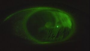
Only 26.2% of recruited patients showed a normal BUT (>10 s); 73.8% had a BUT lower than 10 s, 83.07% lower than 15 s (Fig. 1).
Corneal sensitivity was absent in four patients (9.8%) and strongly decreased compared to the other eye in two patients (7.3%). The lower tear meniscus was thin (<0.2 mm) in 21 patients (51.2%).
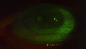
According to the Oxford scale, fluorescein staining of the cornea was positive in 56% of patients: 16 (69.5% of which was grade I), seven (30.5%) of grade II (Fig. 2); while lissamine green staining of the conjunctiva was positive in 73.2% of patients: 17 (56.6%) of grade I, nine (30%) of grade II and four (13.4%) of grade III.

Figure 1: Significant tear instability with low BUT, 1 year after Macular Pucker surgery.

Figure 2: Corneal and conjunctival surface staining in a patient after 12 months since vitrectomy surgery.
Total | Female | Male | |
Number of patients | 41 | 17 | 24 |
Average age (years) | 65 | 64 | 67 |
At least 1 symptom (%) | 80 | 9.75 | 12.2 |
At least 2 symptoms (%) | 58.5 | 29.26 | 29.26 |
BUT <15 s (%) | 83 | 62 | 38 |
BUT <10 s (%) | 71 | 60 | 40.2 |
Corneal fluorescein staining ≥2 | 17% | 43% (9.7% of total number of subjects observed) | 57% (7.3% of total number of subjects examined) |
Conjunctival lissamine green staining ≥2 | 31.7% | (14.5% of the total number of subjects observed) | (17% of total number of subjects observed |
BUT: Break-up time. | |||
Table 1: Characteristics of the patients and the results.
Discussion
Due to the recent amelioration of the technique, the quality of the results of vitrectomy and epiretinal membrane removal has largely increased the anatomical success of the surgery. This brings a high level of satisfaction to the surgeons but also makes patients undergoing vitrectomy, especially those undergoing surgery for epi-retinal membranes, have great expectations. The tear film, the epithelia of the cornea and conjunctiva, the lacrimal glands and the eyelids integrated by vascular, nervous, endocrine and immune activities form an anatomic and functional unit working as a system aimed to protect the eye from the external environment and provide for a high-quality refractive surface of the cornea [1-3].
Its mission to protect the integrity of the eye bulb and provide clear vision is dependent on several integrated activities of the various components of the ocular surface system, which, through epithelial cell renewal, quick nerve and vascular reactions and the production of an efficient tear film, guarantee the proper homeostasis [9,10].
Because of the nature of its function and anatomic location, the ocular surface is vulnerable to environmental, as well as direct factors. The ocular surface is a highly dynamic system capable of fast and continuous adaptive reactions to environmental, toxic, infective, traumatic and inflammatory conditions that tend to interfere with its homeostasis. When the adaptive response is not able to cope with external or internal aggressions, a persisting dysfunction may develop.
Pathogenic events can disturb the system homeostasis, whatever their cause (infective, immune, traumatic, surgical or toxic), if not promptly neutralized by appropriate reactions of the ocular surface system, will create, within time, a vicious cycle of events leading to the development of chronic disease [11].
This self-stimulated loop is characterized by the presence of:
- A progressive failure of tear film stability [12]
- Morpho-functional changes of the epithelial structure and its secretory abilities [13]
- The build-up of sub-clinic or clinically apparent inflammatory processes involving the bulbar surface, as well as the lid margin inducing changes to the Meibomian glands and the nerve functions [14]
DED, the most frequent consequence of the loss of homeostasis of the ocular surface system, is a common condition around the world, increasing with age, affecting around 6-16% of the adult population in western European countries [15]. With its long-standing and bothering symptoms, dry eye is a problem for patients and their eye care providers and has a significant impact on the quality of vision and QoL [15]. It can affect daily functions and has been reported to influence the outcomes of refractive surgery [16] and cataracts if ever needed [17].
It has been reported that DED affects QoL to a similar degree as class III/IV angina, which is widely recognized as a chronic disease with negative QoL impact; a situation that 16% of patients would give up years of life to ameliorate [17].
Dry eye also represents a significant social cost with an impact on work productivity. The direct cost of DED has been estimated between $300 and $1,100 per patient annually, with additional costs due to impaired work productivity and its economic impact on healthcare systems is similar to rheumatoid arthritis [18,19].
Our study shows that more than 6 months after the vitreoretinal surgery, 58% of the studied population had at least two symptoms of ocular surface dysfunction, 31% had lissamine green conjunctival staining and 71% had a pathological BUT (<10 s), a condition clinically diagnosed as a DED.
The high frequency of short BUT (74% <10 seconds; 83% <15 seconds”) is particularly interesting, indicating a significant presence of tear film instability and the high risk of developing a DED, with signs and symptoms, if the ocular surface system is stressed, loses its ability to adapt and enter the vicious cycle.
In Japan, a short BUT dry eye has been described as a very frequent cause of symptoms and depression of the QoL [20]. The possible causes of tear instability with short BUT may be many and all of them can cause a break in ocular surface system homeostasis and the possible beginning of a vicious cycle leading to chronic DED. As we know, several patients may have an unstable but still compensated ocular surface system, which if exposed to significant stress, can lose its ability to compensate and give start to the vicious cycle of dry eye.
Most patients undergo cataract emulsification with an intraocular lens implant at the same time as vitrectomy and it is well known that cataract surgery is associated with a certain frequency (nearly 10% in the first 3 months) of dry eye-like symptoms and signs for several months [21-24]. The presence of some loss of cornea sensitivity in a considerable number of patients long term after surgery is intriguing. Corneal nerves are responsible for sensations of touch, pain and temperature and play an important role in blink reflex, wound healing, tear production and secretion. After surgery involving the ocular surface, nerve function can be reduced, which may be due to neurotrophic factor deprivation from loss of corneal epithelium integrity [25]. Damage of corneal nerves will result in decreased tear production and epithelial cell qualitative and quantitative turnover. The impact of vitrectomy on cornea innervation is not completely known [26].
It is known that diabetic patients after plana vitrectomy for proliferative diabetic retinopathy tend to show nerve problems delayed healing of epithelial defects [27]. In addition, cornea nerve malfunctions have been reported after encircling band and extensive laser photocoagulation [28,29].
A reduction in cornea sensitivity was described after vitreoretinal surgery in both diabetic and non-diabetic patients (diabetic patients being much more frequently involved) allowing the authors to state that the vitrectomy procedure reduces the cornea sensitivity whether a 20-G or 23-G vitrectomy systems were used. Because corneal sensitivity is a major player in tear secretion, both basal and reflex [30] reductions in cornea sensitivity, even if temporary, could be a starting point to the build-up of a vicious cycle leading towards a chronic dry eye [31].
A decrease in cornea sensitivity has been described also after cataract surgery, which we know is often performed associated with vitrectomy [32].
The elevation of inflammatory factors in tear film due to ocular surface irritation, use of PVP-I at concentrations other than 5% and/or not formulated for ophthalmic use [33], eye drops administered postoperatively and their preservatives [34,35] can be another cause of dry eye after vitreoretinal surgery in addition to the trauma and resection of corneal nerves and the damage to epithelial cells and the long-term microscopy light exposure. It has also been demonstrated that vigorous irrigation of the tear film and manipulation of the ocular surface intraoperatively may reduce the goblet cell density and result in shortened BUT postoperatively [36].
Furthermore, it is possible that the stretch of orbicular muscle fibers exerted by the speculum to keep the eye open during surgery will change the ability of the lids to spread a smooth, uniform mucus layer on the ocular surface inducing tear film instability and dry spots with epithelial exposure.
The ocular surface symptoms are not only reported to be present in the immediate postoperative period, as it is expected as a consequence of surgical trauma [8], but in a considerable number of patients tend to last longer and become chronic. Invalidating the effect of an apparently anatomically wise, successful surgery.
As we know, several patients have an unstable, but still barely compensated, ocular surface system, which, if exposed to significant stress, can lose its equilibrium and give rise to a chronic ocular surface problem [37]. Importantly, findings of studies evaluating signs of DED (eg., tear film BUT and tear volume) suggest there are individuals with dry eye who are not aware of their condition [38].
This fact is congruent with our findings of a higher frequency of dry eye signs in the smaller female group of our patients. Since it’s well known that the frequency of the disease and the sensibility to its risk factors is higher in females compared to males [39,40].
Conclusion
In conclusion, the patients who underwent vitreoretinal surgery for removal of epiretinal membranes showed in the long-term signs and symptoms of ocular surface dysfunction (dry eye) with a frequency that is more than double the expected frequency of the disease.
Because of the selection of patients who had surgery at least 6 months before the evaluation of the ocular surface and being our clinical setting a second-level reference center for vitreoretinal diseases, many patients were lost to the retinal follow-up because they had no further retinal problems and were checked by their first ophthalmologist. This is the reason why the number of subjects of this study is limited and prevents the ability to give extensive indications. However, it is relevant that none of these patients reported ocular surface discomfort or dysfunction before vitreoretinal surgery.
Realizing the risk of developing a postoperative dry eye will lead surgeons to check for patients with ocular surface features at high risk of developing tear films and ocular surface problems and treat them before surgery. To reduce possible stress during surgery, it would be recommended to follow the correct antisepsis procedure (i.e. to use the 5% PVP-I eye drops formulated for ophthalmic use, for the cleaning of the periocular surface and installation on the ocular surface for 2 minutes of contact and after removing PVP-I 5% from an ocular surface with sterile saline irrigation), to reduce conjunctival desiccation and manipulative trauma, to limit the use of potentially toxic drugs (such as mydriatic), to avoid exaggerated tension of the orbicular fibers by the speculum and the high-frequency use of toxic, preserved drugs in the postoperative period. On the other hand, a delayed diagnosis prevents patients from receiving the correct treatment that could alleviate their DED-related discomfort and the reduction in QoL.
Understanding the risk factors for DED, its possible etiologies and the pathophysiologic mechanisms by which it develops and progresses can help clinicians dealing with patients who are going to have or have had vitreoretinal surgery when they approach the diagnosis and management of this complication. Its recognition will lead to prompt treatment with the improvement of symptoms and possibly with the interruption of the vicious cycle and stabilization of the ocular surface system homeostasis. In this way, trying to provide a more satisfactory surgery for the doctor and the patient.
Conflict of Interests
The authors have no conflict of interest to declare.
Acknowledgments
Editorial assistance was provided by Aashni Shah (Polistudium SRL, Milan, Italy).
Study Approval Statement
Study approved by the Ethic Committee, Ospedale San Martino, Genoa, Italy.
Consent to Participate Statement
Written informed consent obtained by all patients before surgery.
Funding Sources
None
Author Contributions
MR, Sl, CD, FB contributed to data collection and surgery, SB and Mc to write and review the paper.
Data Availability Statement
Data are available from MR.
References
- Sandali O, El Sanharawi M, Lecuen N, Barale PO, Bonnel S, Basli E, et al. 25, 23 and 20-gauge vitrectomy in epiretinal membrane surgery: a comparative study of 553 cases. Graefes Arch Clin Exp Ophthalmol. 2011;249(12):1811-9
- Rizzo S, Genovesi-Ebert F, Murri S, Belting C, Vento A, Cresti F, et al. 25-gauge, sutureless vitrectomy and standard 20-gauge pars plana vitrectomy in idiopathic epiretinal membrane surgery: a comparative pilot study. Graefes Arch Clin Exp Ophthalmol. 2006;244(4):472-9.
- Okamoto F, Okamoto Y, Fukuda S, Hiraoka T, Oshika T. Vision-related quality of life and visual function after vitrectomy for various vitreoretinal disorders. Invest Ophthalmol Vis Sci. 2010;51(2):744-51.
- Brightbill FS, Myers FL, Bresnick GH. Postvitrectomy keratopathy. Am J Ophthalmol. 1978;85(5):651-5.
- Perry HD, Foulks GN, Thoft RA, Tolentino FI. Corneal complications after closed vitrectomy through the pars plana. Arch Ophthalmol. 1978;96(8):1401-3.
- Chiambo S, Baílez Fidalgo C, Pastor Jimeno JC, Coco Martín RM, Rodríguez de la Rúa Franch E, De la Fuente Salinero MA, et al. Complicaciones corneales epiteliales de las vitrectomías: estudio retrospectivo [Corneal epithelial complications after vitrectomy: a retrospective study]. Arch Soc Esp Oftalmol. 2004;79(4):155-61.
- Barabino S, Labetoulle M, Rolando M, Messmer EM. Understanding symptoms and quality of life in patients with dry eye syndrome. Ocul Surf. 2016;14(3):365-76.
- Yu JG, Ni F, Xiang Y, Feng YF, Wang J, Fu XA. A prospective study on postoperative discomfort after 20-gauge pars plana vitrectomy. Clin Ophthalmol. 2015;9:1379-84.
- Tseng SC, Tsubota K. Important concepts for treating ocular surface and tear disorders. Am J Ophthalmol. 1997;124(6):825-35.
- Gipson IK. The ocular surface: the challenge to enable and protect vision: the Friedenwald lecture. Invest Ophthalmol Vis Sci. 2007;48(10):4391-8.
- Rolando M, Zierhut M. The ocular surface and tear film and their dysfunction in dry eye disease. Surv Ophthalmol. 2001;45:S203-10.
- Holly F, Lemp MA. Formation and rupture of the tear film. Exp Eye Res. 1973;15:515-54.
- Rolando M, Terragna F, Giordano G, Calabria G. Conjunctival surface damage distribution in keratoconjunctivitis sicca. An impression cytology study. Ophthalmologica. 1990;200(4):170-6.
- McDermott AM, Perez V, Huang AJ, Pflugfelder SC, Stern ME, Baudouin C, et al. Pathways of corneal and ocular surface inflammation: A perspective from the cullen symposium. Ocul Surf. 2005;3(4 Suppl):S131-8
- Rolando M, Iester M, Macrí A, Calabria G. Low spatial-contrast sensitivity in dry eyes. Cornea. 1998;17(4):376-9.
- De Paiva CS, Chen Z, Koch DD, Hamill MB, Manuel FK, Hassan SS, et al. The incidence and risk factors for developing dry eye after myopic LASIK. Am J Ophthalmol. 2006;141(3):438-45.
- Cho YK, Kim MS. Dry eye after cataract surgery and associated intraoperative risk factors. Korean J Ophthalmol. 2009;23(2):65-73.
- Clegg JP, Guest JF, Lehman A, Smith AF. The annual cost of dry eye syndrome in France, Germany, Italy, Spain, Sweden and the United Kingdom among patients managed by ophthalmologists. Ophthalmic Epidemiol. 2006;13(4):263-74.
- Yamada M, Mizuno Y, Shigeyasu C. Impact of dry eye on work productivity. Clinicoecon Outcomes Res. 2012;4:307-12.
- Yamamoto Y, Yokoi N, Higashihara H, Inagaki K, Sonomura Y, Komuro A, et al. Clinical characteristics of short tear film breakup time (BUT)-type dry eye]. Nippon Ganka Gakkai Zasshi. 2012;116(12):1137-43.
- Khanal S, Tomlinson A, Esakowitz L, Bhatt P, Jones D, Nabili S, et al. Changes in corneal sensitivity and tear physiology after phacoemulsification. Ophthalmic Physiol Opt. 2008;28(2):127-34.
- Barabino S, Solignani F, Rolando M. Dry eye-like symptoms and signs after cataract surgery. Invest Ophthalmol Vis Sci. 2010;51(13):6254.
- Mencucci R, Boccalini C, Favuzza E, Paladini I, Menchini U. Ocular discomfort after cataract surgery: effect of a Hyaluronic acid and Carboxymethylcellulose ophthalmic solution. Invest Ophthalmol Vis Sci. 2014;55(13):1503.
- Kasetsuwan N, Satitpitakul V, Changul T, Jariyakosol S. Incidence and pattern of dry eye after cataract surgery. PLoS One. 2013;8(11):e78657.
- Shaheen BS, Bakir M, Jain S. Corneal nerves in health and disease. Surv Ophthalmol. 2014;59(3):263-85.
- Chung H, Tolentino FI, Cajita VN, Acosta J, Refojo MF. Reevaluation of corneal complications after closed vitrectomy. Arch Ophthalmol. 1988;106(7):916-9.
- Chen WL, Lin CT, Ko PS, Yeh PT, Kuan YH, Hu FR, et al. In-vivo confocal microscopic findings of corneal wound healing after corneal epithelial debridement in diabetic vitrectomy. Ophthalmology. 2009;116(6):1038-47.
- Kaufman PL, Rohen JW. Parasympathetic denervation of the ciliary muscle following panretinal photocoagulation. Curr Eye Res. 1991;10:437-55.
- Lincoff H, Stopa M, Kreissig I, Madjarov B, Sarup V, Saxena S, et al. Cutting the encircling band. Retina. 2006;26(6):650-4.
- Mahgoub MM, Macky TA. Changes in corneal sensation following 20 and 23 G vitrectomy in diabetic and non-diabetic patients. Eye (Lond). 2014;28(11):1286-91.
- Aragona P, Rolando M. Towards a dynamic customised therapy for ocular surface dysfunctions. Br J Ophthalmol. 2013;97(8):955-60.
- Kohlhass M. Corneal sensation after cataract and refractive surgery. J Cataract Refract Surg. 1998;24:1399-409.
- Mac Rae SM, Brown B, Edelhauser HF. The corneal toxicity of presurgical skin antiseptics. Am J Ophthalmol. 1984;97(2):221-32.
- Zabel RW, Mintsioulis G, MacDonald IM, Valberg J, Tuft SJ. Corneal toxic changes after cataract extraction. Can J Ophthalmol. 1989;24:311-6.
- Roberts CW, Elie ER. Dry eye symptoms following cataract surgery. Insight. 2007;32:14-21.
- Li XM, Hu L, Hu J, Wang W. Investigation of dry eye disease and analysis of the pathologic factors in patients after cataract surgery. Cornea. 2007;26(9)S16-20.
- Brewitt H, Sistani F. Dry eye disease: the scale of the problem. Surv Ophthalmol. 2001;45(Suppl 2):S199-202.
- Baudouin C, Aragona P, Van Setten G, Rolando M, Irkeç M, Benítez del Castillo J, et al. Diagnosing the severity of dry eye: a clear and practical algorithm. Br J Ophthalmol. 2014;98(9):1168-76.
- Schaumberg DA, Sullivan DA, Buring JE, Dana MR. Prevalence of dry eye syndrome among US women. Am J Ophthalmol. 2003;136(2):318-26.
- The epidemiology of dry eye disease: report of the Epidemiology Subcommittee of the International Dry Eye WorkShop (2007). Ocul Surf. 2007;5(2):93-107.
Article Type
Research Article
Publication History
Received Date: 22-02-2024
Accepted Date: 17-03-2024
Published Date: 25-03-2024
Copyright© 2024 by Barabino S, et al. All rights reserved. This is an open access article distributed under the terms of the Creative Commons Attribution License, which permits unrestricted use, distribution, and reproduction in any medium, provided the original author and source are credited.
Citation: Barabino S, et al. Dry Eye-Like Ocular Surface Dysfunction in Post-Vitreoretinal Surgery Eyes. J Ophthalmol Adv Res. 2024;5(1):1-10.

Figure 1: Significant tear instability with low BUT, 1 year after Macular Pucker surgery.

Figure 2: Corneal and conjunctival surface staining in a patient after 12 months since vitrectomy surgery.
| Total | Female | Male |
Number of patients | 41 | 17 | 24 |
Average age (years) | 65 | 64 | 67 |
At least 1 symptom (%) | 80 | 9.75 | 12.2 |
At least 2 symptoms (%) | 58.5 | 29.26 | 29.26 |
BUT <15 s (%) | 83 | 62 | 38 |
BUT <10 s (%) | 71 | 60 | 40.2 |
Corneal fluorescein staining ≥2 | 17% | 43% (9.7% of total number of subjects observed) | 57% (7.3% of total number of subjects examined) |
Conjunctival lissamine green staining ≥2 | 31.7% | (14.5% of the total number of subjects observed) | (17% of total number of subjects observed |
BUT: Break-up time. | |||
Table 1: Characteristics of the patients and the results.


