Bile PEFK1*, Diabate Z1, Diomande GF1, Djiguimde WP2, Koffi KF-H1, Gode LE1, Goule AM1, Babayeju OR1, Ouattara Y1, Diomande IA1
1Ophthalmology Department, University Hospital Center, Bouaké, Cote d’ivoire
2Ophthalmology Department, University Hospital Center, Bogodogo, Ouagadougou, Burkina Fas, Cote d’ivoire
*Correspondence author: Philippe EFK BILE, Ophthalmology Department, University Hospital Centre, Bouake, 01 BP 1174 Bouake 01, Cote d’Ivoire; Email: [email protected]
Published Date: 30-04-2024
Copyright© 2024 by Bile PEFK, et al. All rights reserved. This is an open access article distributed under the terms of the Creative Commons Attribution License, which permits unrestricted use, distribution, and reproduction in any medium, provided the original author and source are credited.
Abstract
Objective: To describe the epidemiological, clinical and histopathological aspects of malignant tumors in the Gbêkê region.
Materials and methods: Retrospective cross-sectional descriptive study conducted in the ophthalmology department of the Bouake University Hospital over a 5-year period [January 1, 2018, to December 31, 2022]. It involved patients seen in consultation for malignant ocular tumors confirmed after clinical and paraclinical examination.
Results: Prevalence was 0.19%. The mean age was 31.52 years, with extremes of 4 months and 72 years. Reasons for consultation were dominated by exophthalmos (41.67%). Most patients (77.08%) had a consultation delay of over 12 months. The conjunctiva was the preferred site of ocular malignancies (41.67%). Squamous cell carcinoma was the most common tumour of all ages, accounting for 41.66%. Squamous cell carcinoma was the most common malignancy in adults, with 55.56%. In children, retinoblastoma remains the most common tumor (58.33%).
Keywords: Tumor; Malignant; Eye; Bouake
Introduction
Malignant orbito-ocular tumors are abnormal cellular proliferations that develop at the expense of the eye and its adnexa [1]. They are rare and serious, posing a public health and management problem in our sub-Saharan regions. In Central Africa, their frequency varied from 0.4% to 2% [2,3,4]. In West Africa, they ranged from 0.7% to 1.33% [5, 6]. In Bouaké, in the absence of recent data on malignant tumors, we conducted this study to describe their epidemiological, clinical and histopathological aspects in the Gbêkê region.
Material and Methods
This was a descriptive retrospective cross-sectional study that took place in the ophthalmology department of Bouake University Hospital over a 5-year period [January 1, 2018, to December 31, 2022]. It involved all patients seen for consultation in whom a diagnosis of malignant ocular tumor had been made and confirmed after clinical and paraclinical examination. Patients with incomplete medical records or a clinical diagnosis of tumor without histological evidence were not included. The survey form provided information on sociodemographic data (age, sex), clinical and paraclinical data (reason for consultation, time elapsed before first consultation, visual acuity, slit-lamp examination, diagnosis retained after histo-anatomopathological examination). This survey form has been drawn up considering the bibliography of studies carried out on malignant ocular tumours.
Results
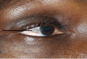
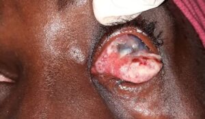
Malignant ocular tumors numbered 48 out of a total of 25472 patients seen during the study period, representing a prevalence of 0.19%. There were 36 adults and 12 children. The majority were male, with 25 cases, giving a sex ratio of 1.09. The average age was 31.52 years, with extremes of 4 months and 72 years. Reasons for consultation were dominated by exophthalmos (41.67%), followed by ulcerating-bourging masses with 25% (Table 1). Most patients (77.08%) had a consultation delay of over 12 months (Table 2). The conjunctiva remained the preferred site of ocular malignancies (41.67%), followed by the orbit and its orbital tissue (37.5%). Histologically, squamous cell carcinomas were the most common tumour type, accounting for 41.66% of all tumours of all ages. They were divided between squamous cell carcinomas of the conjunctiva (35.41%) and those of the eyelid (6.25%). Angiosarcoma followed at 16.67% (Table 3). Squamous cell carcinoma was the most common malignancy in adults, accounting for 55.56%. In children, retinoblastoma remained the most common tumour (58.33%) (Table 4,5, Fig. 1-3).
Reasons for Consultation | Number | Percentage |
Exophthalmos | 20 | 41.67 |
Ulcerative conjunctival mass | 12 | 25 |
Conjunctival ulceration | 10 | 20.83 |
Palpebral swelling | 4 | 8.33 |
Exorbitis | 2 | 4.17 |
Total | 48 | 100 |
Table 1: Breakdown of patients by reason for consultation.
Consultation Period (Months) | Number | Percentage |
| [0 – 3] | 3 | 6.25 |
| [3 – 6] | 1 | 2.08 |
| [6 – 9] | 6 | 12.25 |
| [9 -12] | 1 | 2.08 |
Sup à 12 | 37 | 77.08 |
Total | 48 | 100 |
Table 2: Breakdown of patients by consultation time.
Anatomical Site | Histological Types | Numbers | Percentage |
Conjunctiva | Squamous cell carcinoma | 17 | 35.41 |
Eyelid
| Squamous cell carcinoma | 3 | 6.25 |
Orbital angiosarcoma | 8 | 16.67 | |
Orbit (orbital tissue)
| Burkitt’s lymphoma | 2 | 4.17 |
Leiomyosarcoma | 3 | 6.25 | |
Rhabdomyosarcoma | 3 | 6.25 | |
Metastasis | 2 | 4.17 | |
Retinoblastoma | 7 | 14.58 | |
Retina | Irian melanoma | 3 | 6.25 |
Iris | Total | 48 | 100 |
Table 3: Distribution by histological type and anatomical site, all ages combined.
Histological Types in Children | Numbers | Percentage |
Retinoblastoma | 7 | 58.33 |
Burkitt’s lymphoma | 3 | 25 |
Rhabdomyosarcoma | 2 | 16.67 |
Total | 12 | 100 |
Table 4: Histological distribution in children.
Histological Types in Children | Numbers | Percentage |
Squamous cell carcinoma | 20 | 55.56 |
Angiosarcoma | 8 | 22.22 |
Leiomyosarcoma | 3 | 16.67 |
Metastasis | 2 | 6.25 |
Irian melanoma | 3 | 9.37 |
Total | 36 | 100 |
Table 5: Histological distribution in adults.

Figure 1: Conjunctival squamous cell carcinoma.

Figure 2: Conjunctival squamous cell carcinoma as an ulcerating, budding mass. 
Figure 3: Metastatic tumor with exophthalmos.
Discussion
Malignant oculo-orbital tumours are relatively uncommon in sub-Saharan Africa, affecting subjects of all ages and genders. The prevalence in our study was 0.19%. Several authors confirm the rarity of these tumors. Indeed, Sylla in Mali, Vonor in Togo and Bra’Eyatcha in Cameroon found relatively low prevalences in their studies, with 0.7, 1.33 and 2% respectively [2,5,6]. The average age of the patients was 31.52 years. These results are similar to those of Akpe in Nigeria, Sanfo in Burkina Faso with mean ages of 31.3 and 33.64 years [7,8]. These mean ages concerned relatively young subjects. However, oculo-orbital malignancies can occur at any age. Most cases were male, with a sex ratio of 1.09. This predominance has been noted by several authors in the literature [4-8]. However, some authors, including Levecq, found a female predominance, with 640 women versus 617 men [9]. Exophthalmos dominated the reasons for consultation, accounting for 41.67% of cases. This protrusion of the globe outside the orbit remains one of the most frequent signs motivating consultation, according to the literature [10]. It was most often related to the delay in consultation, which was most often greater than twelve months. The most common anatomical location was the conjunctiva (41.67%). These results are like those of several authors throughout the literature, who found the conjunctiva to be the most involved structure in their respective studies. Sanfo, Sylla and Bra’ Eyatcha [2,5,7]. Several other authors also reported predominant conjunctival localization, in proportions of 3% 75% and 88.5% respectively. In terms of histological type, all ages combined, squamous cell carcinoma was the most frequent malignancy (41.67%). It concerned both the conjunctiva and the eyelid. The same observation was made by Sylla in Mali, who found squamous cell carcinoma to be the most frequent malignant tumour of the eye and its appendages in his series, with 39.7% [5]. The same finding was made by Vonor with 20% [6]. Other authors, such as Scat and Templeton also found squamous cell carcinomas to be the predominant malignancy [11,12]. Several risk factors are thought to contribute to this high prevalence, including infectious factors such as HIV infection and HPV 6 infection [13,14]. Mechanical factors such as prolonged exposure to sunlight, dust and a dry atmosphere have also been incriminated [15-17]. Retinoblastoma was the most frequently observed tumor after squamous cell carcinoma. However, it remains the leading malignancy in children with 58.33%. Similar findings were reported by Bella and Bra’ Eyatcha in Cameroon, with 48.44% and 75% respectively [2,15]. Sanfo also found retinoblastoma to be the most frequently observed malignant tumour, at 52.17%. According to Doz and Mac-carty, it accounts for 3% of all tumors in children [18-20].
Conclusion
The conjunctiva was the preferred site of ocular malignancies (41.67%). Squamous cell carcinoma was the most common tumour of all ages, accounting for 41.66%. Squamous cell carcinoma was the most common malignancy in adults, with 55.56%. In children, retinoblastoma remains the most common tumor (58.33%).
Conflict of Interests
The authors have no conflict of interest to declare.
References
- Jaubert F. General pathological anatomy. Masson, Paris. 1984;286.
- Bra’ Eyatcha Bimingo N, Dohvoma VA, Njoya Mare J, Bella AL, Ebana Mvogo C. Clinical and therapeutic aspects of tumors of the eye and its adnexa in northern Cameroon: about 70 cases. Health Sci Dis. 2022;3((2)1):73-6.
- Luembe Kasongo D, Iye Abial S, Kintadi Luyingila G, Maloba Ngoy V, Chenge Borasisi G. Ocular tumors: diagnosis and treatment at the University Clinics of Lubumbashi, DRC. JSMO. 2021;30(1):22-6.
- Mendimi Nkodo JM, Kagmeni G, Haman Nassourou O, Kabeyene Okono CA, Epee E, Moukouri E, et al. Morpho-epidemiological aspects of oculo-orbital tumors at the Yaoundé university hospital – Cameroun. Health Sci Dis. 2014;15(1):1-6.
- Sylla F, Kamate B, Traore CB, Traore M, Diallo D, Coulibaly B, et al. Epidemiological and histopathological study of tumors of the eye and its adnexa: about 63 cases. Scientific Journal SOAO. 2016;1:45-50.
- Vonor K, Banla M, Darre T, Ayena KD, Amegbor K, Dzidzinyo K, et al. Tumeurs oculaires au Togo: epidemiological, clinical, and histopathological aspects of forty-five cases observed at the Sylvanus-Olympio University Hospital in Lomé. Médecine et Sant Tropicales 2015;25:105-6.
- Akpe BA, Omoti AE, Iyasele ET. Histopathology of ocular tumor specimens in Benin city, Nigeria. J Ophthalmic Vis Res. 2009;4:232-7.
- Sanfo M, Millogo M, Coulibaly A, Dargani M F, Konsem T. Epidemiological, clinical, and therapeutic aspects of oculo-orbital tumors at the Yalgado Ouédraogo University Hospital. Scientific journal Col Odonto-Stomatol Afr Chir Maxillo-fac. 2021;28(4):41-6.
- Levecq L, De Potter P, Guagnini AP. Epidemiology of ocular and orbital lesions referred to an ocular oncology center. JF Ophtalmol. 2005;28(8):840-4.
- Ducasse A, Merol Jc, Bonnet F, Litre F, Arndt C, Larre I. Orbital tumors in adults. J Fr Ophtalmol. 2016;39(4):387-99.
- Scat Y, Liotet S, Carre F. Epidemiological study of 1705 malignant tumors of the eye and adnexa. J Fr Ophtalmol. 1996;19(2):83-8.
- Templeton AC. Tumors of the eye and adnexa in African of Uganda. Cancer. 1967;20:1687-98.
- Nagaiah G, Stotler C, Orem J, Mwanda WO, Remick SC. Ocular surface squamous neoplasia in patients with HIV infection in sub-Saharan Africa. Curr Opin Oncol. 2010;22:437-42.
- Guirou N, Gouda M, Abba KHY, Napo A, Sissoko M, Wangara N, et al. Histopathological and genetic aspects of oculo-orbital tumors at HUC-IOTA. Scientific Journal SOAO. 2021;1:9-14.
- Waddell K, Kwehangana J, Johnston WT, Lucas S, Newton R. A case control study of ocular surface squamous neoplasia in Uganda. Int J Cancer. 2010;127:427-32.
- Mahale A, Alkantan H. Altered gene expression in conjunctival squamous cell carcinoma. Mod Pathol. 2016;29(5):452-60.
- Berete CR, Dejardins L, Kouassi LJ, Coulibaly F. Relationship between acquired immunodeficiency virus (HIV-AIDS) and Conjunctival Squamous Cell carcinoma (CSC): epidemioclinical study of 26 cases in the ophthalmology department of Treichville University Hospital (Abidjan- Cote d’ivoire). JFr Ophtalmol. 2016;39(5):467-76.
- Bella LA, Evina TA, Eballe AO. Primary ocular tumors in children: epidemiological and histopathological aspects at the Yaoundé Gyneco-Obstetric and Pediatric Hospital. Cahiers Sante. 2010;20:139-41.
- Dos F. Retinoblastoma: recent aspects. Arch Pediatrie. 2006;13(10):1329-37.
- Mc carthy JH, Barry M, Crwley D, Bronson RT. Genetic ablation of av integrins in epithelial cells of eyelid skin and cunjunctiva leads to squamus cell carcinoma. Am J Pathol. 2008;172(6):1740-7.
Article Type
Research Article
Publication History
Received Date: 03-02-2024
Accepted Date: 23-04-2024
Published Date: 30-04-2024
Copyright© 2024 by Bile PEFK, et al. All rights reserved. This is an open access article distributed under the terms of the Creative Commons Attribution License, which permits unrestricted use, distribution, and reproduction in any medium, provided the original author and source are credited.
Citation: Bile PEFK, et al. Epidemioclinical and Histological Profile of Malignant Ocular Tumors at University Hospital Center of Bouake. J Ophthalmol Adv Res. 2024;5(1):1-5.

Figure 1: Conjunctival squamous cell carcinoma.

Figure 2: Conjunctival squamous cell carcinoma as an ulcerating, budding mass. 
Figure 3: Metastatic tumor with exophthalmos.
Reasons for Consultation | Number | Percentage |
Exophthalmos | 20 | 41.67 |
Ulcerative conjunctival mass | 12 | 25 |
Conjunctival ulceration | 10 | 20.83 |
Palpebral swelling | 4 | 8.33 |
Exorbitis | 2 | 4.17 |
Total | 48 | 100 |
Table 1: Breakdown of patients by reason for consultation.
Consultation Period (Months) | Number | Percentage |
| [0 – 3] | 3 | 6.25 |
| [3 – 6] | 1 | 2.08 |
| [6 – 9] | 6 | 12.25 |
| [9 -12] | 1 | 2.08 |
Sup à 12 | 37 | 77.08 |
Total | 48 | 100 |
Table 2: Breakdown of patients by consultation time.
Anatomical Site | Histological Types | Numbers | Percentage |
Conjunctiva | Squamous cell carcinoma | 17 | 35.41 |
Eyelid
| Squamous cell carcinoma | 3 | 6.25 |
Orbital angiosarcoma | 8 | 16.67 | |
Orbit (orbital tissue)
| Burkitt’s lymphoma | 2 | 4.17 |
Leiomyosarcoma | 3 | 6.25 | |
Rhabdomyosarcoma | 3 | 6.25 | |
Metastasis | 2 | 4.17 | |
Retinoblastoma | 7 | 14.58 | |
Retina | Irian melanoma | 3 | 6.25 |
Iris | Total | 48 | 100 |
Table 3: Distribution by histological type and anatomical site, all ages combined.
Histological Types in Children | Numbers | Percentage |
Retinoblastoma | 7 | 58.33 |
Burkitt’s lymphoma | 3 | 25 |
Rhabdomyosarcoma | 2 | 16.67 |
Total | 12 | 100 |
Table 4: Histological distribution in children.
Histological Types in Children | Numbers | Percentage |
Squamous cell carcinoma | 20 | 55.56 |
Angiosarcoma | 8 | 22.22 |
Leiomyosarcoma | 3 | 16.67 |
Metastasis | 2 | 6.25 |
Irian melanoma | 3 | 9.37 |
Total | 36 | 100 |
Table 5: Histological distribution in adults.


