Haafiz Hashim1, Hongyi Zhang1, Haris Hashim2, Karanjit Kooner1,3*
1University of Texas Southwestern Medical Center, 5323 Harry Hines Blvd, Dallas, TX 75390, USA
2University of Texas at Dallas, 800 W Campbell Rd, Richardson, TX 75080, USA
3VA North Texas Health Care System, 4500 S Lancaster Rd, Dallas, TX, 75216, USA
*Correspondence author: Karanjit S Kooner, MD, Department of Ophthalmology, University of Texas Southwestern Medical Center, 5323 Harry Hines Blvd, Dallas, TX 75390-9057, USA; Email: [email protected]
Published Date: 02-12-2024
Copyright© 2024 by Hashim H, et al. All rights reserved. This is an open access article distributed under the terms of the Creative Commons Attribution License, which permits unrestricted use, distribution, and reproduction in any medium, provided the original author and source are credited.
Abstract
Purpose: To characterize the ability of ImageJ-derived measurements of Optical Coherence Tomography Angiography (OCTA) to diagnose healthy vs Primary Open-Angle Glaucoma (POAG) Eyes.
Methods: This retrospective study analyzed 85 healthy and 81 POAG eyes. Initially, demographics, historical data, intraocular pressure, cup/disc ratio and retinal nerve fiber layer thickness were collected for all patients. Thereafter, quantitative vascular parameters including Vessel Density (VD), Vessel Length Density (VLD) and Fractal Dimension (FD) were obtained by analyzing OCTA scans using the open-source software ImageJ. Measurements were obtained from the Radial Peripapillary Capillary (RPC) layer of the optic nerve head and the superficial and deep capillary plexuses of the macula. Fifty healthy and fifty POAG eyes (training set) were randomly selected to train two diagnostic models: one based on OCTA parameters (model A) and the other based on clinical and structural data (model B). These models were tested on the remaining 35 healthy and 31 POAG eyes and receiver operating curves were constructed to compare their ability to identify POAG.
Results: VD, VLD and FD as obtained by ImageJ were all significantly reduced in the POAG group (p < 0.0001). The RPC layer was the most effective at classifying glaucoma (AUC = 0.9184, CI: 0.85-0.98). Model A (AUC = 0.917, CI: 0.847-0.986) slightly outperformed model B (AUC = 0.863, CI: 0.776-0.949), albeit not to the level of statistical significance (p = 0.111)
Conclusion: Our pilot study indicates that OCTA vascular parameters are similar in effectiveness to clinical exam and structural features at diagnosing glaucoma.
Keywords: Glaucoma; Optic Neuropathy; Optical Coherence Tomography Angiography; Retina; Imagej; Superficial Retinal Capillaries; Deep Retinal Capillaries; Radial Peripapillary Capillaries
Introduction
Primary Open Angle Glaucoma (POAG) is a chronically progressive optic neuropathy and one of the most common causes of irreversible blindness worldwide [1,2]. It is a multifactorial disease defined by damage to the retinal ganglion cells. Traditionally, glaucoma has been thought to be caused by two primary mechanisms: direct damage to the optic nerve fibers at the lamina cribrosa due to increased Intraocular Pressure (IOP, mechanical theory) and ischemic injury to retinal ganglion cells due to poor vasculature in the retina, choroid and optic nerve head (vascular theory) [2-5].
Optical Coherence Tomography Angiography (OCTA) is an imaging technique that provides detailed, three-dimensional visualization of the retinal and optic nerve head microvasculature [6,7]. Various metrics have been devised to quantify vascular changes captured by OCTA. Vessel Density (VD) measures the proportion of the scan occupied by vessels, while Vessel Length Density (VLD) assesses the aggregate length of the imaged vasculature. Fractal Dimension (FD) measures the spatial organization of the vasculature, quantifying the complexity of the branching pattern. All three of these parameters have been shown to decrease in eyes with glaucoma, reflecting reduced microvascular perfusion which may contribute to glaucoma pathogenesis [2-5,8-16].
Past OCTA research in POAG has focused primarily on VD, finding it to have similar diagnostic efficacy to structural parameters such as Retinal Nerve Fiber Layer thickness (RNFL) and Ganglion Cell Complex thickness (GCC) [7,17-20]. However, only a few studies have examined VLD and FD in detail, generally finding them to be inferior to structural metrics [12,21,22]. Furthermore, there is scant research on the combined diagnostic ability of the aforementioned vascular parameters (VD, VLD and FD) compared to demographic and clinical features or structural measurements such as RNFL.
Therefore, the purpose of our study is twofold: first, we aimed to characterize the ability of VD, VLD and FD to diagnose glaucoma and second, we attempted to determine the combined ability of these vascular metrics to diagnose POAG compared to clinical and structural data. We hypothesized that VD, VLD and FD would all be equally effective markers for diagnosing glaucoma. In addition, these combined vascular parameters will have similar accuracy to the clinical and structural features used today in POAG diagnosis.
Methodology
Study Population
This was an Institutional Review Board (IRB) approved, retrospective, cross-sectional study performed at the University of Texas Southwestern (UTSW) Medical Center. We adhered to the ethical principles described in the Declaration of Helsinki as well as the privacy standards outlined in the 1996 Health Insurance Portability and Accountability Act (HIPAA). Informed consent was waived by the IRB due to the retrospective nature of the study.
We reviewed the electronic medical records of all consecutive patients seen at the UTSW tertiary care ophthalmology clinic between January 2020 and January 2021, selecting 166 total patients. All patients had undergone a full ophthalmologic exam, including visual acuity, automated visual field exam (24-2 Humphrey Field Analyzer, Carl Zeiss, Inc., Oberkochen, Germany), dilated fundus imaging, gonioscopy, Goldman applanation tonometry and OCTA imaging (RTVue XR Avanti version 2018.0.0.18, Visionix Inc, Lombard, Illinois).
We included all patients over the age of 18 with open angles on gonioscopy. Healthy eyes were defined as those with normal optic discs on fundoscopy without visual field defects or elevated intraocular pressure > 21 mmHg. POAG eyes were those with characteristic glaucomatous optic disc changes (increased vertical disc cupping, thinning of the neuroretinal rim, notching, disc hemorrhages, displacement of retinal vessels) combined with corresponding visual field changes, with or without elevated IOP. Exclusion criteria were moderate to severe myopia or hyperopia (defined as spherical equivalent > 3 diopters), secondary or angle closure glaucoma, severe vitreoretinal disease, previous optic neuropathy, ocular trauma, Alzheimer’s disease, dementia, stroke, image quality index < 4 and signal strength index < 45. One eye was chosen at random from each patient and classified as healthy or POAG.
Data Acquisition
After defining our study population, we collected the following data: age, gender, race, ethnicity, family history of glaucoma and history of Hypertension (HTN), Diabetes (DM) or Cardiovascular Disease (CVD, defined here as coronary artery disease, peripheral arterial disease or congestive heart failure). In addition, we collected the following ocular data relevant to the clinical diagnosis of POAG: IOP, RNFL and Cup/Disc Ratio (CDR), determined by the OCTA imaging software.
Image Acquisition
For OCTA imaging, each patient underwent a 3 × 3 mm macular scan centered on the fovea and a 4.5 × 4.5 mm scan centered on the Optic Nerve Head (ONH). Optimal centering was performed with the aid of proprietary eye-tracking software to minimize decentration and motion artifacts. OCTA scans were constructed using isotropically spaced consecutive B-scans (400/304 B-scans for the macula/ONH, respectively), which in turn were created from multiple A-scans (400/304 A-scans for the macula/ONH, respectively). For our investigation, the areas of interest were the Superficial Capillary Plexus (SCP) and Deep Capillary Plexus (DCP) in the macula and the Radial Peripapillary Capillary layer (RPC) in the Optic Nerve Head (ONH). These layers were segmented by the Angiovue software using the following pre-defined criteria: from the Internal Limiting Membrane (ILM) to 10 microns superficial to the Inner Plexiform Layer (IPL) for the SCP, 10 microns superficial to the IPL to 10 microns deep to the outer plexiform layer for the DCP and from the ILM to the deep boundary of the RNFL layer in a 3.45 mm diameter circle centered on the optic nerve for the RPC.
Image Processing and Quantification
For each scan, we calculated VD, VLD and FD (Fig. 1). All image processing was performed using the open-source software ImageJ (version 1.54a) developed at the National Institutes of Health. Following import of the scans, a simple global thresholding algorithm was applied, assigning each pixel a value of 0 (white, vessel) or 255 (black, background) based on whether that pixel’s original value was above or below a threshold applied to all pixels in the image. The threshold was computed for each scan using the default ‘IsoData’ algorithm. This method starts with an initial threshold, dividing the image into objects and background. The threshold is then changed to the composite average of the background and object pixels and this process is repeated iteratively until the threshold is larger than the composite average [23]. The binary image generated from this procedure was used to calculate VD by dividing the total number of pixels making up vessels (i.e. white pixels) by the total number of pixels in the image. Next, Image-J’s built-in skeletonization algorithm was applied to the binarized images, making each vessel 1 pixel wide. VLD was then calculated by dividing the total length of the vasculature (i.e. the total number of pixels making up the skeletonized vessels) by the total number of pixels in the image. Finally, the skeletonized vessel map was used to calculate FD using ImageJ’s built-in box-counting algorithm. This algorithm splits the image into equal sized boxes and counts how many of the boxes (N) contain part of the vascular pattern. This process is repeated for smaller and smaller box sizes, increasing the magnification (r), which is the inverse of the box size. FD is calculated by taking the logarithm of N and r and calculating their slope as r increases [24].
Generalized Linear Modelling
To quantify the diagnostic performance of the parameters of study, we created generalized linear models. Each model is a linear equation that shows the relationship between the explanatory variables (i.e. what we are analyzing) and the outcome variables (healthy vs POAG). By creating models using different sets of data as explanatory variables (e.g. vascular parameters vs combined demographic and structural data), we can compare the effectiveness of various sets of data at diagnosing glaucoma.
To construct our models, we utilized the penalized likelihood method proposed by Firth [25]. The equations were modelled using the R (Version 4.3.0, The R Foundation, Vienna, Austria) ‘brglm’ package, also titled “Bias Reduction in Binomial Response Generalized Linear Models.” To create the models, a set of 50 healthy and 50 POAG eyes were selected at random (training set). The models were then tested on the remaining 35 healthy and 31 POAG eyes (testing set). Receiver operating curves (ROC) were constructed based on the testing data.
Similarly, the three vascular parameters (VD, VLD and FD) were analyzed separately using generalized linear modelling. These models were compared to each other to determine which parameter had the best performance at diagnosing glaucoma. All 6 models are depicted in Fig. 2.
To determine the effectiveness of utilizing multiple vascular parameters, we designed two combined models. Model A utilized all the vascular data, namely VD, VLD and FD from the SCP, DCP and RPC. Model B was created as a point of comparison and used clinical and structural data relevant in glaucoma diagnosis today, namely age, sex, race, ethnicity, family history, HTN, DM, CVD, IOP, RNFL and CDR. By comparing these two models, we approximated how effectively vascular measurements (model A) can diagnose POAG compared to the current standard of practice (model B).
Statistical Analysis
Demographic characteristics and OCTA parameters were compared between the healthy and POAG groups, utilizing Student’s t-test for continuous variables (age, IOP, RNFL, CDR, VD, VLD, FD) and the chi-squared test for categorical variables (sex, race, ethnicity, family history, HTN, DM, CVD and eye studied). We assessed the goodness-of-fit of the generalized linear models with the Hosmer-Lemeshow test. Model performance was determined by generating receiver operating curves (ROC) for each model and comparing the Areas Under the Curves (AUC) via the De Long test. Demographic analysis was run on Microsoft Excel (Version 16.0, Microsoft Inc.), while logistic regression, ROC and AUC analysis were carried out with the software R. The threshold for significance was set at a two-sided p-value of <0.05.
Results
A total of 85 healthy and 81 POAG eyes were selected after all exclusions. Significant differences were found between healthy and POAG patients on the basis of age, gender, HTN, RNFL and CDR. The POAG group was significantly older, had a higher prevalence of hypertension, more males, increased CDR and thinner RNFL. No significant differences between groups were observed with regards to race, family history, DM, CVD, eye studied or IOP. Characteristics of both study groups are summarized in Table 1 (Fig. 3).
Variables | Healthy n = 85 (%) | Glaucoma n = 81 (%) | p-Value |
Eye Studied | 0.421 | ||
OS | 43 (50.6%) | 46 (56.8%) | |
OD | 42 (49.4%) | 35 (43.2%) | |
Age | 62.18 ± 2.56 | 71.38 ± 2.26 | < 0.001 |
Sex | 0.013 | ||
Male | 32 (37.6%) | 46 (56.8%) | |
Female | 53 (62.4%) | 35 (43.2%) | |
Race | 0.420 | ||
White | 38 (44.7%) | 35 (43.2%) | |
Black | 30 (35.3%) | 34 (42.0%) | |
South Asian | 7 (8.2%) | 6 (7.4%) | |
East Asian | 8 (9.4%) | 2 (2.5%) | |
American Indian or Alaskan Native | 0 (0%) | 1 (1.2%) | |
Unknown | 2 (2.4%) | 3 (3.7%) | |
Ethnicity | 0.627 | ||
Hispanic | 11 (12.9%) | 8 (9.9%) | |
Not Hispanic | 65 (76.5%) | 61 (75.3%) | |
Unknown | 9 (10.6%) | 12 (14.8%) | |
Family History | 0.357 | ||
Yes | 47 (55.3%) | 38 (46.9%) | |
No | 30 (35.3%) | 30 (37.0%) | |
Not Available | 8 (9.4%) | 13 (16.0%) | |
Hypertension | 0.005 | ||
Yes | 46 (54.1%) | 61 (75.3%) | |
No | 34 (40.0%) | 20 (24.7%) | |
Unknown | 5 (5.9%) | 0 (0%) | |
Diabetes Mellitus | 0.267 | ||
Yes | 23 (27.1%) | 28 (34.6%) | |
No | 56 (65.9%) | 51 (63.0%) | |
Unknown | 6 (7.1%) | 2 (2.5%) | |
Cardiovascular Disease | 0.269 | ||
Yes | 50 (58.8%) | 55 (67.9%) | |
No | 29 (34.1%) | 24 (29.6%) | |
Unknown | 6 (7.1%) | 2 (2.5%) | |
IOP | 14.78 ± 0.75 | 15.31 ± 0.83 | 0.355 |
RNFL | 100.45 ± 2.25 | 74.32 ± 2.88 | < 0.001 |
CDR | 0.40 ± 0.04 | 0.62 ± 0.03 | < 0.001 |
Table 1: Characteristics of study participants.
Average values for the OCTA parameter analysis are included below in Table 2. Significant differences between healthy and POAG patients were found for all three parameters (VD, VLD and FD) in all three layers analyzed (p < 0.001).
Healthy | Glaucoma | p-Value | |
Superficial Capillary Layer | |||
VD | 0.24 ± 0.01 | 0.20 ± 0.01 | < 0.001 |
VLD | 0.11 ± 0.004 | 0.09 ± 0.004 | < 0.001 |
FD | 1.65 ± 0.01 | 1.58 ± 0.02 | < 0.001 |
Deep Capillary Layer | |||
VD | 0.33 ± 0.01 | 0.27 ± 0.01 | < 0.001 |
VLD | 0.18 ± 0.01 | 0.14 ± 0.01 | < 0.001 |
FD | 1.79 ± 0.01 | 1.72 ± 0.02 | < 0.001 |
Optic Nerve Head | |||
VD | 0.31 ± 0.01 | 0.23 ± 0.01 | < 0.001 |
VLD | 0.13 ± 0.003 | 0.09 ± 0.01 | < 0.001 |
FD | 1.69 ± 0.01 | 1.56 ± 0.02 | < 0.001 |
ROC analysis for the three vascular layers is illustrated in Fig. 3. The RPC layer performed the best (AUC = 0.9184, CI: 0.85-0.98) and was significantly different from the SCP (AUC = 0.7682, CI: 0.65-0.88, p = 0.004) and DCP (AUC = 0.8038, CI: 0.69-0.92, p = 0.03). | |||
Table 2: Comparison of OCTA parameters between healthy and glaucoma groups.
ROC analysis for the vascular parameters VD, VLD and FD is illustrated in Fig. 4. FD performed the best (AUC = 0.9141, CI: 0.84-0.98), followed by VLD (AUC = 0.8898, CI: 0.80-0.98) and VD (AUC = 0.8646, CI: 0.77-0.96). No significant differences were found between the three vascular parameters.
Finally, ROC analysis for the combined diagnostic models A (vascular model) and B (clinical/structural model) are depicted in Fig. 5. Model A (AUC = 0.9167, CI: 0.85-0.99) outperformed model B (AUC = 0.8625, CI: 0.78-0.95), although not to the level of statistical significance (p = 0.11).
Discussion
In this retrospective study, we analyzed the retinal and optic nerve vascular morphology of 85 healthy and 81 POAG eyes by using VD, VLD and FD to quantitatively characterize scans of the SCP, DCP and RPC. Significant reductions in VD, VLD and FD were noted in POAG compared to healthy eyes for each scan layer (SCP, DCP, RPC, p < 0.001). We also compared the diagnostic value of VD, VLD and FD using generalized linear models and no significant difference was found between them. Interestingly, the RPC layer of the optic nerve was more effective at identifying POAG than the SCP (p = 0.004) and DCP (p = 0.03) of the macula. Finally, we assessed the combined ability of the vascular parameters to diagnose POAG by comparing the multivariate models A and B and found that both models had similar performance.
OCTA Vascular Parameters
In line with previous literature, POAG eyes showed significant reductions in vascular OCTA parameters [2,5-12,14-17]. VD is by far the most investigated variable, with prior work showing pronounced differences between healthy and glaucomatous patients [8-11]. Given the importance of retinal microvasculature on glaucoma progression, this is unsurprising [2,26]. VLD has been examined less frequently relative to VD, but investigations have noted similar performance and variability to VD, consistent with our results [12,13]. The parameter which performed the best for our data was FD. One possible explanation is that FD captures the spatial organization of the retinal vessels, as opposed to VD and VLD, which only examine the total amount of vasculature [14]. Multiple investigators have found FD to be reduced in POAG patients [15,21,22]. However, the data is conflicted, with one meta-analysis finding no association between FD and glaucoma [14,27]. Our results are markedly positive in comparison, with POAG patients demonstrating significant reductions in FD. Our AUC analysis even showed that FD slightly outperformed both VD and VLD, albeit not to the level of statistical significance. Our results suggest the potential of using alternatives such as FD and VLD to diagnose POAG alongside more commonly used parameters such as VD.
Retinal Layers
We also analyzed the diagnostic efficacy of different retinal capillary layers. Vascular analysis of the ONH has shown that VD reductions in the RPC are highly correlated with POAG progression, matching the diagnostic performance of RNFL [8]. However, macular VD has been less sensitive in comparison, being outperformed by analogous structural metrics such as GCC [28]. Our investigation agreed with these findings, with the RPC measurements significantly outperforming those from the macular scans. This highlights the role of the vascular theory in glaucoma, which postulates that glaucomatous neuropathy is caused by microangiopathy, especially in the distribution of the posterior ciliary arteries, creating poor perfusion most pronounced in the region of the ONH [29]. In addition, we found that the RPC-only model (Fig. 3) had slightly better performance than model A (Fig. 5), which combined both RPC and macular layers (AUC 0.918 vs 0.914, respectively). This further highlights the central role of the ONH and peripapillary vasculature in the pathogenesis of glaucoma.
Another finding worth mentioning is that our data demonstrated the DCP to have a marginally higher AUC than the SCP, in contrast to most studies which find the SCP to be superior [30-32]. Interestingly, the DCP is also a superior indicator of vascular pathology in other diseases like diabetic retinopathy [33]. Given that the DCP is primarily a capillary network while the SCP contains both arterioles and venules, the DCP may be more susceptible to elevated IOP, resulting in poor flow [34]. Further research into the role of the SCP vs DCP in glaucoma patients of varying severities may better characterize this relationship.
OCTA vs Clinical/OCT Model
Our final major finding was that models based only on vascular parameters have similar performance at diagnosing POAG compared to those utilizing clinical and structural data. This emphasizes the importance of vascular health on glaucoma pathogenesis and indicates that VD, VLD and FD may serve as useful markers in the diagnosis of glaucoma. While prior investigations have compared VD, VLD and FD to CDR and RNFL individually to our knowledge, we are the first to examine the combined effectiveness of VD, VLD and FD [11,21,35]. By combining these parameters, our vascular model A (Fig. 5) was able to identify changes in both the quantity (VD) and structure of the vessels (VLD and FD). The difference between model A and B was not statistically significant (p = 0.11). However, the difference in AUC was relatively substantial (0.9167 vs 0.8625), suggesting that a larger study may show the vascular model to be superior.
Strengths/Limitations
Our study had several strengths. First, we had a well-diversified study population. Second, we analyzed the diagnostic efficacy of multiple variables, characterizing not only the individual vascular parameters, but also the capillary layers and combined vascular data. Third, our use of the open-source software ImageJ increases the accessibility and reproducibility of our findings. Finally, the use of one randomly selected eye per patient increased our study’s strength.
There were also some limitations that warrant consideration. First, as this was a cross-sectional study, no claims can be made regarding the causation of vascular changes in POAG. Second, as noted prior, there were several significant differences between healthy and POAG groups including age, sex and prevalence of hypertension, potentially serving as confounding variables. Third, we analyzed 3 × 3 mm macular scans, while others have suggested that 6 × 6 mm scans may have greater diagnostic accuracy, especially in cases of mild glaucoma [36]. Fourth, we did not categorize the POAG patients in terms of disease severity; therefore, the effect of disease progression is not clear. Finally, we excluded approximately 40% of scans due to poor image quality which may limit the overall generalizability of our findings.
Conclusion
Our study has demonstrated the combined ability of vascular parameters to diagnose glaucoma, underscoring the importance of the vascular theory of glaucoma pathogenesis and demonstrating the utility of examining multiple parameters. Based on our results, we suggest that a combined objective vascular metric termed retinal vessel index derived from VD, VLD and FD may allow for superior detection of POAG.
Conflict of Interest
The authors declare no potential conflicts of interest with respect to the research, authorship and/or publication of this article.
Funding
Funded in part by NIH/NEI Core Grant for Vision Research P30EY030413 and a Challenge Grant from Research to Prevent Blindness, New York, NY.
Ethical Approval and Consent to Participate
Ethical approval was obtained from the UT Southwestern Medical Center Institutional Review Board (IRB number STU 052011-145). Informed consent requirement was waived for the purposes of this study.
Data Availability
All data is available from the authors upon reasonable request.
References
- Allison K, Patel D and Alabi O. Epidemiology of glaucoma: the past, present and predictions for the future. Cureus 2020;12:e11686.
- Weinreb RN, Aung T and Medeiros FA. The pathophysiology and treatment of glaucoma: a review. JAMA. 2014;311:1901-11.
- Müller H. Anatomische beiträge zur ophthalmologie. Archiv für Ophthalmologie. 1858;4:1-54.
- GRAEFE V. Amaurose mit sehnervenexcavation. Arch Ophthalmol. 1857;3:484.
- Jaeger E. Ueber glaucom und seine Heilung durch Iridectomie. Druck von C. Gerold’s Sohn. 1858.
- Rao HL, Pradhan ZS, Suh MH. Optical coherence tomography angiography in glaucoma. J Glaucoma. 2020;29:312-21.
- WuDunn D, Takusagawa HL, Sit AJ, Rosdahl JA, Radhakrishnan S, Hoguet A, et al. OCT angiography for the diagnosis of glaucoma: a report by the American Academy of Ophthalmology. Ophthalmol. 2021;128(8):1222-35.
- Yip VCH, Wong HT, Yong VKY. Optical coherence tomography angiography of optic disc and macula vessel density in glaucoma and healthy eyes. J Glaucoma. 2019;28:80-7.
- Rao HL, Pradhan ZS, Weinreb RN. Regional comparisons of optical coherence tomography angiography vessel density in primary open-angle glaucoma. Am J Ophthalmol. 2016;171:75-83.
- Lommatzsch C, Rothaus K, Koch JM. OCTA vessel density changes in the macular zone in glaucomatous eyes. Graefe’s Archive for Clinical and Experimental Ophthalmol. 2018;256:1499-508.
- Akil H, Huang AS, Francis BA. Retinal vessel density from optical coherence tomography angiography to differentiate early glaucoma, pre-perimetric glaucoma and normal eyes. Plos One. 2017;12:e0170476.
- Li Z, Xu Z, Liu Q. Comparisons of retinal vessel density and glaucomatous parameters in optical coherence tomography angiography. Plos One 2020;15:e0234816.
- Lei J, Durbin MK, Shi Y. Repeatability and reproducibility of superficial macular retinal vessel density measurements using optical coherence tomography angiography en face images. JAMA Ophthalmol. 2017;135:1092-8.
- Yu S, Lakshminarayanan V. Fractal dimension and retinal pathology: a meta-analysis. Applied Sci. 2021;11:2376.
- Song CH, Kim SH and Lee KM. Fractal dimension of peripapillary vasculature in primary open-angle glaucoma. Korean J Ophthalmol. 2022;36:518-26.
- Song Y, Cheng W, Li F. Ocular factors of fractal dimension and blood vessel tortuosity derived from OCTA in a healthy chinese population. Transl Vis Sci Technol. 2022;11:1.
- Rao HL, Kadambi SV, Weinreb RN. Diagnostic ability of peripapillary vessel density measurements of optical coherence tomography angiography in primary open-angle and angle-closure glaucoma. Br J Ophthalmol. 2017;101:1066-70.
- Rao HL, Pradhan ZS, Weinreb RN. A comparison of the diagnostic ability of vessel density and structural measurements of optical coherence tomography in primary open angle glaucoma. PLoS One. 2017;12:e0173930.
- Geyman LS, Garg RA, Suwan Y. Peripapillary perfused capillary density in primary open-angle glaucoma across disease stage: an optical coherence tomography angiography study. Br J Ophthalmol. 2017;101:1261-8.
- Chung JK, Hwang YH, Wi JM. Glaucoma diagnostic ability of the optical coherence tomography angiography vessel density parameters. Curr Eye Res. 2017;42:1458-67.
- Lee KM, Song CH and Kim SH. Fractal dimension of peripapillary intervascular space in primary open-angle glaucoma. Investigative Ophthalmology and Visual Sci. 2024;65:1214.
- Shen R, Wang Y, Cheung C. Comparison of optical coherence tomography angiography metrics in primary angle-closure glaucoma and normal-tension glaucoma. Scientific Rep. 2021;11:23136.
- Ridler TW and Calvard S. Picture thresholding using an iterative selection method. IEEE Transactions on Systems, Man and Cybernetics. 1978;8:630-2.
- Panigrahy C, Seal A, Mahato NK. Differential box counting methods for estimating fractal dimension of gray-scale images: A survey. Chaos, Solitons and Fractals. 2019;126:178-202.
- Firth D. Bias reduction of maximum likelihood estimates. Biometrika. 1993;80:27-38.
- Chan KKW, Tang F, Tham CCY. Retinal vasculature in glaucoma: a review. BMJ Open Ophthalmol. 2017;1:e000032.
- Tatham AJ, Tan B, Cheng K. Vessel density and multispectral fractal dimensions in normal tension glaucoma. Investigative Ophthalmology and Visual Sci. 2020;61:4791.
- Wang Y, Xin C, Li M. Macular vessel density versus ganglion cell complex thickness for detection of early primary open-angle glaucoma. BMC Ophthalmol. 2020;20:17.
- Chen CL, Bojikian KD, Gupta D. Optic nerve head perfusion in normal eyes and eyes with glaucoma using optical coherence tomography-based microangiography. Quant Imaging Med Surg. 2016;6:125-33.
- Lee YJ, Sun S, Kim YK. Diagnostic ability of macular microvasculature with swept-source OCT angiography for highly myopic glaucoma using deep learning. Scientific Rep. 2023;13:5209.
- Hwang HS, Lee EJ, Kim H. Relationships of macular functional impairment with structural and vascular changes according to glaucoma severity. Investigative Ophthalmology and Visual Sci. 2023;64:5-5.
- El-Nimri NW, Manalastas PIC, Zangwill LM. Superficial and deep macula vessel density in healthy, glaucoma suspect and glaucoma eyes. J Glaucoma. 2021;30:e276-84.
- Ashraf M, Sampani K, Clermont A. Vascular density of deep, intermediate and superficial vascular plexuses are differentially affected by diabetic retinopathy severity. Investigative Ophthalmology and Visual Sci. 2020;61:53.
- Hormel TT, Jia Y, Jian Y. Plexus-specific retinal vascular anatomy and pathologies as seen by projection-resolved optical coherence tomographic angiography. Prog Retin Eye Res. 2021;80:100878.
- Arnould L, Haddad D, Baudin F. Repeatability and reproducibility of retinal fractal dimension measured with swept-source optical coherence tomography angiography in healthy eyes: a proof-of-concept study. Diagnostics. 2022;12:1769.
- Penteado RC, Bowd C, Proudfoot JA. Diagnostic ability of optical coherence tomography angiography macula vessel density for the diagnosis of glaucoma using difference scan sizes. J Glaucoma. 2020;29:245-51.
Article Type
Research Article
Publication History
Received Date: 09-11-2024
Accepted Date: 25-11-2024
Published Date: 02-112-2024
Copyright© 2024 by Hashim H, et al. All rights reserved. This is an open access article distributed under the terms of the Creative Commons Attribution License, which permits unrestricted use, distribution, and reproduction in any medium, provided the original author and source are credited.
Citation: Hashim H, et al. Glaucoma Diagnosis Using Multivariate Analysis of Optical Coherence Tomography Angiography Compared to Clinical and Structural Retinal Data. J Ophthalmol Adv Res. 2024;5(3):1-11.
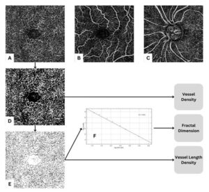
Figure 1: OCTA scan analysis. The upper row depicts raw scans of the deep capillary plexus (A), superficial capillary plexus (B) and radial peripapillary capillaries (C). ImageJ processing steps include conversion of the raw images (A-C) to a black-and-white binary map (D), which is then skeletonized (E). Next, the box-counting algorithm (F) is applied to the skeletonized map (E) to calculate the fractal dimension. Similarly, vessel density is calculated from the binary image (D), while vessel length density is calculated using the skeletonized map (E).
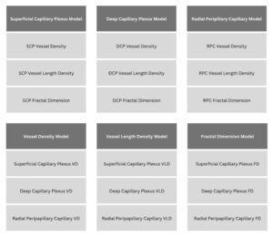
Figure 2: Layer and parameter analysis models.
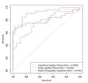
Figure 3: Vascular layer ROC curves. ROC curves for the superficial capillary plexus (dotted line), deep capillary plexus (dashed line) and RPC (alternating dots and dashes) layers are illustrated above. Vessel density, vessel length density and fractal dimension were used to generate these curves.
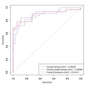
Figure 4: OCTA parameter ROC curves. ROC curves for vessel density (dotted line), vessel length density (dashed line) and fractal dimension (alternating dots and dashes) are displayed above. Data from the superficial capillary plexus, deep capillary plexus and radial peripapillary capillaries were used to generate these curves.
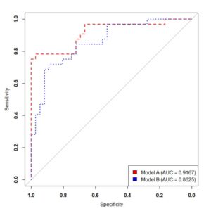
Figure 5: ROC curves of OCTA model vs clinical data model. The ROC curves of the combined diagnostic models are depicted above. Model A (dashed line) was trained using vessel density, vessel length density and fractal dimension from all three capillary layers, while model B (dotted line) was trained with intraocular pressure, retinal nerve fiber layer thickness and cup/disc ratio, as well as age, race, sex, ethnicity, family history of glaucoma, hypertension, diabetes and cardiovascular disease history.
Variables | Healthy n = 85 (%) | Glaucoma n = 81 (%) | p-Value |
Eye Studied | 0.421 | ||
OS | 43 (50.6%) | 46 (56.8%) | |
OD | 42 (49.4%) | 35 (43.2%) | |
Age | 62.18 ± 2.56 | 71.38 ± 2.26 | < 0.001 |
Sex | 0.013 | ||
Male | 32 (37.6%) | 46 (56.8%) | |
Female | 53 (62.4%) | 35 (43.2%) | |
Race | 0.420 | ||
White | 38 (44.7%) | 35 (43.2%) | |
Black | 30 (35.3%) | 34 (42.0%) | |
South Asian | 7 (8.2%) | 6 (7.4%) | |
East Asian | 8 (9.4%) | 2 (2.5%) | |
American Indian or Alaskan Native | 0 (0%) | 1 (1.2%) | |
Unknown | 2 (2.4%) | 3 (3.7%) | |
Ethnicity | 0.627 | ||
Hispanic | 11 (12.9%) | 8 (9.9%) | |
Not Hispanic | 65 (76.5%) | 61 (75.3%) | |
Unknown | 9 (10.6%) | 12 (14.8%) | |
Family History | 0.357 | ||
Yes | 47 (55.3%) | 38 (46.9%) | |
No | 30 (35.3%) | 30 (37.0%) | |
Not Available | 8 (9.4%) | 13 (16.0%) | |
Hypertension | 0.005 | ||
Yes | 46 (54.1%) | 61 (75.3%) | |
No | 34 (40.0%) | 20 (24.7%) | |
Unknown | 5 (5.9%) | 0 (0%) | |
Diabetes Mellitus | 0.267 | ||
Yes | 23 (27.1%) | 28 (34.6%) | |
No | 56 (65.9%) | 51 (63.0%) | |
Unknown | 6 (7.1%) | 2 (2.5%) | |
Cardiovascular Disease | 0.269 | ||
Yes | 50 (58.8%) | 55 (67.9%) | |
No | 29 (34.1%) | 24 (29.6%) | |
Unknown | 6 (7.1%) | 2 (2.5%) | |
IOP | 14.78 ± 0.75 | 15.31 ± 0.83 | 0.355 |
RNFL | 100.45 ± 2.25 | 74.32 ± 2.88 | < 0.001 |
CDR | 0.40 ± 0.04 | 0.62 ± 0.03 | < 0.001 |
Table 1: Characteristics of study participants.
Healthy | Glaucoma | p-Value | |
Superficial Capillary Layer | |||
VD | 0.24 ± 0.01 | 0.20 ± 0.01 | < 0.001 |
VLD | 0.11 ± 0.004 | 0.09 ± 0.004 | < 0.001 |
FD | 1.65 ± 0.01 | 1.58 ± 0.02 | < 0.001 |
Deep Capillary Layer | |||
VD | 0.33 ± 0.01 | 0.27 ± 0.01 | < 0.001 |
VLD | 0.18 ± 0.01 | 0.14 ± 0.01 | < 0.001 |
FD | 1.79 ± 0.01 | 1.72 ± 0.02 | < 0.001 |
Optic Nerve Head | |||
VD | 0.31 ± 0.01 | 0.23 ± 0.01 | < 0.001 |
VLD | 0.13 ± 0.003 | 0.09 ± 0.01 | < 0.001 |
FD | 1.69 ± 0.01 | 1.56 ± 0.02 | < 0.001 |
ROC analysis for the three vascular layers is illustrated in Fig. 3. The RPC layer performed the best (AUC = 0.9184, CI: 0.85-0.98) and was significantly different from the SCP (AUC = 0.7682, CI: 0.65-0.88, p = 0.004) and DCP (AUC = 0.8038, CI: 0.69-0.92, p = 0.03). | |||
Table 2: Comparison of OCTA parameters between healthy and glaucoma groups.


