Konstantinos AA Douglas1, Marios D Tibilis1, Georgios S Dimtsas1, Vivian Paraskevi Douglas2, Konstantinos Nanopoulos1, Marilita M Moschos1*
11st Department of Ophthalmology, National and Kapodistrian University of Athens, “G. Gennimatas” General Hospital, Athens, Greece
2Athens Naval Hospital, Department of Ophthalmology, Athens, Greece
*Corresponding Author: Marilita M Moschos, 1st Department of Ophthalmology, National and Kapodistrian University of Athens, “G. Gennimatas” General Hospital, Athens, Greece;
Email: [email protected]
Published Date: 27-05-2022
Copyright© 2022 by Moschos MM, et al. All rights reserved. This is an open access article distributed under the terms of the Creative Commons Attribution License, which permits unrestricted use, distribution, and reproduction in any medium, provided the original author and source are credited.
Abstract
Foveal hypoplasia is a term commonly used to describe the lack of development or underdevelopment of the foveal pit which anatomically and functionally correlates with a region of high visual acuity. Despite the fact that, fovea facilitates more precise and greater visual processing, the absence of foveal pit does not mean, a priori, poor visual acuity. While foveal hypoplasia has been usually associated with variable ocular abnormalities, isolated foveal hypoplasia though rare, has been also described. We present a case of a 31 year-old Caucasian male with an isolated foveal hypoplasia and the clinical evaluation in combination with the imaging findings, documented by multifocal ERG for the first time in literature.
Keywords
Isolated Foveal Hypoplasia; Foveal Pit; Optical Coherence Tomography; Fluorescein Angiography; Fundus Autofluorescence; Electroretinogram
Introduction
Fovea is an excavated area of the retina that contributes to the visual acuity. The development of the fovea starts at week 25 of gestation while it fully completes between 15th and 45th month after birth. Human fovea is composed of cone photoreceptors and Müller cells, while the absence of the rod photoreceptors is typical [1,2]. Foveal hypoplasia refers to the clinical finding when the foveal pit is poorly demarcated as well as there is a lack of foveal pigmentation, absence of the Foveal Avascular Zone (FAZ) and continuity of the inner retinal layers in the fovea [3]. It has been associated with a large number of ocular disorders such as albinism, aniridia, microphthalmos and achromatopsia but it has also been reported as an isolated finding [4].
Thomas, et al. have developed a grading system for the assessment of foveal hypoplasia using Ultra high-Resolution Optical Coherence Tomography (UHR- OCT) [5]. Four grades were distinguished based on the absence of foveal pit, Outer Segment (OS) lengthening and Outer Nuclear Layer (ONL) widening and they noticed a progressive VA deterioration which was significant in each grade [5].
Herein, we report the clinical findings and the diagnostic approach of a rare case of isolated foveal hypoplasia of both eyes in an adult man, assessed by OCT and multifocal Electroretinogram (mfERG) recording.
Case Presentation
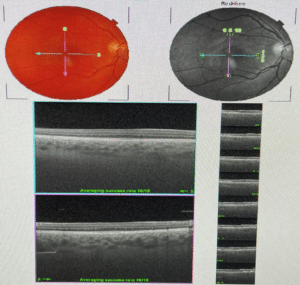
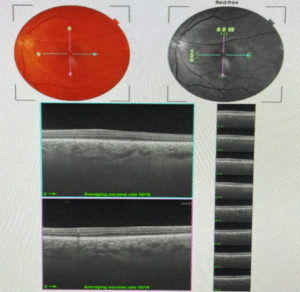
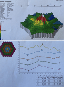
A 31 year-old Caucasian male was referred to our department complaining of moderate bilateral visual impairment since early childhood (~2 years old). His present and past medical history were unremarkable and his family history was non-contributory. On examination, Best Corrected Visual Acuity (BCVA) was 1/10 in both eyes (OU) and intra-ocular pressure was 12mmHg. Extraocular movements were full without presence of nystagmus. No iris trans- illumination defects were noted in both eyes on anterior chamber examination, while the chamber angle was wide open. Color vision was normal. On dilated fundus examination ill-defined maculofoveal areas without foveal reflexes were demonstrated bilaterally. The retinal vessels appeared to be normal OU. Spectral-domain optical coherence tomography (Heidelberg Engineering, Heidelberg, Germany) showed grade 3 foveal hypoplasia, which was characterized by the absence of foveal pit, persistence of inner retinal layers, absence of OS layer lengthening and retained widening of the ONL OU (Fig. 1). There was no foveal attenuation on fundus autofluorescence due to lack of macular pigmentation and on fluorescein angiogram reduction of the avascular zone was noted bilaterally. Full-field ERG was normal but the multifocal ERG was reduced bilaterally in both foveal (area 1) and parafoveal (area 2) areas. The values were 90 nv/deg2 in the foveal area and 71 nv/deg2 in the parafoveal area of the right eye and 89 nv/deg2 and 66 nv/deg2 in the left eye respectively (Fig. 2, 3).


Figure 1: Spectral-domain optical coherence tomography showing grade 3 foveal hypoplasia in both eyes (a, Right eye; b, Left eye).

Figure 2: Multifocal ERG of the right eye showing signal depression in both foveal and parafoveal areas (90 nv/deg2 in the foveal area and 71 nv/deg2 respectively).

Figure 3: Multifocal ERG of the left eye showing signal depression in both foveal and parafoveal areas (89 nv/deg2 in the foveal area and 66 nv/deg2 respectively).
Discussion
Foveal pit morphology is a feature of all human retinas. During the development of the fovea two remarkable processes take place; the bidirectional displace of inner and outer layers and the cone specialization. The inner layers are moved centrifugally and the foveal pit is formed, while the cones are moved centripetally to increase their density at the foveal. At the age of 4, the cone density at the fovea is 108,000/mm2 [2]. As far as the cone specialization is concerned, it refers to the lengthening of the cones outer segment and the increase of its density [6].
It is commonly believed that the presence of the foveal pit means high visual acuity. However, the cone specialization is not correlated with neither the presence nor absence of the pit nor with the avascular zone. And that could partially justify the high visual acuity maintenance in several cases of foveal hypoplasia, either the ones that are isolated or the ones associated with other ocular disorders. As a result, the absence of a well demarcated foveal pit should always be evaluated and interpreted in conjunction with the history and clinical examination [6,7]. Thomas, et al., have developed a grading system for the assessment of foveal hypoplasia using UHR-OCT. Four grades were distinguished based on the absence of foveal pit, OS lengthening and ONL widening and they noticed a progressive VA deterioration in grades 2, 3 and 4 [5]. Whilst the OCT demonstrates the anatomical impairment, the mfERG gives us the opportunity to understand the functional impairment, the foveal hypoplasia leads to the mfERG allows simultaneously the derivation of 61 or 103 local ERG signals in a visual field of 40°-50° of diameter around the fovea. Thus, the decrease of retinal function due to regional disorders in the outer retinal layers can be described in details, which allows the functional mapping of the retina, separated in five concentric rings. Isolated foveal hypoplasia does not appear to have a clear genetic pattern. Missense mutations of the PAX6 gene, which is associated with normal eye configuration, could be noticed among patients [8,9]. However, the gene seems to have low degree of penetrance. The data suggest that the etiology derives from not only genetic but also non-genetic factors [8-10].
In conclusion, we present a rare case of isolated foveal hypoplasia in a young adult. His vision was significantly impaired since early childhood without nystagmus or any other clinical findings related to other ocular disorders noted. We suggest that MF-ERG could be an objective method of assessing the function of the macula. Additionally, isolated FH should always be included in the differential diagnosis in cases presenting with bilateral visual impairment, especially when there is lack of foveal reflex.
Conflict of Interest
The authors declare that there is no conflict of interest.
Informed Consent
The patient has consented to the submission of the case report to the journal.
Statements of Human Rights
All procedures performed in studies involving human participants were in accordance with the ethical standards of the institutional and/or national research committee and with the 1964 Declaration of Helsinki and its later amendments or comparable ethical standards.
Statement on the Welfare of Animals
This article does not contain any studies with animals performed by any of the authors.
Acknowledgements
The authors thank the patient for participating in this report.
References
- Dubis AM, Costakos DM, Subramaniam CD, Godara P, Wirostko WJ, Carroll J, et al. Evaluation of normal human foveal development using optical coherence tomography and histologic examination. Arch Ophthalmol. (Chicago, Ill : 1960). 2012;130:1291-300.
- Kondo H. Foveal hypoplasia and optical coherence tomographic imaging. Taiwan J Ophthalmol. 2018;8:181.
- Karaca EE, Çubuk MÖ, Ekici F, Akçam HT, Waisbourd M, Hasanreisoǧlu M. Isolated foveal hypoplasia: clinical presentation and imaging findings. Optometry and vision science : official publication Am Acad Optometry. 2014;91.
- Vincent A, Kemmanu V, Shetty R, Anandula V, Madhavarao B, Shetty B. Variable expressivity of ocular associations of foveal hypoplasia in a family. Eye (London, England). 2009;23:1735-9.
- Thomas MG, Kumar A, Mohammad S, Proudlock FA, Engle EC, Andrews C, et al. Structural grading of foveal hypoplasia using spectral-domain optical coherence tomography a predictor of visual acuity? Ophthalmol. 2011;118:1653-60.
- Marmor MF, Choi SS, Zawadzki RJ, Werner JS. Visual insignificance of the foveal pit: reassessment of foveal hypoplasia as fovea plana. Arch Ophthalmol. 2008;126:907-13.
- Mota Á, Fonseca S, Carneiro Â, Magalhães A, Brandão E, Falcão-Reis F. Isolated foveal hypoplasia: tomographic, angiographic and autofluorescence patterns. Case Rep in Ophthalmological Med. 2012.
- Azuma N, Yamaguchi Y, Handa H, Hayakawa M, Kanai A, Yamada M. Missense mutation in the alternative splice region of the PAX6 gene in eye anomalies. Am J Human Genetics. 1999;65:656-63.
- Azuma N, Nishina S, Yanagisawa H, Okuyama T, Yamada M. PAX6 missense mutation in isolated foveal hypoplasia. Nature Genetics. 1996;13:141-2.
- Oliver MD, Dotan SA, Chemke J, Abraham FA. Isolated foveal hypoplasia. Br J Ophthalmol. 1987;71:926-30.
Article Type
Case Report
Publication History
Received Date: 22-04-2022
Accepted Date: 20-05-2022
Published Date: 27-05-2022
Copyright© 2022 by Moschos MM, et al. All rights reserved. This is an open access article distributed under the terms of the Creative Commons Attribution License, which permits unrestricted use, distribution, and reproduction in any medium, provided the original author and source are credited.
Citation: Moschos MM, et al. Isolated Foveal Hypoplasia Assessed by Multi-Focal Electroretinogram: A Clinical Presentation. J Ophthalmol Adv Res. 2022;3(2):1-6.


Figure 1: Spectral-domain optical coherence tomography showing grade 3 foveal hypoplasia in both eyes (a, Right eye; b, Left eye).

Figure 2: Multifocal ERG of the right eye showing signal depression in both foveal and parafoveal areas (90 nv/deg2 in the foveal area and 71 nv/deg2 respectively).

Figure 3: Multifocal ERG of the left eye showing signal depression in both foveal and parafoveal areas (89 nv/deg2 in the foveal area and 66 nv/deg2 respectively).


