Sudarshan Kumar Khokhar1, Amber Amar Bhayana1*, Priyanka Prasad1
1- Senior Resident, Dr. RP Centre for Ophthalmic Sciences, AIIMS, New Delhi, India
*Corresponding Author: Amber Amar Bhayana, Senior Resident, Dr. Rajendra Prasad Centre for Ophthalmic Sciences, All India Institute of Medical Sciences, New Delhi, India; Email: [email protected]
Published Date: 21-11-2020
Copyright© 2020 by Khokhar SK, et al. All rights reserved. This is an open access article distributed under the terms of the Creative Commons Attribution License, which permits unrestricted use, distribution, and reproduction in any medium, provided the original author and source are credited.
Clinical Image
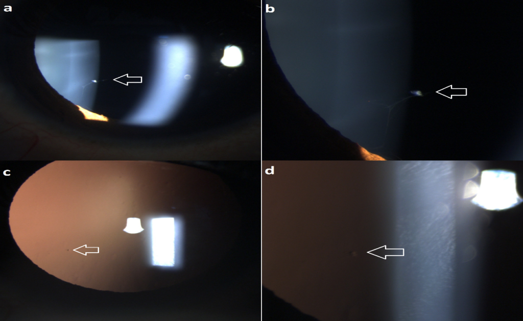
Persistent fetal vasculature can have a wide spectrum of presentations varying from Bergmeister’s papillae to persistent pupillary membrane (from posterior to anterior) [1]. Just like Mittendorf’s dot which is seen as an opacity on the posterior capsule and represents attachment of hyaloid artery to the lens, we would like to describe another form of persistent fetal vasculature which we would like to call Khokhar’s dot after the author which represents crumpled mass of opacity seen trapped in the network of persistent pupillary membrane anterior to the crystalline lens (Fig. 1) [1].


Figure 1: (a): oblique illumination x10 showing persistant pupillary membrane which has crumpled to form an opacity (white arrow); (b): x25 magnified view of (a); (c): retroillumination x10 of (a); (d): x25 magnification of (c).
References
1. Khokhar S, Dhull C. Commentary: Understanding angiogenic factors in pathogenesis of persistent fetal vasculature. Indian J Ophthalmol. 2019;67:1622-3.
Article Type
Clinical Image
Publication History
Received Date: 23-10-2020
Accepted Date: 14-11-2020
Published Date: 21-11-2020
Copyright© 2020 by Khokhar SK, et al. All rights reserved. This is an open access article distributed under the terms of the Creative Commons Attribution License, which permits unrestricted use, distribution, and reproduction in any medium, provided the original author and source are credited.
Citation: Bhayana AA, et al. Khokhar’s Dot. J Ophthalmol Adv Res. 2020;1(1):1-2.


Figure 1: (a): oblique illumination x10 showing persistant pupillary membrane which has crumpled to form an opacity (white arrow); (b): x25 magnified view of (a); (c): retroillumination x10 of (a); (d): x25 magnification of (c).


