Kohler’s Disease Case Report: Treatment with Regenerative Distraction Arthroplasty Technology
Gordon Slater1*, Tandose Sambo1, Tayla Slater1, Valeria Pozharova1
1New South Head Rd, Double Bay NSW 2028, Australia
*Correspondence author: Gordon Slater, MBBS (UNSW) FRACS, FA, Ortho A, New South Head Rd, Double Bay NSW 2028, Australia; Email: Gordonjakll@gmail.com
Citation: Slater G, et al. Kohler’s Disease Case Report: Treatment with Regenerative Distraction Arthroplasty Technology. Jour Clin Med Res. 2023;4(2):1-12.
Copyright© 2023 by Slater G, et al. All rights reserved. This is an open access article distributed under the terms of the Creative Commons Attribution License, which permits unrestricted use, distribution, and reproduction in any medium, provided the original author and source are credited.
| Received 01 May, 2023 | Accepted 22 May, 2023 | Published 29 May, 2023 |
Abstract
Kohler’s disease is a rare condition which in most cases responds to non-operative management. This report presents an unusual case where the thinning of the navicular resulted in a symptomatic pathological fracture. The patient was treated initially by Open Reduction and Internal Fixation (ORIF). The fracture united and the patient’s symptoms initially resolved. Three years later, symptomatic peri-navicular arthritis developed which was treated by distraction arthroplasty leading to symptom resolution. This case presents a novel solution which otherwise would have resulted in a complex midfoot fusion in a young patient. The methodology demonstrates the utility of new technological advances in joint regenerative technology.
Keywords: Regenerative Technology; Kohler’s Disease; Avascular Necrosis; Distraction Arthroplasty
Introduction
Kohler’s disease was first observed and documented by Alban Kohler in 1908[1,2]. The condition is classified as belonging to the family of osteochondrosis. Osteochondrosis involves Avascular Necrosis (AVN) of primary or secondary centres of ossification. In Kohler’s disease, the navicular bone is affected. The primary candidates for Kohler’s disease are young children, who exhibit the rare condition during their childhood. Stress-related compression is found to be a predominant factor in the development of Kohler’s disease [3-6]. Medical science continues to investigate the root cause of the formation of Kohler’s disease.
Current orthopaedic theory is that stress-related compression in the vicinity of the navicular bone may develop inducing a delay in bone formation or ossification in young children. The development phases of bone formation in children usually take place in the range of 18-30 months. The rates of cartilage and bone formation may induce structural weakness to the bone. The navicular bone enables movement in the body. As stresses are placed on the bone, its development can be hindered. Weight gain in children can cause excessive strain on the navicular bone and affect the blood supply. With a decrease in nutrients, bone formation will be hindered leading to collapse of the bone. This report aims to shed light on the nature of Kohler’s disease and describe a novel treatment, distraction arthroplasty.
Signs and Symptoms
Kohler’s disease predominantly occurs in young male children between the ages of 1-10 years old. The peak range is between 3-7 years old [4]. The root cause, whether genetic or environmentally induced, is still being investigated by medical researchers. It is initially detected via limping and painful swelling of the feet [4]. According to the pain felt by the child, he/she may often start walking on the side of the foot to minimize the sensation. Other symptoms experienced by children include [4]:
- Pain or tenderness along the length of the arch of the foot
- Pain during walking or standing
- Navicular bone blood supply is affected, resulting in bone degradation
Female children have significantly less odds of developing Kohler’s disease. Medical statistics indicate that male children are four times more likely to develop the condition [5].
Osteochondritis Dissecans of the Navicular Bone
To fully understand the mechanism of development of Kohler’s disease, an understanding of the pathology via Osteochondritis Dissecans (OCD) of the navicular will be described [3]. OCD is often seen in children. This rare condition frequently impacts the c onvex articular surfaces of the ankle joint. Other joint sites such as the elbow and the knee are also affected. Less than 2% of the population is affected by Kohler’s disease [4]. The aetiology of osteochondrosis includes three major root causes: vascular accidents, heredity, and coagulation anomalies [5].
- Vascular Accidents: When sudden compressive forces are placed on the feet, there may be a restriction in the blood supply to the critical areas such as the navicular bone. As nutrient supply is depleted there may be a loss of tissue, and reduction in the development of the bones during maturity [6]
- Heredity: Medical science is still attempting to determine if there is a specific gene that causes Kohler’s disease. Genetic predispositions are currently thought to be an impactor [7]
- Coagulation Anomalies: Coagulation Anomalies are also found to be impactors to the formation of Kohler’s disease [8]
The navicular bone can also undergo a process known as spontaneous osteonecrosis, resulting in death of the navicular bone cells and deformation of the bone. This condition is known as Mueller-Weiss disease. Medical science is currently debating on the best treatment for this condition. Two methods of treatment include open triple fusion and talonavicular-cuneiform arthrodesis. The treatments are normally applied when the disease has progressed to Stage 4 [9]. Under the right conditions, both triple fusion and TNC arthrodesis have proven to be suitable methods for the treatment of Mueller-Weiss disease [10].
Population Prevalence and Diagnosis
Kohler’s Disease is a rare bone disorder that affects only 2% of the population. Males are more likely to be affected than females at a ratio of 5:1. There are various disorders in the osteochondrosis family that are similar to Kohler’s disease. These include Friedberg Disease and Grierson-Gopalan syndrome [11,12]. Whenever there is a persistent pain in the foot and ankle region, it should be assessed by an orthopaedic specialist. During the consultation, X-rays of the feet will be taken, to have a better understanding of the inner state of the feet. In the affected foot, the X-ray will normally show flattening, sclerosis, and fragmentation of the navicular bone. Normal feet will not have this condition, and the skeletal structure will be healthy.
Standard Treatment Protocols
Kohler’s disease will naturally resolve itself if the right conditions are induced for healing. In acute cases, the symptoms will last for a few days. In more severe cases, symptoms can last for six months to two years. Treatment plans often involve alleviating pain initially, and then enabling the foot to heal by having patients eliminate pressure on the feet. Orthopaedic specialists may prescribe the utilization of casts to keep the feet stabilized. They may also recommend that patients wear special shoes that can support the feet [13]. With the appropriate treatment plan, full recovery, and restoration of the function of the feet is possible. Additional treatments include a walking short leg cast equipped with toe extension [14]. The patient will wear the cast for approximately 6-8 weeks, and subsequent treatment may include arch supports. The cast is placed in a position that will ensure that the navicular is relaxed. By alleviating posterior tibialis strain, the optimum healing will be achieved.
Case Report
This report presents an extremely rare case where Kohler’s resulted in a pathological fracture with breach of the mid foot arch. A 17-year-old female patient presented with a pathological fracture from advanced Kohler’s disease to the senior editor. The patient did not respond to conservative measures. Pain was severe including activity related pain, use of painkillers and knee pain. Mobility and activity were severely limited. The X-ray presented also suggested intertarsal instability induced by separation of the navicular (Fig. 1,2).
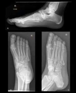
Figure 1: Initial Patient X-rays May 2017 (A) lateral, (B) anteroposterior, (C) oblique.
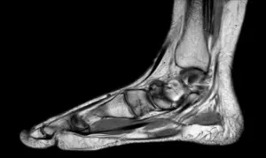
Figure 2: MRI June 2017. Findings: A two-part fracture through the body of the navicular was evident. At the site of the fracture there is patchy subacute marrow oedema. Superior fragment of the fracture was seen extruding superiorly into the talonavicular joint space. Medially the talonavicular joint showed a moderate effusion. There was loculated effusion in the posterior subtalar joint. No stress fractures identified in the navicular cuneiforms and metatarsals. Achilles tendon normal. Plantar fascia returned a normal signal. Anterior extensor tendons normal. No tenosynovitis in the peroneus brevis and longus. Medial flexor tendons normal.
Conclusion of MRI Scan (Fig. 2): There is a possible stressed two-part fracture through the body of the navicular. Remaining mid tarsal bones and metatarsals normal. Reactive effusion noted in the talonavicular joint and posterior subtalar joint.
Conservative measures fail to relieve symptoms. Boot and NSAIDs.
Initial Surgical Treatment July 2017
Patient initially underwent a navicular ORIF + bone graft as well as an insertion of arthroereisis plug and bridging plate. Post operatively patient was progressing well (Fig. 3).
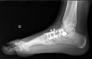
Figure 3: Right Foot X-ray August 2017, lateral.
At 4 weeks:
- X-ray demonstrated satisfactory reduction in deformity
- Patient advised to commence Touch Weight Bearing (TWB) at 6 weeks post-op
At 3 months:
- Total weight bearing in CAM boot with mild pain.
- Ankle ROM: DF: 15o, PF 60o, eversion 15o, inversion 5o
Gait: independent in boot, no aids. Trendelenburg type gait due to boot height
- Treatment included ankle PROM, gentle isometric calf, calf massage and gait retraining
Subsequent Removal of Plate and Screws February 2018
X-ray showed break of bridging plate. Overall foot alignment was good. Arthroereisis plug and bridging plate were removed without difficulty. One screw was left to secure the fracture. Arthroscopy of subtalar and talonavicular joint was performed revealing satisfactory joint surfaces. Patient was discharged with standard post-operative protocol; limb obs, elevation and consultation 7-10 days post operatively. Patient continued with physio and use of CAM boot, had minimal pain 9 days post-op. Advised to progress out of boot.
After 3 months post-op patient had progressed out of the boot. Foot was well aligned, minimal tenderness and reduced symptoms. Advised to gradually return to activity as tolerated and scheduled for review 15 months after operation date.
Review April 2020
Patient presented to the senior editor in April 2020 with pain at the medial aspect of the ankle joint. She was prescribed Celebrex and referred for X-ray /MRI. Upon review the signs of advancing arthritis. Senior editor prescribed NSAIDs and CAM boot. Upon review 4 weeks later, it was concluded that the patient needed either distraction arthroplasty or a medial column fusion as well as an injection with la and celestone. Patient was advised to continue physiotherapy. Patient was still experiencing pain (Fig. 4).
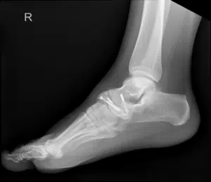
Figure 4: X-ray April 2020, lateral.
Review July 2021
Follow-up MRI scan showed incongruity of the T/N N/C and options were discussed with regards to surgery. The option of medial column fusion vs ankle plus midfoot distraction arthroplasty was discussed. Ankle distraction arthroplasty scheduled as patient did not want to go straight to fusion.
Ankle Distraction Arthroplasty August 2021
Positioning
Patient was placed supine on the operating table. A tourniquet was applied and leg was exsanguinated. Standard prepping and draping occurred. IV antibiotics given with induction and a regional block administered.
Method
A non-invasive distractor was applied and a 2.7 mm three chip arthroscope was introduced through a medial portal. Joint line synovitis was encountered in the anterior, lateral, and medial gutters and this was resected using a 4.5 mm full radius chondrotome. Good clearance of the synovitic material was obtained.
Medial Gutter
MCL:ok
Talus:1-2
Tibia:1
Lateral Gutter
Lateral Ligament
Talus:1
Tibia:1
Central Joint
Talus:1
Tibia:1
Trifurcation Zones
Noted to be hypertrophied and marked synovitis was resected without difficulty.
Tib-Fib Joint: Stable
Guided injection to T/N and N/C. Platelet rich plasma injected and arthroscopic inspection performed. cartilage gd 2. marked synovitis.
- Frame assembly: Two ring proximal fixateur selected and assembled with a foot ring. 6x telescopic arms were applied to the base plate and the distal ring in a standard hexagonal system. TSF. 4x threaded bolts used to bridge the rings with 8x bolts.
Size adjusted to leg shape and telescopic arms set to minimum length and locked.
- Placement: Linen placed under the thigh and the ring placed to allow contact of the foot onto the ground. Ring applied with proximal ring such that the foot transosseous wire placement through safe zones could be performed. Careful measurement of the frame positioning made so there was no contact with posterior aspect of the frame by the calf. Initial stab incision made at the medial tibial cortex. Safe zones for wire placement identified. Diamond tip drill 2.5 mm used to drill the medial tibial cortex. 2.7 mm guide wire used to pass through the tibia. Locked into place using slotted nuts and blocks. Calcaneus wire.Medial tibial neurovascular bundle palpated, medial tibial wire inserted posterior to the bundle. Lateral placement to avoid the peroneal tendons. 2.7 wire attached with slotted bolts and blocks on the foot ring. Process repeated a further 5 times securing further 5 x 2.7 mm wires onto the frame securing it into position. Wires inserted through anatomical safe zones. Skin protection. Wires with bolts attached to rings. 80 Nuts and bolts checked and tightened. Initial distraction made.
Telescopic arms locked in initial distraction. Distal ring applied. Two wires passed through the first and second metatarsals and then secured to the frame. Circular with telescopic arms. Initial distraction performed.
- Closure: Portals dressed with gelinette. Injection with local and adrenaline using a 10 ml syringe and spinal needle
- Post-op: Elevation and limb obs conducted. Patient advised to mobilise as tolerated, permitted to TWB. Anti-biotics were continued. Patient discharged after 1 day with knee scooter and education on pin site management (Fig. 5,6).
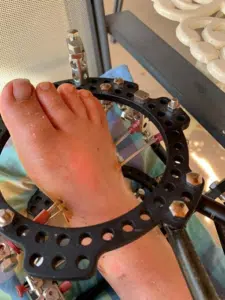
Figure 5: Photograph of applied coupler frame, immediately post-surgery.
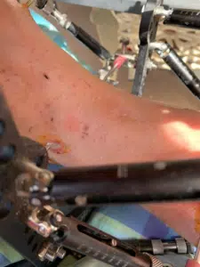
Figure 6: Close-up photograph of pin site medial ankle, immediately post-surgery.
Review 1: August 2021
At 2 weeks post ankle distraction arthroplasty:
- Ankle progressing well
- Palexia IR given
- X-ray ordered
- Proximal ring and the foot ring lengthened providing distraction at the midfoot
- Patient advised how to distract the joints daily
Review 2: August 2021
At 3 weeks post ankle distraction arthroplasty
- Right foot and ankle X-ray going well
- Distraction was progressing ok
- Patient had some calf pain, an ultrasound was ordered to check for a DVT. Subsequently cleared
- Review for frame adjustment scheduled for 4 days’ time
- X-ray and CT scan scheduled for 12 days’ time
- Satisfactory distraction noted (Fig. 7)
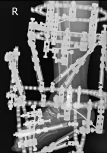
Figure7: Patient X-ray post application of coupler frame, lateral.
Right Foot Removal of Illizarov Frame September 2021
After 8 weeks the external fixator was removed.
Positioning
Patient was placed supine on the operating table. A tourniquet was applied and leg was exsanguinated. Standard prepping and draping occurred. IV antibiotics given with induction and a regional block administered.
Method
An injection with local anaesthetic and steroid was administered with a 10ml syringe spinal needle.
Frame removed and multiple pin sites debrided.
3 mm screw targeted and removed.
Ostectomy of dorsal navicular to remove the screw was performed.
On fluroscan the joints have regenerated.
Closure
The wounds were closed in layers using rapide. A Jelonet and compression dressing was applied.
Post-Operative
The patient was discharged to the ward for standard post-operative protocol; limb obs, elevation and consultation 7-10 days post operatively. Patient continued with physiotherapy, able to mobilize without pain within 12 days post removal with CAM walker boot and crutches.
Review November 2021
42 days post frame removal X-rays showed increase in intertarsal joint space inferring successful regeneration of the mid tarsal joints. Pain receding. Recommended gait retraining in high lace boot and stationary bike exercise (Fig. 8).
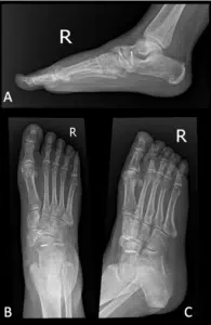
Figure 8: Patient X-ray 42 days post frame removal, right foot (A) lateral, (B) AP, (C) oblique.
Review December 2021
3 months post removal, the right foot was progressing well. Patient no longer in pain and walking without the CAM boot. Recommended to progress to treadmill exercise.
Review February 2022
Good progression.
Review May 2022
Patient was able to resume normal activity, working full time and wearing an ankle brace. Pain killers no longer needed and night-time resolved.
Discussion
Kohler’s disease is a rare condition which in the natural history is resolution with observation and non-operative management [15-17]. We found a previous case report which had been treated with a medial column fusion. This appeared to lead to a satisfactory resolution of the patient symptoms. Medial column fusion does lead to a significant increase in the stiffness of the foot [10]. In the last 7 years the senior author has been treating some cases of ankle, hindfoot and midfoot arthritis and cartilage injury with distraction arthroplasty. This is seen as a potential step before more definitive measures such as fusion or joint replacement. It does not “burn bridges” as other options can be used if there is failure of the procedure. The advantage for the patient is that if there is successful regeneration then a return to age matched activities is possible rather than merely an improvement in the current symptom profile.
Principles of Regeneration [18-22]:
- Debridement
- Stimulation of joint surfaces
- Distraction
- Insertion of triggering agents
- Adequate time for distraction
- Use of potentiators such as Hyperbaric oxygen therapy
- Use of disease modulators such as prednisone (or potentially pentosan in the future)
It would appear there is a greater chance of success with younger patients, a shorter period of time since the initiating event and a stable well aligned joint. It would also appear that regeneration is a much more complex decision-making process for patient selection than for fusion or joint replacement. This we would propose as being the reason that there has been such a wide range of results in joint regeneration technology. Just as with amputation which we removes disease process and limb completely, fusion and joint replacement replace the joint “organ”. As such they can be described as organ loss procedures [20]. Regeneration falls into organ salvage/preservation and as such is more akin to dentistry where the salvation of the tooth and ongoing maintenance is paramount [23,24].
Regeneration Success Factors
For the surgeon and patient, a constellation of factors need to be considered to predict the chance of success and the time that success will likely take [20,21].
- Patient age
- Time since initiating injury
- Disease extent/bipolar disease
- Joint congruity
- Subarticular cyst formation
- Realignment/other procedure requirement
- Type of disease
- Obesity
- Intercurrent disease/diabetes/anti coagulants
- Number of joints to be regenerated
- Smoking
- Sex
- Previous intra-articular infection/gout
- Hyper elasticity/CMT/Charcot disease
- Compliance
- Immunosuppressed
- Joint stability
- Number of joints to be regenerated
In essence, you regenerate when you can still regenerate. Thus, it is surgery which is performed early rather than late; it is not surgery of desperation. This patient demonstrates a number of positive attributes for regeneration with her young age giving priority over other factors such as multiple joint involvement, joint incongruity, bipolar disease etc. We believe, particularly in younger patients, that distraction arthroplasty can provide an alternative to fusion. Nil further use of adjuvant treatments was required in this case. Our initial experience with patients above the age of 70 has been poor. Currently distraction is no longer offered except in acute situations.
Further case analyses are required and follow up needed. Initial results have been very encouraging with regeneration. Importantly, failure of regeneration of a joint will ‘prime the joint’ leading to fixation to occur quickly. We will continue to explore this. Further regeneration of adjacent joint appears to be beneficial in minimising the stiffness that inevitably occurs after surgery.
Disclaimer
Dr Gordon Slater is a medical director of Integrant Pty Ltd, a biotechnology and surgical equipment company. He also has a pecuniary interest in RegenU, Australia and MD Hyperbaric.
Conflict of Interest
The authors have no conflict of interest to declare.
References
- John’s Hopkins Medicine. Ankle Fracture Open Reduction and Internal Fixation. Johns Hopkins Medicine. 2022. [Last accessed on May 22, 2023]
- Laissue JA, Blattmann H, Slatkin DN. Alban Köhler (1874-1947): Inventor of grid therapy. Zeitschrift Fur Medizinische Physik. 2011;22(2):90-9.
- Beil FT, Bruns J, Habermann CR, Rüther W, Niemeier A. Osteochondritis dissecans of the tarsal navicular bone: a case report. J Am Podiatr Med Assoc. 2012;102(4):338-42.
- Kohler Disease – NORD. National Organization for Rare Disorders. 2022. [Last accessed on May 22, 2023]
https://rarediseases.org/rare-diseases/kohler-disease
- Krueger C. Kohler’s Disease. 2022. [Last accessed on May 22, 2023]
https://www.orthobullets.com/pediatrics/4071/kohlers-disease
- Rovensky J, Payer J. Köhler’s disease. 2009. [Last accessed on May 22, 2023]
https://www.researchgate.net/publication/285332939_Kohler%27s_disease
- Perform Podiatry. Kohler Disease. 2022. [Last accessed on May 22, 2023]
https://www.performpodiatry.co.nz/kohler-disease/
- Trammell AP. Kohler Disease. National Library of Medicine. 2022. [Last accessed on May 22, 2023]
https://www.ncbi.nlm.nih.gov/books/NBK507831/
- Zhang H. Open triple fusion versus TNC arthrodesis in the treatment of Mueller-Weiss disease. BioMed Central. 2017. [Last accessed on May 22, 2023]
https://josr-online.biomedcentral.com/articles/10.1186/s13018-017-0513-3
- Volpe A, Monestier L, Malara T, Riva G, La Barbera G, Surace MF. Müller-Weiss disease: Four case reports. World J Orthopedics. 2020;11(11):507.
- Freiberg’s disease – about the disease – genetic and rare diseases information center. genetic and rare diseases information center. 2021. [Last accessed on May 22, 2023]
https://rarediseases.info.nih.gov/diseases/2380/freibergs-disease
- Burning feet syndrome (Grierson-Gopalan syndrome). Cleveland Clinic. 2018. [Last accessed on May 22, 2023]
https://my.clevelandclinic.org/health/symptoms/17773-burning-feet-syndrome-grierson-gopalan-syndrome
- Patel SB, Diamond HS. Avascular necrosis: practice essentials, pathophysiology, etiology. Medscape. 2022. [Last accessed on May 22, 2023]
https://emedicine.medscape.com/article/333364-overview#
- Vascular disease: types, causes, symptoms and treatment. Cleveland Clinic. 2022. [Last accessed on May 22, 2023]
https://my.clevelandclinic.org/health/diseases/17604-vascular-disease
- Kohler’s disease (avascular necrosis of the navicular) – ankle, foot and orthotic centre. Ankle, Foot and Orthotic Centre. 2022. [Last accessed on May 22, 2023]
https://ankleandfootcentre.com.au/kohlers-disease-avascular-necrosis-navicular/
- Vargas B. Kohler disease treatment and management: Medical therapy, complications, long-term monitoring. Diseases and Conditions – Medscape Reference. 2022. [Last accessed on May 22, 2023]
- Vargas B, Panchbhavi VK. Kohler disease: practice essentials, pathophysiology, etiology. Medscape. 2022. [Last accessed on May 22, 2023]
https://emedicine.medscape.com/article/1234753-overview
- Viladot A, Sodano L, Marcellini L. Joint debridement and microfracture for treatment late-stage Freiberg-Kohler’s disease: long-term follow-up study. Foot and Ankle Surgery. 2019;25(4):457-61.
- Slater G, Malley MO, Slater T, Sambo T. Hyperbaric Oxygen Therapy: An Overview. J Regen Biol Med. 2022;4(3):1-15.
- Slater G. Regenerative medicine requires a paradigm shift in outcome measures. J Stem Cell Res. 2021;2(1):1-18.
- Slater G, Mathen L. Current thinking in pin-site management in external hexagonal frames. J Orthop Study Sports Med. 2021;1(1):1-9.
- Slater GL, Javadian S, Mathen L. A review of distraction arthroplasty vs ankle arthrodesis vs ankle replacement. J Regen Biol Med. 2022;4(1)1-28.
- Zhao R, Yang R, Cooper PR, Khurshid Z, Shavandi A, Ratnayake J. Bone grafts and substitutes in dentistry: A review of current trends and developments. Molecules. 2021;26(10):3007.
- Haben Fesseha M. Bone grafting, its principle and application: A review. Osteol Rheumatol Open J. 2020;1:43-50.
This work is licensed under a Creative Commons Attribution 2.0 International License.


