Juan Cano Parra1, Francesc Artigas2,3,4*
1Policlínica ICOA, Barcelona, Spain
2Department of Neuroscience and Experimental Therapeutics, Institut d’Investigacions Biomèdiques de Barcelona, IIBB‐CSIC; Barcelona, Spain
3Institut d’Investigacions Biomèdiques August Pi i Sunyer (IDIBAPS), Barcelona, Spain
4Centro de Investigación Biomédica en Red de Salud Mental (CIBERSAM), Instituto de Salud Carlos III, Madrid, Spain
*Correspondence author: Francesc Artigas, Institut d’Investigacions Biomèdiques de Barcelona, Rosselló 161, 08036 Barcelona, Spain;
Email: francesc.artigas@iibb.csic.es
Published Date: 18-11-2023
Copyright© 2023 by Artigas F, et al. All rights reserved. This is an open access article distributed under the terms of the Creative Commons Attribution License, which permits unrestricted use, distribution, and reproduction in any medium, provided the original author and source are credited.
Abstract
Purpose: To examine the therapeutic potential of the amino acid taurine in the non‐ neovascular or ∙dry” form of Age‐Related Macular Degeneration (AMD), one of the main causes of vision loss in the elderly, which still lacks effective treatments. Taurine is the most abundant amino acid in the retina and exerts trophic and neuroprotective actions in cellular and animal models. Likewise, a recent Science paper indicates that taurine deficiency is a driver of aging and that taurine supplementation may be an effective treatment for age‐related diseases.
Case Description: A dry AMD patient (F.A., male, 62‐yr old) presented a Snellen best corrected visual acuity of 0.15 on the Right Eye (RE) and 0.2 on the Left Eye (LE) in June 2013, together with central retina atrophy and reduction of central macular thickness to 144 µm (RE) and 159 µn (LE) (OCT analysis; Topcon 3D OCT1000). Oral taurine intake (600 mg t.i.d.) arrested macular degeneration over a 5.5‐yr period and moderately improved visual acuity and macular thickness after doubling the dose from 1.8 g/day to 3.6 g/day in December 2018. This improvement remained stable until last control visit in January 2023.
Conclusion: Taurine may have disease‐modifying properties in dry AMD at the dose used. The present observations add to existing literature to foster proof‐of‐concept clinical trials using a high oral dose of taurine for the treatment of AMD.
Keywords: Age‐Related Macular Degeneration; Photoreceptors; Retinal Pigment Epithelium; Taurine; Visual Acuity
Introduction
Age‐related Macular Degeneration (AMD) is one of the leading causes of vision loss and blindness in older adults worldwide [1,2]. Its prevalence is higher among Europeans and North Americans, than among Asians and Africans, suggesting the involvement of gene x environment interactions [1]. Due to the growth in elderly population, AMD prevalence is expected to increase worldwide, from 196 million people in 2020 to 288 million people in 2040 [1].
AMD is associated to dysfunction and loss of Retinal Pigment Epithelium (RPE) and has a still poorly understood pathogenesis, likely involving a complex interaction of genetic, functional and environmental factors [2‐4]. This makes it difficult he identification of therapeutic targets and the subsequent development of treatments to prevent/arrest RPE neurodegeneration.
Very few therapies area available for the treatment of AMD. Among them, antioxidants are frequently prescribed [5,6] under an unclear rationale. Their effectiveness is more than controversial, and their use has not been recommended [7]. Oligonucleotide‐based therapies (aptamers) have been developed for the treatment of the neovascular or “wet” form of the disease. Their intraocular administration inhibits the synthesis of the Vascular Epithelial Growth Factor (VEGF), to prevent an excessive blood‐vessel formation in the retina [8,9]. Likewise, anti‐VEGF therapies based on monoclonal antibodies have also been developed, both strategies improving visual outcomes in neovascular AMD [9].
However, the non‐neovascular or “dry” form of the disease, more prevalent than the neovascular form, still lacks appropriate treatment. Besides to antioxidants, with poor or no effectiveness, novel stem cell‐based strategies have been developed in order to promote retinal cell growth yet their complexity prevents a widespread use [7,10].
Taurine is the most abundant amino acid in the retina and plays a major role in retinal function, including photoreceptor survival [11]. Taurine depletion impairs vision in animal models whereas its administration prevents retinal excitotoxicity induced by antagonists of N‐methyl‐D‐aspartate (NMDA) receptors in rodents [12,13]. Trophic effects of taurine have also been reported, such as the promotion of cell growth in retinal cell cultures and neurite outgrowth and synapse formation in the central nervous system [14,15]. Likewise, its systemic administration provides neuroprotection against retinal photoreceptor degeneration and visual function impairments [16].
Unfortunately, very little information is available on the potential neuroprotective/trophic effects of taurine in humans, although a few studies published indicate a positive role. Hence, individuals with a genetic deficiency of the SLC6A6 taurine transporter ‐which leads to a dramatic fall of its efficacy and of taurine levels (to ~15% of control values) ‐ show childhood cardiomyopathy together with retinal degeneration and vision impairment. Interestingly, a 2‐4‐month high‐dose taurine treatment (100 mg/kg/day p.o.) corrected cardiomyopathy, arrested retinal degeneration and improved vision [17].
Likewise, a recent Science paper shows that circulating levels of taurine markedly decline with age in mice, monkeys and humans and that taurine supplementation enhanced the health span (the period of healthy living) and life span in mice and health span in monkeys [18]. This suggests that taurine supplementation may be an effective treatment for age‐related diseases.
Here we report on a long‐lasting stabilization and subsequent moderate improvement of vision and macular thickness and retinal pigmentation by oral taurine intake in a case of dry AMD. One of us (FA; https://orcid.org/0000‐0002‐5880‐5720) was diagnosed of dry AMD in 2013.
Having a long‐lasting research experience in Translational Neuroscience, and being aware of the role of taurine in retinal function, the patient started to take oral taurine as a dietary supplement in 2013, under the careful supervision and control of his ophthalmologist (JCP).
Case Report
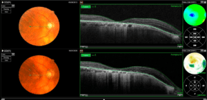
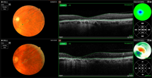
A 62‐year‐old male patient attended ICOA consultation on June 2013 presenting a Snellen best corrected visual acuity of 0.15 on the Right Eye (RE) and 0.2 on the Left Eye (LE). The Ocular Coherence Tomography (OCT) analysis (Topcon 3D OCT1000) of fundus eye revealed a bilateral dry AMD with central retina atrophy and a reduction of central macular thickness to 144 µm (RE) and 159 µn (LE) (Fig. 1,2). The patient started to take taurine supplement 600 mg t.i.d. p.o. (1.8 g/day in total; 27 mg/kg/day; Bonusan Labs (Alicante, Spain) and followed annual control. In December 2018, the dose was doubled to 1.800 mg b.i.d. (3.6 g/day in total; 54 mg/kg/day; the night dose was suppressed), without any side effect. In January 2020, the patient had an objective improvement on Snellen visual acuity to 0.2 (RE) and 0.3 (LE), together with an increase of macular thickness to 166 µm (RE) and 206 µm (LE) (Fig. 1,2), which remained stable until last control in January 2023. Likewise, an enhanced pigmentation of the whole retina in both eyes was also observed (Fig. 1,2).

Figure 1: Topcon 3D OCT1000 image of the fundus of RE. Upper and lower row panels correspond to June 2013 and January 2020, respectively. Macular thickness was 144 µm in June 2013 and 166 µm January 2020, showing an increase of 11 µm of macular thickness in central map difference.

Figure 2: Topcon 3D OCT1000 image of the fundus of LE. Upper and lower row panels correspond to June 2013 and January 2020, respectively. Macular thickness was 159 µm in June 2013 and 206 µm in January 2020, showing an increase of 79 µm of macular thickness in central map difference.
Discussion
AMD is a highly prevalent disease, being the main cause of vision loss in older adults [1,2]. Due to the growth in elderly population, the number of affected individuals is expected to rise globally, from 198 million people in 2020 to 288 million in 2040 [1]. AMD involves a progressive decrease in visual acuity and macular thickness resulting in in severe vision problems that dramatically worsen the quality of life of affected individuals. The neovascular form of the disease can be treated with oligonucleotide (aptamers) and monoclonal antibody therapies to reduce the expression of VEGF, thus preventing excessive blood‐vessel formation and rupture within the retina [8,9]. However, there are no effective treatments for the dry form of the disease, more prevalent than the neovascular or “wet” form.
Despite being a single case observation, the present results suggest that a high dose oral taurine may be a useful disease‐modifying therapy in dry AMD, given the remarkable arrest of macular degeneration and vision loss for over a 10‐yr period. This was accompanied by a mild improvement in visual acuity and macular thickness after doubling the dose from 1.8 to 3.6 g/day (from 27 mg/kg/day to 54 mg/kg/day). Likewise, taurine markedly increased pigmentation in the whole retina. These observations are in accordance with accumulated preclinical evidence indicating a neuroprotective and tropic role of taurine in the retina (see Introduction). Likewise, they parallel the remarkable improvement of vision after a high oral taurine dose (100 mg/kg/day) of individuals with a genetic deficiency of the SLC6A6 taurine transporter, resulting in low taurine levels, cardiomyopathy, retinal degeneration and vision impairment [17]. Moreover, the present observations fully agree with a very recent report showing that circulating levels of taurine markedly decline with age and that taurine supplementation at high doses increases health span and life span (when examined) in mice, monkeys and humans [18]. Taurine reduced cellular senescence, protected against telomerase deficiency, suppressed mitochondrial dysfunction and decreased DNA damage, among other functions, suggesting that taurine supplementation can be an effective treatment for age‐ related diseases [18].
Within this overall context, the present study is the first one showing that a high taurine dose may have disease‐modifying properties in AMD. To our knowledge, no similar observations have been reported elsewhere, let alone for such an extended period of time (10 yr). A much lower dose of taurine (400 mg/day) was used in the TOZAL study, which reported moderate but significant increases of the visual acuity in 37 patients with dry AMD [19]. That study used a combination of antioxidants in addition to taurine (Omega‐3 Fatty Acids, Zinc antioxidant, Lutein), which makes it difficult to clarify the exact role of each individual component and of their possible interactions. Given the improvement of visual acuity and macular thickness observed after doubling the dose (from 18.8 g/day to 3.6 g/day) observed in the present case report, doses higher than 400 mg/day are likely required for the treatment of dry AMD.
Hence, given the excellent tolerability, lack of side effects and easy use of oral taurine, the present observations add to existing literature to foster the development of a proof‐of‐ concept, phase IIa clinical trials on taurine-particularly at high doses‐ as a therapeutic agent in AMD [11,18]. Taurine may be particularly beneficial in “dry” form of AMD, lacking effective treatments. Moreover, unlike expensive oligonucleotide or monoclonal antibody strategies, taurine can be an affordable treatment worldwide.
Funding Information
The authors have not received financial support for the present study. F.A. acknowledges overall group support from Centro de Investigaciones Biomédicas en Red de Salud Mental (CIBERSAM).
Conflict of Interest
The authors have no conflict of interest to declare.
References
- Wong WL, Su X, Li X, Cheung CM, Klein R, Cheng CY, et al. Global prevalence of age‐ related macular degeneration and disease burden projection for 2020 and 2040: a systematic review and meta‐analysis. Lancet Glob Health. 2014;2:e106‐16.
- Smith W, Assink J, Klein R, Mitchell P, Klaver CC, Klein, et al. Risk factors for age‐related macular degeneration: Pooled findings from three continents. Ophthalmol. 2001;108:697‐704.
- Nowak JZ. Age‐related Macular Degeneration (AMD): pathogenesis and therapy. Pharmacol Rep 2006;58:353‐63.
- Haddad S, Chen CA, Santangelo SL, Seddon JM. The genetics of age‐related macular degeneration: a review of progress to date. Surv Ophthalmol 2006;51:316‐63.
- Schmidl D, Garhöfer G, Schmetterer L. Nutritional supplements in age‐related macular degeneration. Acta Ophthalmol. 2015;93:105‐21.
- Dziedziak J, Kasarełło K, Cudnoch‐Jędrzejewska A. Dietary antioxidants in age‐related macular degeneration and glaucoma. Antioxidants 2021;10:1743.
- Banerjee M, Chawla R, Kumar A. Antioxidant supplements in age‐related macular degeneration: are they actually beneficial? Ther Adv Ophthalmol 2021; 13:1‐14.
- Whitehead KA, Langer R, Anderson DG. Knocking down barriers: advances in siRNA delivery. Nat Rev Drug Discov. 2009;8:129‐38.
- Daien V, Finger RP, Talks JS, Mitchell P, Wong TY, Sakamoto T, et al. Evolution of treatment paradigms in neovascular age‐related macular degeneration: a review of real‐world evidence. Br J Ophthalmol. 2021;105:1475‐79.
- Kashani AH. Stem cell‐derived retinal pigment epithelium transplantation in age‐ related macular degeneration: recent advances and challenges. Curr Opin Ophthalmol. 2022;133:211‐8.
- Militante JD, Lombardini JB. Taurine: evidence of physiological function in the retina. Nutr Neurosci. 2002;5:75‐90.
- Militante J, Lombardini JB. Age‐related retinal degeneration in animal models of aging: possible involvement of taurine deficiency and oxidative stress. Neurochem Res. 2004;29:151‐60.
- Lambuk L, Iezhitsa I, Agarwal R, Bakar NS, Agarwal P, Ismail NM. Antiapoptotic effect of taurine against NMDA‐induced retinal excitotoxicity in rats. Neurotoxicol. 2019;70:62‐71.
- Cubillos S, Lima L. Taurine trophic modulation of goldfish retinal outgrowth and its interaction with the optic tectum. Amino Acids. 2006;31:325‐31.
- Mersman B, Zaidi W, Syed NI, Xu F. Taurine promotes neurite outgrowth and synapse development of both vertebrate and invertebrate central neurons. Front Synaptic Neurosci. 2020;22;12:29
- Tao Y, He M, Yang Q, Ma Z, Qu Y, Chen W, et al. Systemic taurine treatment provides neuroprotection against retinal photoreceptor degeneration and visual function impairments. Drug Des Devel Ther. 2019;13:2689‐702.
- Ansar M, Ranza E, Shetty M, Paracha SA, Azam M, Kern I, et al. Taurine treatment of retinal degeneration and cardiomyopathy in a consanguineous family with SLC6A6 taurine transporter deficiency. Hum Mol Genet. 2020;29:618‐23.
- Singh P, Gollapalli K, Mangiola S, Schranner D, Yusuf MA, Chamoli M, et al. Taurine deficiency as a driver of aging. Science. 2023;380:9257.
- Cangemi FE. TOZAL Study: an open case control study of an oral antioxidant and omega‐3 supplement for dry AMD. BMC Ophthalmol. 2007;26;7:3.
Article Type
Case Report
Publication History
Received Date: 19-10-2023
Accepted Date: 07-11-2023
Published Date: 18-11-2023
Copyright© 2023 by Artigas F, et al. All rights reserved. This is an open access article distributed under the terms of the Creative Commons Attribution License, which permits unrestricted use, distribution, and reproduction in any medium, provided the original author and source are credited.
Citation: Artigas F, et al. Long‐Lasting Stabilization and Improvement of Dry Age‐Related Macular Degeneration by a High Oral Taurine Dose. J Ophthalmol Adv Res. 2023;4(3):1-5.

Figure 1: Topcon 3D OCT1000 image of the fundus of RE. Upper and lower row panels correspond to June 2013 and January 2020, respectively. Macular thickness was 144 µm in June 2013 and 166 µm January 2020, showing an increase of 11 µm of macular thickness in central map difference.

Figure 2: Topcon 3D OCT1000 image of the fundus of LE. Upper and lower row panels correspond to June 2013 and January 2020, respectively. Macular thickness was 159 µm in June 2013 and 206 µm in January 2020, showing an increase of 79 µm of macular thickness in central map difference.


