MR Imaging in Unilateral Hydrocephalus in Adults
Lokesh Rana1*, Dinesh Sood2, Naresh Rana1, Deepak Singh3
1-Assistant Professor, Department of Radio-diagnosis, Dr RPGMC, Kangra at Tanda, Himachal Pradesh, India
2-Professor, Department of Radio-diagnosis, Dr RPGMC, Kangra at Tanda, Himachal Pradesh, India
3-Resident, Department of Radio-diagnosis, Dr RPGMC, Kangra at Tanda, Himachal Pradesh, India
*Corresponding Author: Lokesh Rana, Assistant Professor, Department of Radio-diagnosis, Dr RPGMC, Kangra at Tanda, Himachal Pradesh, India; Email: poojalokesh2007@gmail.com
Citation: Rana L, et al. MR Imaging in Unilateral Hydrocephalus in Adults. J Clin Med Res.
2020;1(2):1-6.
Copyright© 2020 by Rana L, et al. All rights reserved. This is an open access article distributed under the terms of the Creative Commons Attribution License, which permits unrestricted use, distribution, and reproduction in any medium, provided the original author and source are credited.
| Received 07 Jun, 2020 | Accepted 24 Jun, 2020 | Published 02 Jul, 2020 |
Abstract
We present a case of a 19-year-old male who presented with history of headache and seizures of 3 years duration. There was unilateral lateral ventricle hydrocephalous. One of the two lateral ventricles was dilated. Idiopathic stenosis of foremen of Monro was found. It is one of the important causes, though it is a rare entity. Delayed clinical presentation may lead to delay in the diagnosis and thus causing delay in surgical planning if required.
Keywords
Neurology; Hydrocephalus; Tumors; Tomography
Introduction
Unilateral hydrocephalous implies to dilation of one of the lateral ventricles which may be progressive in nature resulting in compression of the cerebral nervous tissues [1,2]. Idiopathic occlusion seen at the level of foramen of Monro is the most common cause, however, underlying cause may be congenital web, haemorrhage, neoplasm, vascular anomalies, infection, trauma etc [3,4]. In this case report, we are reporting radiological features of idiopathic unilateral foramen of Monro stenosis with hydrocephalus.
Case Report
A 19- year-old male presented to the neurology OPD with chief complaints of progressive headache and seizures for three years duration. After clinical assessment the patient was referred to our department for MRI brain and find out cause of fourth nerve palsy. We did routine MRI scan in GE 1.5 Tesla machine which included axial T1W, FLAIR, DWI, FIESTA, GRADIENT, axial and coronal T2W and T1W post contrast sequences. The MR images showed presence of uniformly dilated right lateral ventricle with peri-ventricle ooze of CSF, the rest of the ventricle system was normal. There was presence of narrowing at the level of foramen of Monro without any abnormal enhancing lesion (Fig. 1-7). Therefore, diagnosis of idiopathic unilateral right-sided foramen of Monro stenosis was made according to others [3-6].
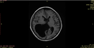
Figure 1: MRI scan of the case in axial T1W.
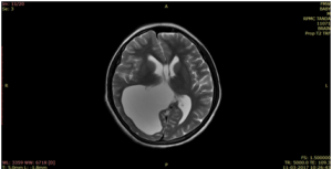
Figure 2: MRI scan of the case in axial T2W.
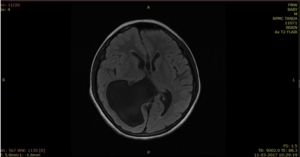
Figure 3: MRI scan of the case in axial FLAIR.
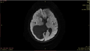
Figure 4: MRI scan of the case in axial DWI.
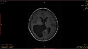
Figure 5: MRI scan of the case in axial.
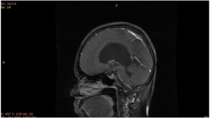
Figure 6: MRI scan of the case in sagittal.
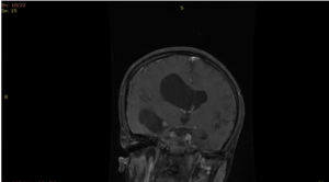
Figure 7: MRI scan of the case in coronal T1W post contrast sequences.
It showed asymmetrical unilateral dilatation of right lateral ventricle without periventricular CSF seepage with normal appearance of rest of ventricular system. Stenosed right foramen of Monro with normal appearance of 3rd and 4th ventricles were noted. No abnormal enhancing lesion was seen in and around stenosed left foramen of Monro in post-gadolinium T1 weighted images.
Discussion
Imaging with magnetic resonance and computed tomography scan are important in timely diagnosis as well as in following-up cases of unilateral obstructed hydrocephalus after treatment. Though cranial neurososngram can be helpful in perinatal period, however, computed tomography and MRI can more accurately detect the foramen of Monro stenosis and rule out other secondary causes of the obstruction as they provide cross sectional anatomical views [5,7-9].
The rare causes of idiopathic obstruction of foramen of Monro might be due to absence or narrowing of the foramen. In the narrowed foramen, a membrane appeared to block the normal sized foramen [10]. Our case of the present report was obviously falling in first group.
The congenital and adult variants of presentation are reported in these cases and congenital hydrocephalus which is mostly bilateral may have variable prenatal causes such as obstruction of aqueduct of Sylvius, congenital malformations of Dandy-Walker or Arnold-Chiari and infection involving the central nervous system, especially congenital toxoplasmosis and cytomegalovirus [3,9,11]. Hegazy and Hegazy stated that distension of brain ventricles of hydrocephalus before cranial suture closure could result in enlargement of the skull size, however, after closure such as in our case, it might press the nervous tissue resulting in neurological manifestations [1,2].
Unilateral hydrocephalus commonly due to postnatal causes such as tumors in the region of the foramen of Monro may grow in size to occlude one of the foramina and usually cause bilateral obstruction. On the other hand, infections such as intrauterine mumps infection and intraventricular hemorrhage can also lead to prenatal obstruction to the flow of CSF at the level of the foramen of Monro [3,4,6].
Conclusion
Unilateral obstructive hydrocephalous of idiopathic cause is a rare entity and potential a treatable condition in which cross-sectional imaging with magnetic resonance and computed tomography plays important role in establishing diagnosis.
References
- Hegazy A. Clinical Embryology for medical students and postgraduate doctors. LAP; Lambert Academic Publishing, Berlin, 2014.
- Hegazy AA, Hegazy MA. Newborns’ Cranial Vault: Clinical Anatomy and Authors’ Perspective. Int J Human Anat. 2018;1(2):21-5.
- Wilberg JEJ, Vertosick FTJ, Vries JK. Unilateral hydrocephalus secondary to congenital atresia of the foramen of Monro. J Neurosurg. 1983;59(5):889-901.
- Bhagwati S. A case of unilateral hydrocephalus secondary to occlusion of one foramen of Monro. J Neurosurg. 1964;21(3):226-9.
- Pribil S, Boone SC, Waley R. Obstructive hydrocephalus at the anterior third ventricle caused by dilatated veins from an arterio-venosus malformation. Surg Neurol. 1983;20(6):487-92.
- Baumann B, Danon R, Weitz R, Blumensohn R, Schonfeld T, Nitzan M. Unilateral hydrocephalus due to obstruction of the foramen of Monro: another complication of intrauterine mumps infection. Eur J Pediatr. 1982;139(2):158-9.
- Weiner Z, Bronshtein M. Transient unilateral ventriculomegaly: sonographic diagnosis during the second trimester of pregnancy. J Clin Ultrasound. 1994;22(1):59-61.
- Nakamura S, Makiyama H, Miyagi A, Tsubokawa T, Ushinohama H. Congenital unilateral hydrocephalus. Childs Nerv Syst. 1989;5(6):367-70.
- Eller TW, Pasternak JF.Isolated ventricles following intraventricular hemorrhage. J Neurosurg. 1985;62(3):357-62.
- Ethyl SB, Yahya MB. Congenital occlusion of the foramen of Monro. Radiology. 1969;2(5):1061-4.
- Gaston BM, Jones BE. Perinatal unilateral hydrocephalus. Pediatr Radiol. 1989;19:328-9.
- Mohanty A, Das BS, Kolluri VS, Hedge T. Neuro-endoscopic fenestration of occluded foramen of Monro causing unilateral hydrocephalus. Pediatr Neurosurg. 1996;25(5):248-51.
- Oi S, Yamada H, Sasaki K, Matsumoto S. Atresia of the foramen of Monro resulting in severe unilateral hydrocephalus with subfalcial herniation and infratentorial diverticulum. Neurosurgery. 1985;16(1):103-6.
This work is licensed under Attribution-NonCommercial-NoDerivs 2.0 Generic (CC BY-NC-ND 2.0) International License. With this license readers are free to share, copy and redistribute the material in any medium or format as long as the original source is properly cited.

