Diomandé GF1, Koffi KF-H1, Bilé PEFK1*, Godé LE1, Goule AM1, Babayeju OR1, Bérété PIJ2, Djémi EM2, Diabaté Z1, Ouattara Y1, Diomandé IA1
1Ophthalmology Department, University Hospital Centre (CHU) Bouaké, 01 BP 1174 Bouaké 01, Côte d’Ivoire
2Stomatology Department, University Hospital Center (CHU) Bouaké, 01 BP 1174 Bouaké 01, Côte d’Ivoire
*Correspondence author: Philippe EFK BILE, Ophthalmology Department, University Hospital Centre (CHU) Bouaké, 01 BP 1174 Bouaké 01, Côte d’Ivoire; Email: [email protected]
Published Date: 27-10-2023
Copyright© 2023 by Bile PEFK, et al. All rights reserved. This is an open access article distributed under the terms of the Creative Commons Attribution License, which permits unrestricted use, distribution, and reproduction in any medium, provided the original author and source are credited.
Abstract
Aim: To contribute to the knowledge and improvement of the management of non-traumatic keratitis.
Materials and methods: Retrospective descriptive study based on the analysis of 105 patient records received at the ophthalmology department of Bouaké University Hospital from January 1, 2018, to June 30, 2019, for non-traumatic corneal lesions with or without associated signs.
Results: The prevalence was 2.41%. The mean age of patients was 38.52 years with extremes of 1 and 86 years. Males predominated (59.05%), with a sex ratio of 1.44. The main general risk factors were atopy (79.54%). History of ocular trauma was the predominant ocular risk factor (54.17%). Non-infectious keratitis predominated (54.29%). The main etiology was allergy (54.38%). Infectious keratitis was dominated by bacterial forms (60.41%). Treatment was predominantly topical, with antiallergic (23.81%) and antibiotic (65.71%) agents.
Conclusion: Non-traumatic keratitis is a serious and life-threatening condition. They occur most frequently in young adult males. The severity of the lesions observed in this condition calls for early management and patient education.
Keywords: Allergy; Antibiotic; Etiology; Non-Traumatic Keratitis
Introduction
Keratitis is an inflammation of the cornea. The etiologies of this condition vary and may be infectious or non-infectious. Clinical expressions are diverse. The frequency of this pathology is high and has been on the increase in recent years due to an increase in the number of contributing factors, which in our context are dominated by trauma and ocular surface pathologies [1]. The increasingly frequent wearing of contact lenses was the primary risk factor, particularly in industrialized countries [2]. Corneal pathologies are a major cause of blindness worldwide. Corneal abscess is a diagnostic and therapeutic emergency that can lead to anatomical and/or functional loss of the eyeball [3]. Sequelae are dominated by corneal scarring, dystrophy and leukoma. With early and appropriate treatment, the disease can be cured without sequelae. Occasionally, however, the course of the disease can be unfavorable, due to the virulence of the germ, delayed diagnosis and the existence of tares [4]. In Côte d’Ivoire, studies of non-traumatic keratitis are rare, hence the interest of this study, the aim of which was to describe the epidemiological, clinical and therapeutic aspects of non-traumatic keratitis at Bouaké University Hospital.
Materials and Methods
This was a retrospective descriptive study carried out in the ophthalmology department of Bouaké University Hospital. It focused on the records of patients received in consultation in the said department during the period from January 1, 2018, to June 30, 2019 for non-traumatic corneal lesions with or without associated signs. The parameters studied were age, gender, occupation, local and general risk factors, Distance Visual Acuity (DVA), characteristics of corneal lesions (their nature, character as well as topography), associated manifestations and treatment undertaken. Data analysis was performed using EPI INFO software version 7.0. Figures were produced in Excel 2016. Tables and data entry were carried out in Word 2016. Quantitative variables were expressed as means and extreme values. Qualitative variables were expressed as proportions.
Results
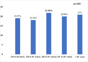
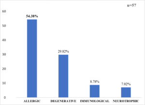
During the study period, 4354 patients consulted our department for all pathologies, 105 of whom presented with a non-traumatic corneal lesion. This represents a prevalence of 2.41%. Corneal damage was unilateral in 81 patients and bilateral in 23, for a total of 127 eyes. The mean age of the patients was 38.52 years, with extremes of 1 and 86 years. The most common age range was 30 to 45, with 21.90% (Fig. 1). Males accounted for the majority (59.05% of cases), with a sex ratio of 1.44. Farmers and students were the most exposed socio-professional strata, with 18.10% of cases in each category (Table 1). Decreased visual acuity was the most frequent reason for consultation (99.04%) (Table 2). General risk factors were dominated by atopy in 79.54% of cases (Table 3). Ocular risk factors were dominated by a history of ocular trauma in 54.17% of cases (Table 4). The right eye was the most affected (42.86%). The damage was bilateral in 21.90% of cases. Visual acuity was greater than or equal to 3/10 in 48.82% of patients. Superficial punctate keratitis was the most common lesion (54.7%). Corneal ulceration followed in 28.12% of cases. Lesions were diffuse in 33.59% of cases and accompanied by conjunctival hyperemia in 71.09% (Table 5). Non-infectious keratitis accounted for 54.29%, dominated by allergic forms (54.38%) (Fig. 2). Outpatient treatment was used in 88.57% of cases (n=105). Specific local medical treatment was dominated by antibiotics (65.71%).

Figure 1: Age distribution of patients.
|
Profession |
Number (n=105) |
Percentage (%) |
|
No profession |
12 |
11,42 |
|
Farmers |
19 |
18,10 |
|
Pupils |
19 |
18,10 |
|
Students |
8 |
7,62 |
|
Ménagères |
17 |
16,20 |
|
Informal sector |
11 |
10,47 |
|
Civil servants |
11 |
10,47 |
|
Shopkeepers |
8 |
7,62 |
|
Total |
105 |
100 |
Table 1: Distribution of patients by profession.
|
Reason for Consultation |
Number |
Percentage (%) |
|
Eye pain |
99 |
94,28 |
|
Eye redness |
103 |
98,09 |
|
DAV |
104 |
99,04 |
|
Tearing |
81 |
77,14 |
|
Ocular pruritus |
45 |
42,85 |
|
Foreign body sensation |
32 |
30,47 |
|
Other |
11 |
10,48 |
Table 2: Breakdown of patients by reason for consultation.
|
General Risk Factors |
Number (n=44) |
Percentage (%) |
|
Atopy |
35 |
79,54 |
|
Immunodepression |
6 |
13,64 |
|
Diabetes |
3 |
6,82 |
|
Total |
44 |
100 |
Table 3: Distribution of patients according to general risk factors.
|
Local Risk Factors |
Numbers (n=48) |
Percentage (%) |
|
History of Ocular Trauma |
26 |
54,17 |
|
Previous Eye Surgery |
21 |
43,75 |
|
Contact Lens Wear |
1 |
2,08 |
|
Total |
48 |
100 |
Table 4: Distribution of patients according to ocular risk factors.
|
Specific Treatment |
Effectif (n=105) |
Percentage (%) |
|
Antibiotic |
69/105 |
65,71 |
|
Antifungical |
4/105 |
3,81 |
|
Antiviral |
9/105 |
8,57 |
|
Anti Allergique |
25/105 |
23,81 |
|
Antiseptic |
22/105 |
20,95 |
|
Total |
105 |
100 |
Table 5: Distribution of patients according to the type of specific local medical treatment used.

Figure 2: Distribution of patients by type of non-infectious keratitis.
Discussion
Non-traumatic keratitis is a serious condition that can lead to loss of ocular function. Their prevalence in this study was 2.41%. This result is comparable to that of Ibrahim in England, whose study on the epidemiological and microbiological aspects of corneal infections found a frequency of 3.3% [5]. The high frequency of non-traumatic keratitis in France could be explained by the fact that the population is predominantly rural and therefore exposed to the many risk factors associated with this condition. Patients aged between 30 and 45 years were the most numerous (21.90%), with an average age of 38.52 years. The average age found in our work is superposable with that of Tasanee in Thailand [6], who found an average age of 45 in his patients with keratitis. In the work of Seck in Senegal and Baklouti in Tunisia, the mean age of their patients was significantly higher than ours, with mean ages of 50 and 59.2 respectively [7,8]. The predominance of this condition in young adults in our study would seem to be linked to their frequent exposure to the risk factors most associated with their occupational activities, such as farming. Males accounted for the majority (59.05%) in our study, with a sex ratio of 1.44. Similar findings were made by Laspina in Paraguay and Sitoula in Nepal, who noted sex ratios of 1.50 and 1.39 respectively [9,10]. Other authors, however, noted a clear female predominance [11,8]. These facts confirm that non-traumatic keratitis can occur in all subjects regardless of gender. Farmers and schoolchildren came first, accounting for 36.20% of cases. The high proportion of farmers and schoolchildren, as well as housewives, could be explained by the presence of local risk factors such as a history of trauma. When these are not perforating, they may favour the inoculation of germs through the break in the cornea. As for the occurrence of this condition in farmers, it could be linked to their working conditions, which are often septic, exposing the ocular surface to infections of all kinds. Decreased Visual Acuity (DVA) was the main reason for consultation (99.04%). Our results are in line with those of Baklouti in Tunisia, who noted a predominance of reduced visual acuity in all his patients [8].
This BAV found in various studies is linked to the fact that the cornea is the first transparent medium which, when altered, impedes the transmission of light impulses. The presence of ocular pain in this condition is thought to be linked to the significant innervation of the cornea, the most innervated structure in the human body. General risk factors were dominated by atopy (79.54%), followed by immunodepression (13.64%) and diabetes (6.82%). The involvement of atopy in the occurrence of non-traumatic keratitis has also been noted by Schaefer in France (42%) and Srinivasan in India (65.50%) [12,14]. Ocular atopia is the most common cause of scratching lesions, leading to fragility of ocular surface structures and thus to the onset of keratitis. Although immunodepression and diabetes are factors that contribute to the development of keratitis, they also aggravate the clinical picture, precipitating the progression to blindness. In fact, unbalanced diabetes and immunodepression can sustain corneal infection and encourage the growth of germs on the corneal surface. A history of ocular trauma was the most common ocular risk factor (54.17%), followed by a history of ocular surgery (43.75%). In his work in Mali, Dr. Bakayoko noted that ocular risk factors were dominated by a history of ocular trauma (65.67%) [15]. These risk factors are thought to be at the root of damage to the epithelial barrier. Indeed, the epithelial barrier is said to be the primary defence of the ocular surface and its weakening because of various risk factors would lead to the inoculation of both saprophytic and exogenous germs. Most patients (48.82%) had good visual acuity (≥ 3/10). The preservation of visual acuity in our patients would appear to be due, on the one hand, to the high rate of patients seen at the early stage of symptomatology and, on the other hand, to the sometimes-eccentric location of the lesion. Severe AVB (<1/20) occurred in 43.3% of cases. Sitoula, India, also found severe VAD in 65% of patients with keratitis [10]. The collapse in visual acuity observed by some authors could be explained by delays in consultation, which are at the origin of late management, the existence of co-morbidity factors in elderly subjects, the virulence of certain germs and the presence of underlying pathologies. Superficial punctate keratitis was the most common lesion in the study, accounting for 54.7%. Ulcerations were a distant second (28.12%). Superficial punctate keratitis, which is generally the result of scratching lesions in patients with keratoconjunctivitis, may be due to the presence of numerous atopic conditions. These scratching lesions can sometimes coalesce if management is delayed or inappropriate. According to Collin J, in the early stages, keratitis is characterized by superficial lesions which subsequently coalesce into large patches of ulceration. These patches of ulceration can sometimes evolve into a noisier and more complicated clinical picture, making this condition even more serious [16]. Most corneal lesions were punctiform (54.68%), followed by rounded (33.59%). Corneal lesions were diffuse (33.59%) in most of our patients, followed by central (29.68%). Our results differ from those of Tasanee and Baklouti, whose studies noted a predominance of central location of corneal lesions [8,6]. The predominance of diffuse localization and punctiform character in our study is in line with the high frequency of superficial punctate keratitis that we observed. However, the central location of the corneal lesions and their rounded appearance reflect the evolution of these lesions towards complications such as ulceration, abscessation and often corneal perforation, sometimes jeopardizing the functional visual prognosis. Conjunctival hyperemia was the most common associated manifestation (71.09%), followed by ocular secretions (17.18%) and papillae and follicles (14.84%). The performance of paraclinical examinations in our working conditions would sometimes be difficult, if not impossible. Lack of financial means on the part of patients and lack of suitable technical facilities could be the main reasons for this. For the collection of etiological data on non-traumatic keratitis, we referred to clinical and anamnestic arguments. The clinical picture of non-traumatic keratitis is dominated by non-infectious forms (54.29%). The predominance of a history of atopy (79.54%), the existence of bilateral ocular pruritus (42.85%) and foreign body sensations (30.47%) were all arguments in favor of non-infectious keratitis. The presence of papillae and follicles under the upper eyelid observed on palpebral eversion (14.84%) pointed to non-infectious keratitis of allergic origin. All of the above shows that allergic keratitis was the most frequent etiology of non-infectious keratitis, accounting for 54.38%. However, our results differ from those of Bouazzaa, who found a predominance of immunological keratitis in his patients with non-traumatic non-infectious keratitis (26.10%) [17]. Although the etiology of non-infectious, non-traumatic keratitis was dominated by allergic causes, degenerative forms (28.82%) and immunological forms (8.78%) were not negligible. Infectious keratitis accounted for 45.71%, with bacterial etiology predominating in 60.41% of cases, followed by viral causes (20.84%). The presence of a rounded corneal ulceration with sharp edges, associated with numerous purulent ocular secretions and an inflammatory reaction in the anterior chamber (tyndall and hypopyon) were the presumptive signs that pointed to bacterial causes. The viral form was strongly suspected in view of the map-like, dendritic or fern-like ulcerations sometimes found on examination of patients’ corneas. The etiologies of infectious keratitis varied and fungal and parasitic aspects were also observed. Most of our patients were treated on an outpatient basis (88.57%). The predominance of outpatient treatment is attributable to early management, the sometimes-benign nature of corneal lesions and the lack of financial resources to cover hospitalization costs. As a result, hospitalization is generally reserved for complicated forms of keratitis that threaten visual function [3]. Antibiotics were the most widely used specific treatment (65.71%), followed by antiallergics (20.95%). These results justify the predominance of bacterial and allergic etiologies for keratitis in this study.
Conclusion
Non-traumatic keratitis is a common condition, occurring most frequently in young adults. It is characterized by the cardinal signs of inflammation, with ocular redness and pain predominating. Certain risk factors, such as atopic conditions and ocular trauma, are incriminated. Treatment remains essentially medical.
Conflict of Interest
The authors have no conflict of interest to declare.
References
- Baldwin HC, Marshall J. Growth factors in corneal wound healing following refractive surgery: a review. Acta Ophthalmologica Scandinavica. 2002;80(3):238-47.
- Lecuona K. Ocular trauma. Prevention, evaluation and management. Revue de Santé Oculaire Communautaire. 2006;3(1):11-4.
- Elien GYRR, Bakayoko S, Simaga AS, Sissoko M, Nioumanta M, Sylla A, et al. Microbiological profile, antimicrobial susceptibility and treatment outcome of corneal abscesses at CHU-IOTA. Rev Mali Infect Microbiol. 2021;16(2):1-5.
- Streilen JW. Ocular immune privilege and the Faustian dilemma. The proctor lectures. Invest Ophthalmol Vis Sci. 1996;37(10):1940-50.
- Ibrahim YW, Boase DL, Cree IA. Epidemiological characteristics, predisposing factors and microbiological profiles of infectious corneal ulcers: the Portsmouth corneal ulcer study. Br J Ophthalmol. 2009;93:1319-24.
- Sirikul T, Prabriputaloong T, Smathivat A, Chuck RS, Vongthongsri A. Predisposing factors and etiologic diagnosis of ulcerative keratitis. Cornea. 2008;27(3):283-7.
- Seck SM, Diakhaté M, Oulfath A, Sow MN, Dieng M, Gueye NN. Severe infectious keratitis in tropical environments: 118 cases collected over 10 years. Médecine et Santé Tropicales. 2019;29(2):151-4.
- Baklouti K, Ayachi M, Mhiri N, Mrabet A, Ben Ahmed N, Ben Turkia R. Presumed corneal abscesses of bacterial origin. Bull. Soc belge Ophtalmol. 2007;305:39-44.
- Laspina F, Samudio M, Cibils D, Ta CN, Fariña N, Sanabria R, et al. Epidemiological characteristics of microbiological results on patients with infectious corneal ulcers: a 13-year survey in Paraguay. Graefe’s Arch Clin Experimental Ophthalmol. 2004;242:204-9.
- Sitoula RP, Singh SK, Maheseth V, Sharma A. Epidemiology and etiological diagnosis of infective keratitis in eastern region of Nepal. Nepal J Ophthalmol. 2015;7(13):10-5.
- Passos RM, Cariello AJ, Yu MC, Höfling-Lima AL. Microbial keratitis in the elderly – a 32-year review. Arq Bras Oftalmol. 2010;73(4):315-9.
- Schaefer F, Bruttin O, Zografos L. Bacterial keratitis: a prospective clinical and microbiological study. Br J Ophthalmol. 2001;85(7):842-7.
- Srinivasan M, Gonzales CA, George C, Cevallos V, Mascarenhas JM, Asokan B, et al. Epidemiology and etiological diagnosis of corneal ulceration in Madurai, South India. Br J Ophthalmol. 1997;81(11):965-71.
- Amel C, Hachicha F, Malek I, Alaya N, Zeghal I, Ben Ayed N, et al. Le profil épidémiologique des abcès de cornée. 2000.
- Colin J. Herpes cornéen. J Fr Ophtalmol. 1993;16:6-9.
- Bakayoko S, Elien Gagnan Yan, Zaou RR, Kole Sidibe M, Dicko M, Nioumanta M Sylla. Epidemiol Severe Abscesses Cornea IOTAHealth Sci Dis. 2020;21(7): 1-4.
- Bouazza M, Amine Bensemlali A, Elbelhadji M, Benhmidoune L, El Kabli H, M’daghri E, et al. Non-traumatic corneal perforations: Therapeutic. J Fr d’Ophthalmol. 2015:38(8)395-402.
Article Type
Research Article
Publication History
Received Date: 10-09-2023
Accepted Date: 20-10-2023
Published Date: 27-10-2023
Copyright© 2023 by Bile PEFK, et al. All rights reserved. This is an open access article distributed under the terms of the Creative Commons Attribution License, which permits unrestricted use, distribution, and reproduction in any medium, provided the original author and source are credited.
Citation: Bile PEFK, et al. Non-traumatic Keratitis: Epidemio-Clinical, Etiological and Therapeutic Aspects at Bouake Hospital. J Ophthalmol Adv Res. 2023;4(3):1-6.

Figure 1: Age distribution of patients.

Figure 2: Distribution of patients by type of non-infectious keratitis.
Profession | Number (n=105) | Percentage (%) |
No profession | 12 | 11,42 |
Farmers | 19 | 18,10 |
Pupils | 19 | 18,10 |
Students | 8 | 7,62 |
Ménagères | 17 | 16,20 |
Informal sector | 11 | 10,47 |
Civil servants | 11 | 10,47 |
Shopkeepers | 8 | 7,62 |
Total | 105 | 100 |
Table 1: Distribution of patients by profession.
Reason for Consultation | Number | Percentage (%) |
Eye pain | 99 | 94,28 |
Eye redness | 103 | 98,09 |
DAV | 104 | 99,04 |
Tearing | 81 | 77,14 |
Ocular pruritus | 45 | 42,85 |
Foreign body sensation | 32 | 30,47 |
Other | 11 | 10,48 |
Table 2: Breakdown of patients by reason for consultation.
General Risk Factors | Number (n=44) | Percentage (%) |
Atopy | 35 | 79,54 |
Immunodepression | 6 | 13,64 |
Diabetes | 3 | 6,82 |
Total | 44 | 100 |
Table 3: Distribution of patients according to general risk factors.
Local Risk Factors | Numbers (n=48) | Percentage (%) |
History of Ocular Trauma | 26 | 54,17 |
Previous Eye Surgery | 21 | 43,75 |
Contact Lens Wear | 1 | 2,08 |
Total | 48 | 100 |
Table 4: Distribution of patients according to ocular risk factors.
Specific Treatment | Effectif (n=105) | Percentage (%) |
Antibiotic | 69/105 | 65,71 |
Antifungical | 4/105 | 3,81 |
Antiviral | 9/105 | 8,57 |
Anti Allergique | 25/105 | 23,81 |
Antiseptic | 22/105 | 20,95 |
Total | 105 | 100 |
Table 5: Distribution of patients according to the type of specific local medical treatment used.


