Hae-in Choi1, Hyung-ju Park1*
1Gangnam Blue Eye Clinic Hospital, Seoul, Republic of Korea
*Correspondence author: Hyung-ju Park, Gangnam Blue Eye Clinic Hospital, Seoul, Republic of Korea; Email: [email protected]
Published Date: 21-10-2024
Copyright© 2024 by Choi H, et al. All rights reserved. This is an open access article distributed under the terms of the Creative Commons Attribution License, which permits unrestricted use, distribution, and reproduction in any medium, provided the original author and source are credited.
Abstract
Objective: Limited Vitrectomy (CV) has a high chance of recurrence of floater symptoms. We evaluated the effectiveness and safety of a novel technique of vitrectomy: limited vitrectomy and selective removal of anterior vitreous opacities using 20 Megahertz (MHz) ocular ultrasound for vitreous floaters.
Methods: We included 175 eyes of 101 participants who underwent the phacoemulsification with multifocal intraocular lens insertion. Participants were divided into 3 groups: novel vitrectomy (SV), CV and control groups. They were followed up at postoperative 1, 3 and 6 months. Visual Acuity (VA), Spherical Equivalent (SE), Intraocular Pressure (IOP), Contrast Sensitivity Function (CSF) and Patient Satisfaction (PS) were compared between the SV and CV groups. PS and national eye institute-validated visual function questionnaire-39 (NEI VFQ-39) composite scores were assessed. Comparison was performed using the independent Student’s t-test and Fisher’s exact test.
Results: Best corrected VA was not significantly different in both groups. SE was significantly better in the SV group than in the CV group at 6 months postoperatively. There was no significant difference in IOP between both groups at 3 and 6 months postoperatively. The SV group had significantly better CSF than the CV group. The PS was higher in the SV group than in the CV group at 3 and 6 months postoperatively, based on the NEI VFQ-39 composite scores. There were no complications in the SV group. Endophthalmitis (n=1) and retinal detachment (n=1) occurred in the CV group.
Conclusion: The novel 25-gauge CV combined with selectively peripheral and anterior vitrectomy using 20 MHz ocular ultrasound is a more effective and safer procedure compared with simple CV.
Keywords: Vitreous Floaters; Limited Vitrectomy; Novel Vitrectomy; 20 Mhz Ocular Ultrasound; Patient Satisfaction; Contrast Sensitivity Function; NEI VFQ-39
Abbreviations
AULCSF: Area Under Log Contrast Sensitivity Function; BCVA: Best Corrected Visual Acuity; CS: Contrast Sensitivity; CSF: Contrast Sensitivity Function; IOP: Intraocular Pressure; CV: Limited Vitrectomy; Logmar: Logarithm of the Minimal Angle of Resolution; NEI VFQ: National Eye Institute-Validated Visual Function Questionnaire; SV: Novel Vitrectomy; OCT: Optical Coherence Tomography; PVD: Posterior Vitreous Detachment; SV: Selective Peripheral Vitrectomy; SE: Spherical Equivalent; SD: Standard Deviation; VA: Visual Acuity
Introduction
Vitreous opacity increases with age and causes a decrease in the quality of life due to decreased vision and reduced contrast sensitivity or in severe cases, mental disorders such as depression [7,22,34]. The most common cause of floaters is Posterior Vitreous Detachment (PVD) with aging. However, incomplete PVD can also cause symptoms. Other causes include vitreous liquefaction, myopia and asteroid hyalosis with aggregation of vitreous collagen [10,15,33].
Floaters can be diagnosed using ocular ultrasound, spectral-domain Optical Coherence Tomography (OCT), scanning laser ophthalmoscopy and dynamic light scattering, among others [6,9,14,16,20,25,26,28,30]. To date, ultrasound examination is the most commonly used method for the diagnosis of floaters [25,30]. The advantage of vitreous imaging using ultrasound is that the vitreous body can be observed regardless of the media opacity of the anterior segment, such as corneal opacity or lens abnormalities and the size of the vitreous cavity and the density of collagen fibers can be measured. For ultrasound examination, various frequency probes are used. Usually, in the case of the low frequency probe, the tissue is projected deeply to observe the posterior part of the eyeball and the resolution is lowered.
In the case of the high frequency probe, the tissue penetration is thin, so it is easy to observe the anterior part of the eyeball and the resolution is excellent. Therefore, with a 10 or 15 MHz probe, it is possible to better observe intraocular images for screening purposes, diseases of the vitreous body, vitreous fibers and turbidity, etc., but it is difficult to observe the vitreous in front of the equator. A 20 MHz probe provides high-resolution images for good observation of the vitreous anterior to the equator, but it is not suitable for observation of the vitreous body in the posterior part of the eyeball [8,18].
Treatments of symptomatic floaters include a follow-up observation, laser treatments and surgical methods. Surgical treatment is the most reliable method [21]. Surgical methods also vary depending on the condition of the patient’s eye, ranging from central core or limited vitrectomy to extensive vitrectomy with PVD induction. Currently, core or limited vitrectomy (CV) is more frequently performed for the treatment of floaters due to the advantage of short operation times and fewer postoperative complications such as cataracts, retinal tears and retinal detachment [11,23,24,27,29,31]. However, the recurrence rates of the symptomatic floaters are high after CV, according to the authors’ experiences. After the first CV, the remnant or newly developing anterior vitreous opacities cause floater symptoms to recur; therefore, additional vitrectomy is needed in some cases. In the extensive vitrectomy, the vitreous is almost totally removed, but it has the disadvantages of more intraoperative and postoperative complications.
Therefore, based on the characteristics of 20 MHz ocular ultrasound that provides good observation of anterior vitreous opacities, we invented the new, hybrid limited vitrectomy that consists of the CV and selectively peripheral and anterior vitrectomy (SV) using 20 MHz ocular ultrasound. It is hypothesized that this novel procedure has a short operation time and fewer postoperative complications. It has advantages of both the limited and extensive vitrectomies and overcomes their disadvantages. In patients with primary symptomatic vitreous opacities, we compared the effect of each vitrectomy using contrast sensitivity function (CSF) and national eye institute-validated visual function questionnaire (NEI VFQ) testing between a group that underwent CV after diagnosis of vitreous floaters with 15 MHz low-frequency ultrasound and a group where additional vitreous floaters in vitreous body anterior to the equator were diagnosed with 20 MHz high-frequency ocular ultrasound, in addition to the 15 MHz ultrasound. The recurrence of symptomatic vitreous floaters after limited vitrectomy was attributed to anterior vitreous opacities. In the latter group, novel vitrectomy (SV) was performed, which involved not only CV but also selective peripheral and anterior vitrectomy for anterior vitreous floaters confirmed with 20 MHz high-frequency ocular ultrasound. Finally, the effectiveness and safety of SV are evaluated by comparing it with CV, which is used as the treatment for symptomatic vitreous floaters.
Ethical Approval
The study followed the tenets of the 7th version of the Declaration of Helsinki and was approved by the Ethics Committee of the Gangnam Blue Eye Clinic Hospital, (No. 014). All participants provided written informed consent.
Methodology
Study Design, Setting and Participants
This was a single-center, comparative retrospective interventional case series. From May 2020 to March 2024 at Gangnam Blue Eye Clinic, this study included 175 eyes of 101 persons (29 males and 72 females; mean age ± standard deviation [SD], 60.9±4.6 years). Among the 101 participants, 71 had vitreous floaters and 30 were healthy age-matched control participants who were volunteers, staff, patients’ family members or friends. All participants were pseudophakic and had Nd:YAG laser capsulotomy. The pseudophakic eyes all had one kind of multifocal intraocular lenses. Among the 71 patients, 115 eyes underwent 25-gauge vitrectomy for symptomatic vitreous floaters that had lasted longer than 3 months. Of these, 54 eyes of 31 patients underwent SV and 61 eyes of 40 patients underwent CV. Patients were followed up for at least 6 months. Patients aged 18 years or older, who complained of floater symptoms that made it difficult to perform daily activities for at least 3 months and who were diagnosed with vitreous floaters by ultrasound examination were included. Those who underwent vitreous surgery previously or who had ocular trauma, diabetes mellitus retinopathy, retinal vessel occlusion, glaucoma, macular diseases such as the epiretinal membrane and age-related macular degeneration or severe dry eyes, etc. that may affect the vision function, were excluded.
Prior to the surgery in all three groups, comprehensive preoperative ophthalmic examinations including Visual Acuity (VA), Spherical Equivalent (SE), Intraocular Pressure (IOP), the AVISO 15 MHz B scan (Quantel Medical, Compact Touch, France), swept source OCT (OCT, Topcon, DRI OCT Tritron, Japan), panoramic ophthalmoscope (Optos, P200T, U.K.) and CSF test (Takagi, CGT-2000, Japan), were performed. In the SV group, the entire 360 degrees of the anterior vitreous was additionally checked using a 20 MHz ocular ultrasound (Quantel Medical, ABSolu, France).
Ultrasound B Scan
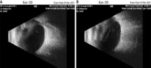
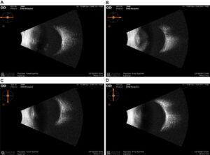
Patients were asked to face the front with eyes closed in a sitting position. After placing the gel on the eyelid, a 15 MHz B scan (Quantel Medical, Compact Touch, France) was placed on the eyelid and vitreous floaters were checked by vertical and transverse axial scan method (Fig. 1). In the SV group, the operating doctor (HJP) directly performed a 360-degree transverse scan using a 20 MHz ocular ultrasound (Quantel Medical, ABSolu, France) after anesthetic drops (AlcaineTM, Alcon, USA) were applied, while the patient was seated with eyes opened. When checking the anterior vitreous of the lower part, the probe was placed on the lower conjunctiva parallel to the limbus while the patient looked up. When checking the upper anterior vitreous, the probe was inserted into the upper conjunctiva parallel to the corneal limbus while the patient looked down. When checking the anterior vitreous of the nasal part, the probe was inserted into the nasal conjunctiva parallel to the corneal limbus while the patient looked towards the ear. When checking the temporal region of the anterior vitreous, the probe was placed on the temporal conjunctiva, parallel to the corneal limbus, while the patient looked towards the nose. The vitreous opacity anterior to the equator was further diagnosed (Fig. 2).

Figure 1: 15 MHz B ocular ultrasound. A: Vertical view; B: Transverse view. There are a lot of vitreous floaters in the vitreous cavity.

Figure 2: Transverse scan of 20 MHz B ocular ultrasound. A: Floaters were observed in the anterior vitreous between 7:00 and 11:00; B, C and D: No opacities were observed in the anterior vitreous.
Contrast Sensitivity (CS)
Contrast sensitivity (CS) was evaluated using the CSF test (Takagi, CGT-2000, Japan). All participants were in a dark-adapted condition for 3 minutes and then tested at a distance of 5 m in a dark room, while wearing correction if ametropic. During mesopic CSF testing, the duration to show the acuity chart was 0.2 seconds and the time interval was set to 1 second. The contrast was obtained using the following formula: Contrast = (Luminancemax – Luminancemin)/(Luminancemax + Luminancemin). The area under log CSF (AULCSF) is the area under the CS curve. It is often used to evaluate CS as a single numerical value, which is obtained by converting degree and contrast to log. Higher numbers indicate better CSF.
Subjective Visual Function Evaluation
The NEI VFQ-39 was created by RAND, sponsored by the National Eye Institute and translated by the Korean Retinal Society. Composite scores were measured before surgery in all groups and at 1, 3 and 6 months postoperatively in all three groups. Satisfaction and recurrence were also evaluated at 1, 3 and 6 months postoperatively in the SV and CV groups.
Surgical Procedures
A 3-port 25-gauge (G) pars plana vitrectomy (Constellation: Alcon Laboratories Inc., Fort Worth, TX) under retrobulbar anesthesia (a 50% mixture of 2% lidocaine and 0.75% bupivacaine) was implemented by one experienced surgeon (HJP). Before surgery, eyelids and periorbital skin were scrubbed with 5% povidone-iodine solution, by the surgeon’s assistant, with the eyelid carefully draped to keep averting eyelashes from the operation field. The sclerotomy sites were made superior at the 10 o’clock and 2 o’clock positions for the vitrectomy probe and endoillumination probe. The infusion was placed between 4 and 5 o’clock positions for left eyes and between the 7 and 8 o’clock positions for right eyes. After displacing the conjunctiva approximately 1 to 2 mm, the sclera was penetrated by trocars, 3 mm posterior to the limbus in pseudophakic eyes at an angle between 20° and 30°, with the bevel up, with a 25-gauge one-step Alcon Laboratory System Kit (Alcon Laboratories, Inc., Fort Worth, TX). All vitrectomy procedures were performed using a surgical microscope (OPMI Lumera ® T, Carl Zeiss Meditec, Dublin, CA) and a wide-angle viewing system with a noncontact lens (Resight 700, Carl Zeiss Meditec, Dublin, CA) was used for visualization of the posterior segment throughout the surgery. In the CV group, the vitrectomy was performed up to the ocular equator. In the SV group, the vitrectomy was performed up to the equator of the eyeball and the operator directly pressed scleral indentation with a depressor to remove the anterior vitreous opacities confirmed with 20 MHz ocular ultrasound under a chandelier light. The chandelier light was installed at 3 mm from the corneal limbus in the same manner as above, at 4 and 11 o’clock in the right eye and 1 and 8 o’clock in the left eye.
In both groups, scleral indentation was performed after vitrectomy to check the peripheral retinal break or retinal degeneration; if found, photocoagulation was performed. At the conclusion of the surgery, the cannulas were removed from the sclera without suturing the sclera or conjunctiva, the conjunctiva was pushed laterally and pressure was applied over the sclerotomy site at the same time, with a cotton tip. If any significant wound leakage was noted, 8-0 vicryl sutures were placed to close the scleral and conjunctival wound sites.
Outcome Measures
Preoperative data, including patient age, gender, the best corrected VA (BCVA) using a Snellen chart, IOP, slit-lamp biomicroscopic findings, panoramic ophthalmoscope (Optos, P200T, U.K.) and swept source OCT (OCT, Topcon, DRI OCT Tritron, Japan), were obtained. In both groups, intraoperative and postoperative complications were checked up. BCVA, SE, IOP, AULCSF and NEI VFQ-39 were measured preoperatively and at each examination 1, 3 and 6 months postoperatively in both groups. IOP was measured by one person using a Goldmann applanation tonometer.
Statistical Analysis
Statistical analysis was performed using SPSS software package (version 28.0: SPSS Inc, Chicago, IL) for Windows. First, Kolmogorov–Smirnov test was performed to test normal distribution of the data. BCVA was measured using a Snellen chart and was converted to logarithm of the minimal angle of resolution (logMAR) for analysis. For descriptive analyses, quantitative data were expressed as mean ± SD and qualitative data as a ratio or percentage. Comparisons of clinical baseline characteristics across all 3 groups was performed using independent Student’s t-test. Categorical variables were compared using Fisher’s exact test when applicable. Postoperative changes in BCVA, SE, IOP, CS and composite score at each time point were analyzed using independent Student’s t-tests between the SV and CV groups. P-value of 0.05 or less were considered statistically significant.
Results
Details of the participants’ baseline characteristics are shown in Table 1. There were no differences in age, gender, eye position, VA, SE and IOP between the three groups. PVDs were more prevalent in participants with vitreous floaters than in healthy control participants (P < 0.001). The NEI VFQ-39 composite score was more prevalent in healthy control participants than in vitreous floaters (P < 0.001). There were no differences between SV and CV groups related to PVD, symptom time, NEI VFQ-39 composite score and follow-up duration. BCVA was not significantly different between the SV and CV groups at 1, 3 and 6 months postoperatively. There was no difference in SE at 1 month postoperatively (P = 0.995) and at 3 months postoperatively (P = 0.104). However, the SV group showed a significantly better result of SE than the CV group (P = 0.023) at 6 months postoperatively. IOP was 16.4±5.0 mmHg in the SV group and 12.8±3.7 mmHg in the CV group at 1 month postoperatively, which was significantly higher in the SV group than CV group (P = 0.002). However, there was no significant difference in IOP between 2 groups at 3 months (P = 0.251) and 6 months postoperatively (P = 0.107). At 1, 3 months and 6 months postoperatively, the SV group had a better CSF than the CV group because the AULCSF of the SV group was significantly more than that of the CV group. The NEI VFQ-39 composite score was not significantly different between the SV and the CV group at 1 month postoperatively (P = 0.320). However, at 3 (P = 0.027) and 6 months postoperatively (P = 0.019), the NEI VFQ-39 composite score in the SV group was significantly more than in the CV group (Table 2).
No intraoperative complications, such as iatrogenic retinal break or detachment and intraocular hemorrhage, were noted in both groups. However, postoperative complications occurred in two eyes in the CV group. Endophthalmitis occurred in one eye (1.6%) on the second day after surgery and thus emergency vitrectomy, antibiotic intraocular injection and silicone oil injection were performed. After the condition was stable, silicone oil was removed at 3 months postoperatively and VA was restored to LogMAR BCVA 0.15 (0.00 preoperatively). In the other eye (1.6%), retinal detachment occurred at 3 months postoperatively due to new retinal detachment and emergency vitrectomy, perfluorocarbon liquids injection, Fluid-Air exchange, subretinal fluid drainage, barrier photocoagulation and Air-Silicone oil exchange were performed. At 3 months postoperatively, after removing silicone oil, VA was restored to LogMAR BCVA 0.22 (0.05 preoperatively). In the CV group, new vitreous floaters occurred in another eye (1.6%) after surgery. The patient was intolerable and thus underwent extensive vitrectomy. The patient was satisfied and there were no further complications. In this case, only the first vitrectomy was included in this study. Additionally, in one eye (1.6%) where PVD did not occur, new vitreous floaters were formed as PVD occurred. However, the patient did not want reoperation. Therefore, laser vitreolysis was performed, which showed good results.
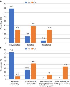
In the SV group at 6 months postoperatively, 94.7% (n = 29) were very satisfied or satisfied with the surgical result and 5.3% (n = 2) were dissatisfied with the surgical result. In the 6-month postoperative CV group, 69.5% (n = 28) were very satisfied or satisfied with the surgical result and 30.4% (n = 12) were dissatisfied (Figure 3A). Patients who were dissatisfied with the surgery were the ones who had postoperative endophthalmitis or residual floaters. In the SV group, all (n = 31, 100%) patients felt the floaters were removed completely or only had an acceptable residual symptom. In the CV group, 52.2 % (n = 21) of the patients felt the floaters were removed completely or only had an acceptable residual, while 47.8% (n = 19) did not feel that they were removed up to 6 months after the vitrectomy surgery (Fig. 3). One patient who developed postoperative endophthalmitis was excluded for this question.

Figure 3: Patient satisfaction in the limited vitrectomy (CV) and novel vitrectomy (SV) groups 6 months postoperatively. A: General degree of patient satisfaction in CV and SV; B: Demand for further vitrectomy after the first vitrectomy for vitreous floaters in CV and SV.
|
Parameters |
SV Group (n = 31, eyes = 54) |
CV Group (n = 40, eyes = 61) |
Control (n = 30, eyes = 60) |
P |
|
Age at operation (years) |
60.5±4.3 |
61.3±2.4 |
58.1±2.8 |
0.433* 0.851† |
|
Gender (male: female) |
8:23 |
12:38 |
9:21 |
0.547† 0.512‡ |
|
Eyes (right: left) |
28:26 |
27:34 |
30:30 |
0.451† 0.745‡ |
|
PVD, n (%) |
12 (22%) |
17 (28%) |
4 (7%) |
<0.001† 0.611§ |
|
Symptom time (months) |
7.9±1.2 |
8.0±0.3 |
0.850* |
|
|
BCVA, logMAR |
0.10±0.17 |
0.07±0.23 |
0.09±0.27 |
0.145* 0.429† |
|
SE (diopter) |
-0.01±0.32 |
-0.05±021 |
-0.03±0.56 |
0.227* 0.565† |
|
IOP (mmHg) |
11.7±2.3 |
10.9±6.1 |
11.2±4.9 |
0.124* 0.421† |
|
AULCSF‡ |
1.061±0.149 |
1.082±0.114 |
1.748±0.049 |
<0.001* 0.650† |
|
NEI VFQ-39 composite score |
72.5±12.6 |
73.1±10.4 |
87.2±7.7 |
<0.001* 0.511† |
|
Follow-up duration (months) |
11.1±7.5 |
10.9±3.8 |
0.650* |
|
|
* Analysis of variance for comparisons across all 3 groups. † Independent Student’s t-test for comparisons between SV and CV groups. ‡ Fisher exact test for comparisons across all 3 groups. § Fisher exact test for comparisons for comparisons between SV and CV groups. AULCSF = area under contrast sensitivity function; BCVA = best-corrected visual acuity; CSF = contrast sensitivity function; logMAR = logarithm of the minimum angle of resolution; CV = core or limited vitrectomy; IOP = intraocular pressure; NEI VFQ-39 = 39-item National Eye Institute Visual Function Questionnaire; PVD = posterior vitreous detachment; SD = standard deviation; SE = spherical equivalent; SV = novel vitrectomy |
||||
Table 1: Baseline demographics and characteristics of SV, CV and control groups.
|
Parameter |
BCVA, logMAR |
Spherical equivalent (diopter) |
IOP (mmHg) |
AULCSF |
NEI VFQ-39 Composite score |
||||||||||
|
|
SV |
CV |
P* |
SV |
CV |
P* |
SV |
CV |
P* |
SV |
CV |
P* |
SV |
CV |
P* |
|
Post-OP (1 month) |
0.09 ± 0.77 |
0.59 ± 0.68 |
0.093 |
-0.14 ± 0.92 |
-0.13 ± 0.33 |
0.995 |
16.4 ± 5.0 |
12.8 ± 3.7 |
0.002 |
1.228 ± 0.237 |
1.140 ± 0.119 |
0.048 |
75.1±16.7 |
74.6±18.9 |
0.320 |
|
Post-OP (3 months) |
0.53 ± 0.56 |
0.04 ± 0.57 |
0.490 |
0.07 ± 0.32 |
-0.13 ± 0.26 |
0.104 |
13.9 ± 3.6 |
12.7 ± 3.7 |
0.251 |
1.601 ± 0.297 |
1.308±0.164 |
<0.001 |
84.7±9.8 |
81.9±11.5 |
0.027 |
|
Post-OP (6 months) |
0.03 ± 0.46 |
0.04 ± 0.55 |
0.748 |
0.11 ± 0.23 |
-0.21 ± 0.27 |
0.023 |
16.0 ± 2.2 |
14.1 ± 3.4 |
0.107 |
1.664 ± 0.254 |
1.397±0.262 |
<0.001 |
85.1±14.8 |
80.4±19.6 |
0.019 |
|
* Independent Student’s t-test AULCSF = area under log contrast sensitivity function; BCVA logMAR = best-corrected visual acuity logarithm of the minimal angle of resolution; CV = core or limited vitrectomy; IOP = intraocular pressure; NEI VFQ-39 = 39-item national eye institute-validated visual function questionnaire; Post-OP = postoperative; SV = novel vitrectomy |
|||||||||||||||
Table 2: Comparison of postoperative outcomes between the SV and CV groups.
Discussion
Vitreous floaters are caused by opacities within the vitreous body and can be imaged, qualified and quantified by ocular ultrasound [14]. Symptomatic vitreous floaters may markedly reduce CSF and in severe cases, affect vision. It is assumed that this is due to floaters interfering with light diffraction patterns, which causes light scattering [15,33]. In this respect, a previous study found it appropriate to refer to clinically significant vitreous floaters as vision degrading vitreopathy [23]. The NEI-VFQ-39 is useful in quantifying the effect on general visual well-being, but floater-specific questionnaires may be more useful [19].
Treatments for symptomatic vitreous floaters include laser treatment and surgery. It was reported in a study that Nd:YAG vitreolysis resulted in moderate symptom improvement in only one-third of patients and worsened in 7.7 % of patients [4]. Therefore, current laser treatment offers only mildly if at all effective treatment for symptomatic improvement. In addition, opacity of the vitreous body too close to the optic nerve and retina for safety reasons limits the procedure [32].
Pars plana vitrectomy, the most effective and reliable method for treating vitreous floaters, is known to improve symptoms. However, this procedure is invasive and can cause complications such as endophthalmitis, retinal tears and detachments, cataracts, glaucoma, vitreoretinal hemorrhage and macular edema. With the use of 25-gauge instruments and sutureless techniques with CV, postoperative complications can be diminished significantly to provide a safe and effective cure for clinically significant floaters [15,23,24].
Recently, the interest in vitreous floaters and their treatment has increased in Korea. In addition, surgery is currently used for treating floaters, which interferes with daily life because of its long duration and inconvenience to patients. When reviewing existing studies, 25-gauge CV has a higher proportion of floaters surgery [24]. However, based on the authors’ experience, when targeting Korean patients, there were significant numbers of cases where the effect of symptom improvement was inferior and unsatisfactory compared to those reported in previous papers. In one patient, CV up to the retinal equator was performed, but the remaining or newly developing vitreous floaters led to the development of this novel surgical method. Accordingly, the purpose of this study was to reduce the incidence and recurrence of complications associated with vitrectomy like CV and to improve symptoms, CSF and further improve VA compared to CV. We proposed a new surgical method to selectively remove opacities in the anterior vitreous and CV using 20 MHz ocular ultrasound.
There was no difference in VA at 1, 3 and 6 months postoperatively between the two groups. There was no difference in IOP at 3 and 6 months postoperatively. There was also no difference in SE at 1 and 3 months postoperatively. However, at 6 months, significant results were obtained in the SV group rather than in the CV group. Removing the anterior vitreous opacities while pressing the periphery of the eyeball with a scleral depressor after performing two additional 25-gauge scleral trocars insertions had no effect on VA and refractive index.
This study was undertaken to determine whether floaters impact CSF of the patients who underwent cataract surgery and whether vitrectomy can improve CSF and alleviate visual dissatisfaction measured with NEI VFQ test in the SV group compared to the CV group. The CSF was improved in the SV group compared to the CV group at 1, 3 and 6 months postoperatively. Particularly, the SV group showed an improvement almost close to the control group. NEI VFQ-39 composite score was significantly higher in the SV group than in the CV group at 3 and 6 months postoperatively. Therefore, many patients in the SV group were more satisfied with the surgical results. There was a difference between the two groups because residual floaters became severe or new floaters were created in the CV group. Another study reported a statistically significant improvement in CSF at 1 month postoperatively [24]. The cause of this difference is not clear, but in this study, all participants underwent one type of multifocal intraocular lens surgery and had laser posterior capsulotomy. It is thought that the CSF was recovered somewhat late in this study because CSF may be lower in people who had cataract surgery with multifocal intraocular lenses in this study than in people in other studies [5,7,17,24]. Therefore, further research on this is necessary.
Endophthalmitis and retinal detachment occurred in the CV group. This may be related to vitreous incarceration in sclerotomies sites as reported by several studies. Other studies reported that beveled incision and non-hollow probes for cannula extraction could partially prevent vitreous incarceration [2,3,12,13,24].
Limitations of this study include the retrospective nature; in particular, the selection of only participants with who underwent phacoemulsification cataract surgery with multifocal intraocular lens insertion, causing potential selection bias; heterogeneity in the intervention group; and CSF measurements that were obtained only in mesopic lightness and a 5 m far target. It is well known that the impact of vitreous floaters varies from negligible to significant, depending on ambient and background lightness. Thus, future studies should examine the relationship between subjective and objective findings in different lightness conditions, using different distances of targets. Further, the small number of patients evaluated prospectively with CSF testing and standardized NEI VFQ testing and the relatively short follow-up in patients having one kind of multifocal intraocular lenses, also limit the findings of this study.
Conclusion
In conclusion, even though the 25-gauge CV has already been reported to be effective and safe in several studies, with a high patient satisfaction, the satisfaction for surgery was somewhat lower in this study. This is partly because the study was conducted on participants who had multifocal intraocular lenses. In addition, it is thought that the vitrectomy for vitreous floaters are negatively perceived in the society of South Korea for a long time. Based on this study, 25-gauge CV combined with selectively peripheral and anterior vitrectomy with the help of 20 MHz ocular ultrasound is more effective and safer than simple CV. We believe that it is a surgical method that can provide significant improvement in CSF and high surgical satisfaction to patients, especially those with reduced CSF who have undergone multifocal intraocular lens cataract surgery and escape unnecessary additional vitrectomy. Further study of this approach is thus warranted.
Conflict of Interest
Authors declare that they have no conflict of interest.
Funding Support
None
References
- Altemir-Gomez I, Millan MS, Vega F, Bartol-Puyal F, Gimenez-Calvo G, Larrosa JM, et al. Comparison of visual and optical quality of monofocal versus multifocal intraocular lenses. Eur J Ophthalmol. 2020;30(2):299-306.
- Benitez-Herreros J, Lopez-Guajardo L, Camara-Gonzalez C, Silva-Mato A. Influence of the interposition of a nonhollow probe during cannula extraction on sclerotomy vitreous incarceration in sutureless vitrectomy. Invest Ophthalmol Vis Sci. 2012;53(11):7322-6.
- Chen SDM, Mohammed Q, Bowling B, Patel CK. Vitreous wick syndrome-a potential cause of endophthalmitis after intravitreal injection of triamcinolone through the pars plana. Am J Ophthalmol. 2004;137(6):1159-60.
- Delaney YM, Oyinloye A, Benjamin L. Nd:YAG vitreolysis and pars plana vitrectomy: surgical treatment for vitreous floaters. Eye (Lond). 2002;16(1):21-6.
- de Silva SR, Evans JR, Kirthi V, Ziaei M, Leyland M. Multifocal versus monofocal intraocular lenses after cataract extraction. Cochrane Database Syst Rev. 2016;12(12):CD003169.
- Fankhauser F. Analysis of diabetic vitreopathy with dynamic light scattering spectroscopy-problems and solutions related to photon correlation. Acta Ophthalmol. 2012;90(3):e173-8.
- Garcia GA, Khoshnevis M, Yee KMP, Nguyen-Cuu J, Nguyen JH, Sebag J. Degradation of contrast sensitivity function following posterior vitreous detachment. Am J Ophthalmol. 2016;172:7-12.
- Hewick SA, Fairhead AC, Culy JC, Atta HR. A comparison of 10 MHz and 20 MHz ultrasound probes in imaging the eye and orbit. Br J Ophthalmol. 2004;88(4):551-5.
- Kennelly KP, Morgan JP, Keegan DJ, Connell PP. Objective assessment of symptomatic vitreous floaters using optical coherence tomography: a case report. BMC Ophthalmol. 2015;15:22.
- Khoshnevis M, Rosen S, Sebag J. Asteroid hyalosis-a comprehensive review. Surv Ophthalmol. 2019;64(4):452-62.
- Kokavec J, Wu Z, Sherwin JC, Ang AJ, Ang GS. Nd:YAG laser vitreolysis versus pars plana vitrectomy for vitreous floaters. Cochrane Database Syst Rev. 2017;6(6):CD011676.
- López-Guajardo L, Benítez-Herreros J. Vitreous incarceration in sclerotomies. Ophthalmology. 2012;119(1):204-5.
- López-Guajardo L, Vleming-Pinilla E, Pareja-Esteban J, Teus-Guezala MA. Ultrasound biomicroscopy study of direct and oblique 25-gauge vitrectomy sclerotomies. Am J Ophthalmol. 2007;143(5):881-3.
- Mamou J, Wa CA, Yee KMP, Silverman RH, Ketterling JA, Sadun AA, et al. Ultrasound-based quantification of vitreous floaters correlates with contrast sensitivity and quality of life. Invest Ophthalmol Vis Sci. 2015;56(3):1611-7.
- Milston R, Madigan MC, Sebag J. Vitreous floaters: etiology, diagnostics and management. Surv Ophthalmol. 2016;61(2):211-27.
- Mojana F, Kozak I, Oster SF, Cheng L, Bartsch DUG, Brar M, et al. Observations by spectral-domain optical coherence tomography combined with simultaneous scanning laser ophthalmoscopy: imaging of the vitreous. Am J Ophthalmol. 2010;149(4):641-50.
- Montés-Micó R, España E, Bueno I, Charman WN, Menezo JL. Visual performance with multifocal intraocular lenses: mesopic contrast sensitivity under distance and near conditions. Ophthalmology. 2004;111(1):85-96.
- Mundt GH, Hughes WF. Ultrasonics in ocular diagnosis. Am J Ophthalmol. 1956;41(3):488-98.
- Nguyen-Cuu J, Nguyen E, Yee KMP, Nguyen J, Sebag J. Self-administered questionnaires correlate with visual function and vitreous structure in patients with vitreous floaters. Invest Ophthalmol Vis Sci. 2017;58(8):1346.
- Pavlin CJ, Sherar MD, Foster FS. Subsurface ultrasound microscopic imaging of the intact eye. Ophthalmology. 1990;97(2):244-50.
- Ryan EH. Current treatment strategies for symptomatic vitreous opacities. Curr Opin Ophthalmol. 2021;32(3):198-202.
- Sebag J. Floaters and the quality of life. Am J Ophthalmol. 2011;152(1):3-4.
- Sebag J, Yee KMP, Nguyen JH, Nguyen-Cuu J. Long-term safety and efficacy of limited vitrectomy for vision degrading vitreopathy resulting from vitreous floaters. Ophthalmol Retina. 2018;2(9):881-7.
- Sebag J, Yee KMP, Wa CA, Huang LC, Sadun AA. Vitrectomy for floaters: prospective efficacy analyses and retrospective safety profile. Retina/ 2014;34(6):1062-8.
- Silverman RH. Focused ultrasound in ophthalmology. Clin Ophthalmol. 2016;10:1865-75.
- Singh IP. Novel OCT application and optimized YAG laser enable visualization and treatment of mid- to posterior vitreous floaters. Ophthalmic Surg Lasers Imaging Retina. 2018;49(10):806-11.
- Sommerville DN. Vitrectomy for vitreous floaters: analysis of the benefits and risks. Curr Opin Ophthalmol. 2015;26(3):173-6.
- Son G, Sohn J, Kong M. Acute symptomatic vitreous floaters assessed with ultra-wide field scanning laser ophthalmoscopy and spectral domain optical coherence tomography. Sci Rep. 2021;11(1):8930.
- Tan HS, Mura M, Lesnik Oberstein SY, Bijl HM. Safety of vitrectomy for floaters. Am J Ophthalmol. 2011;151(6):995-8.
- Thijssen JM. The history of ultrasound techniques in ophthalmology. Ultrasound Med Biol. 1993;19(8):599-618.
- Thompson JT. Much ado about nothing (or something): what is the role of vitrectomy and yttrium-aluminum-garnet laser for vitreous floaters? Ophthalmol Retina. 2018;2(9):879-80.
- Toczołowski J, Katski W. Use of Nd:YAG laser in treatment of vitreous floaters. Klin Oczna. 1998;100(3):155-7.
- Tozer K, Johnson MW, Sebag J. Vitreous aging and posterior vitreous detachment. In: Sebag J, editor Vitreous. Health and Disease. New York: Springer Science+Business Media; 2014:131-50.
- Wu R-H, Jiang JH, Gu YF, Moonasar N, Lin Z. Pars plana vitrectomy relieves the depression in patients with symptomatic vitreous floaters. Int J Ophthalmol. 2020;13(3):412-6.
Article Type
Research Article
Publication History
Received Date: 26-09-2024
Accepted Date: 14-10-2024
Published Date: 21-10-2024
Copyright© 2024 by Choi H, et al. All rights reserved. This is an open access article distributed under the terms of the Creative Commons Attribution License, which permits unrestricted use, distribution, and reproduction in any medium, provided the original author and source are credited.
Citation: Choi H, et al. Novel Vitrectomy Technique Using Ocular Ultrasound for Vitreous Floaters. J Ophthalmol Adv Res. 2024;5(3):1-10.

Figure 1: 15 MHz B ocular ultrasound. A: Vertical view; B: Transverse view. There are a lot of vitreous floaters in the vitreous cavity.

Figure 2: Transverse scan of 20 MHz B ocular ultrasound. A: Floaters were observed in the anterior vitreous between 7:00 and 11:00; B, C and D: No opacities were observed in the anterior vitreous.

Figure 3: Patient satisfaction in the limited vitrectomy (CV) and novel vitrectomy (SV) groups 6 months postoperatively. A: General degree of patient satisfaction in CV and SV; B: Demand for further vitrectomy after the first vitrectomy for vitreous floaters in CV and SV.
Parameters | SV Group (n = 31, eyes = 54) | CV Group (n = 40, eyes = 61) | Control (n = 30, eyes = 60) | P |
Age at operation (years) | 60.5±4.3 | 61.3±2.4 | 58.1±2.8 | 0.433* 0.851† |
Gender (male: female) | 8:23 | 12:38 | 9:21 | 0.547† 0.512‡ |
Eyes (right: left) | 28:26 | 27:34 | 30:30 | 0.451† 0.745‡ |
PVD, n (%) | 12 (22%) | 17 (28%) | 4 (7%) | <0.001† 0.611§ |
Symptom time (months) | 7.9±1.2 | 8.0±0.3 |
| 0.850* |
BCVA, logMAR | 0.10±0.17 | 0.07±0.23 | 0.09±0.27 | 0.145* 0.429† |
SE (diopter) | -0.01±0.32 | -0.05±021 | -0.03±0.56 | 0.227* 0.565† |
IOP (mmHg) | 11.7±2.3 | 10.9±6.1 | 11.2±4.9 | 0.124* 0.421† |
AULCSF‡ | 1.061±0.149 | 1.082±0.114 | 1.748±0.049 | <0.001* 0.650† |
NEI VFQ-39 composite score | 72.5±12.6 | 73.1±10.4 | 87.2±7.7 | <0.001* 0.511† |
Follow-up duration (months) | 11.1±7.5 | 10.9±3.8 |
| 0.650* |
* Analysis of variance for comparisons across all 3 groups. † Independent Student’s t-test for comparisons between SV and CV groups. ‡ Fisher exact test for comparisons across all 3 groups. § Fisher exact test for comparisons for comparisons between SV and CV groups. AULCSF = area under contrast sensitivity function; BCVA = best-corrected visual acuity; CSF = contrast sensitivity function; logMAR = logarithm of the minimum angle of resolution; CV = core or limited vitrectomy; IOP = intraocular pressure; NEI VFQ-39 = 39-item National Eye Institute Visual Function Questionnaire; PVD = posterior vitreous detachment; SD = standard deviation; SE = spherical equivalent; SV = novel vitrectomy | ||||
Table 1: Baseline demographics and characteristics of SV, CV and control groups.
Parameter | BCVA, logMAR | Spherical equivalent (diopter) | IOP (mmHg) | AULCSF | NEI VFQ-39 Composite score | ||||||||||
| SV | CV | P* | SV | CV | P* | SV | CV | P* | SV | CV | P* | SV | CV | P* |
Post-OP (1 month) | 0.09 ± 0.77 | 0.59 ± 0.68 | 0.093 | -0.14 ± 0.92 | -0.13 ± 0.33 | 0.995 | 16.4 ± 5.0 | 12.8 ± 3.7 | 0.002 | 1.228 ± 0.237 | 1.140 ± 0.119 | 0.048 | 75.1±16.7 | 74.6±18.9 | 0.320 |
Post-OP (3 months) | 0.53 ± 0.56 | 0.04 ± 0.57 | 0.490 | 0.07 ± 0.32 | -0.13 ± 0.26 | 0.104 | 13.9 ± 3.6 | 12.7 ± 3.7 | 0.251 | 1.601 ± 0.297 | 1.308±0.164 | <0.001 | 84.7±9.8 | 81.9±11.5 | 0.027 |
Post-OP (6 months) | 0.03 ± 0.46 | 0.04 ± 0.55 | 0.748 | 0.11 ± 0.23 | -0.21 ± 0.27 | 0.023 | 16.0 ± 2.2 | 14.1 ± 3.4 | 0.107 | 1.664 ± 0.254 | 1.397±0.262 | <0.001 | 85.1±14.8 | 80.4±19.6 | 0.019 |
* Independent Student’s t-test AULCSF = area under log contrast sensitivity function; BCVA logMAR = best-corrected visual acuity logarithm of the minimal angle of resolution; CV = core or limited vitrectomy; IOP = intraocular pressure; NEI VFQ-39 = 39-item national eye institute-validated visual function questionnaire; Post-OP = postoperative; SV = novel vitrectomy | |||||||||||||||
Table 2: Comparison of postoperative outcomes between the SV and CV groups.


