Phulen Sarma1, Hardeep Kaur2, Subodh Kumar2, Jaimini Bhattacharyya3, Dibbabani Harikrishna Reddy4, Bikash Medhi2, Prasad Thota5, Mythili Hazarika6, Jitender Gairolla7, Dipankar Das8, Dilip Vaishnav9, Manisha Prajapat2, Pramod Avti10, Ajay Prakash2, Anusuya Bhattacharyya11*
1Department of Pharmacology, AIIMS, Guwahati, India
2Department of Pharmacology, PGIMER, Chandigarh, India
3Department of Management Studies, IIT Madras, Chennai, India
4Department of pharmacology, Central University Punjab, Bathinda, India
5Indian Pharmacopoeia commission, Ghaziabad, Utter Pradesh, India
6Department of Clinical Psychology, Gauhati medical College and Hospital, Assam, India
7Department of Microbiology, AIIMS, Rishikesh, India
8Department of Ophthalmology, Sri Sankaradeva Nethralaya, Guwahati, Assam, India
9Department of Forensic Medicine and Toxicology, Amaltash Institute of Medical Science, Madhya Pradesh, India
10Department of Biophysics, PGIMER, Chandigarh, India
11Department of Ophthalmology, Government Medical College and Hospital, Chandigarh, India
*Correspondence author: Anusuya Bhattacharyya, MBBS, DO, DNB Ophthalmology, Department of Ophthalmology, Government Medical College and Hospital, Sector 32, Chandigarh, India; Email: [email protected]
Published Date: 19-04-2023
Copyright© 2023 by Bhattacharyya A, et al. All rights reserved. This is an open access article distributed under the terms of the Creative Commons Attribution License, which permits unrestricted use, distribution, and reproduction in any medium, provided the original author and source are credited.
Abstract
Background: Respiratory viruses have a tendency for ocular-tropism and SARS-CoV-2 is one of them. In this context, we have undertaken this systematic literature-review and metaanalysis for systemic evaluation of ocular symptoms of COVID-19.
Material and Method: We have screened 14 literature databases applying key-words “Ocular”, “Ophthalmic”, “Conjunctiva”, “Cornea”, “Retina”, “Sclera”, “Uvea”, “2019-nCov”, “2019 novel corona virus”, “COVID-19”, “corona virus disease-2019”. Studies published till 26th April 2020 were included. Studies conducted in COVID-19 population and reporting ocular ma infestations were included. Case reports, series, observational studies were included in the systematic review part, while only observational studies were included in the metaanalysis part. Pooled proportions were evaluated and reported along with 95% confidence interval. Heterogeneity was evaluated using I2 statistics and random or fixed-effect model was selected to evaluate the pooled proportions based upon presence of extent of heterogeneity.
Result: A total of 14 studies (total 2259 patients) were included in our systematic-review and metaanalysis which reported occurrence of ocular symptoms in COVID-19. In our study, prevalence of conjunctivitis/red eye in COVID-19 was 2.8% (95% CI 1.3% to 4.2%, I2=72.41%). Conjunctivitis/red eye was the first symptom of COVID-19 in 1.08% of patients (95% CI 0.37% to 2.44%, I2=0%). Tear sample PCR positivity rate in COVID-19 was 2.6% (1.3% to 4.5%, I2=47%). However, among COVID-19 patients with conjunctivitis/red eye, the PCR positivity rate in tear sample was 20.6% (6/29 cases). Again, among patients who were positive for the virus in tear sample by PCR, the proportion of conjunctivitis/red eye was 33.3% (4/12 cases). We also evaluated the association between occurrence of ocular manifestations and disease severity (mild and moderate vs. severe and critical). The odds was 0.28 (95% CI 0.12-0.67, I2=0%). This highlights that the mild to moderate severity disease had significantly lower occurrence of the ocular symptoms compared to the severe and critical group. However, we couldn’t find any association between tear sample PCR positivity and disease severity [odds ratio 0.46 (95% CI 0.06-3.45, I2=0%)].
Conclusion: In patients with COVID-19, 2.8% of patients show ocular manifestation. However, among patients with ocular manifestation, only 20.6% cases showed tear/conjunctival swab RT-PCR positivity. So, ocular symptoms may warrant a COVID-19 screening test during thie epidemic of COVID-19.
Abbreviations:
NCP: Novel Corona Virus Pneumonia; 2019-nCoV: 2019 Novel Corona Virus; IHC: Immuno Histo Chemistry, SARS-COV-2: Severe Acute Respiratory Virus-2
Keywords: Ocular Manifestation; Ocular Complication; 2019-Ncov; 2019 Novel Corona Virus; COVID-19; Corona Virus Disease 2019
Introduction
Many of the respiratory viruses e.g., rhino virus, respiratory syncytial virus, corona virus etc. shows ocular tropism [1]. The anterior most surface of the eye, i.e., conjunctiva serves as an inoculation-site for the SARS-CoV-2. The ocular surface may get exposed to the virus directly and may lead to a systemic disease state following exposure to the virus i.e., SARS-CoV2. The virus than moves subsequently through the NLD (Naso Lacrimal Duct) and this hypothesis is also supported by results from in-vitro and in-vivo studies. The eye serves as dual purpose of portal of entry and also as a site of replication for the virus [1-3]. Many cellular proteins e.g., α2-6-linked SA (present in trachea and nasal-mucosa), α2-3-linked SA (which serve as a link between upper respiratory tract and ocular tissue) and NLD express both, interaction site of diverse influenza viruses and adenovirus, CD46 (adenovirus), desmoglein-2 (adenovirus), or the coxackie virus or adenovirus receptor (adenovirus), GD1a glycans (adenovirus), ACE2 (SARS-CoV) 1 and CD147 (SARS-CoV 2) [4]. These molecular links further help in the process of ocular tropism of the respiratory-viruses.
The first point of contact between the SARS-CoV-2 and ACE2 receptor plays a major-role in the entry of the virus. The S1 protein plays a major role in the initial contact, which is followed by subsequent S2 mediated fusion and entry of the viral materials inside the cells [5,6]. The ACE2 receptors and TRPMRSS2 are already demonstrated in different parts of human eyes including cornea, conjunctiva and retina, vessels of retina and choroid [7-9]. Interestingly the expression of ACE2 was found to be higher among eyes of patients with glaucoma [10]. CD147 serves as an important role in host virus interactions and the presence of CD147 was well demonstrated by IHC studies in conjunctiva, cornea and retinal pigment epithelium [11]. Among patients with dry eye, CD147 expression in tear samples was higher [12].
The first documented ocular transmission of SARS-CoV-2 was noted by Chinese ophthalmologist Dr. Li Wenliang [13-15]. This observation was followed by many observational studies evaluating the ocular manifestation of COVID-19 infection [16-20]. In this context, we have conducted this systematic-review and meta-analysis to estimate the pooled estimate of proportions of patients showing of different ocular complications/symptoms in case of SARS-CoV 2.
Objectives
Objectives:
- Determination of prevalence of different ocular symptoms in COVID-19 patients
- Proportion of patients presenting conjunctivitis/red eye as first symptom of the disease
- Estimation of incidence of PCR positivity among conjunctival or tear samples
- PCR positivity among COVID-19 patients with conjunctivitis
- Proportion of PCR positive patients showing symptoms of conjunctivitis/red eye
- Relation of ocular manifestation with severity of disease
Inclusion Criteria
Published studies (From inception to 26th April 2020) reporting “ocular manifestation/complication of laboratory confirmed COVID-19” were included without language restriction in systematic review (case report and any types of studies) and metaanalysis (only case series and studies).
Database search: Three independent reviewers AB, HDK and SK searched the PubMed, Google Scholar, Cochrane Central Library, Wiley Online Library, Science Direct, Web-of-Science, OVID, Embase, Scopus, CINHAL PLUS, CNKI, SSRN, BioRixv and Medrixv using appropriate keywords used were: “Ocular”, “Ophthalmic”, “Ophthal*”, “Conjunctiva”, “Cornea’’, “Retina”, “Sclera”, “Uvea”, “2019-nCov”, 2019 “Novel corona virus”, “COVID-19” and “corona virus disease-2019”. Screening of relevant articles: AB and HDK screened the study titles, abstract and study-design, the full text of relevant articles were retrieved and evaluated as per inclusion/exclusion criteria. Any discrepancy raised during the process was resolved by discussing with PS and SK.PS and AB participated in data extraction from the included studies.
Statistical Analysis
“Medcalc statistical software” was used for the meta-analysis [21]. Pooled mean difference with 95% confidence interval was calculated in case of continuous data. In case of dichotomous data, risk-ratio was calculated. I2 was used a measure of statistical heterogeneity among the included studies. P˂0.05 considered as criteria for statistical threshold.
Publication bias: Publication bias was evaluated by plotting the Funnel plot [22].
Risk of Bias Assessment
“Newcastle Ottawa scale” (for cross sectional studies) was used for risk of bias of included studies [23]. “Risk of bias” was assessed under three domains i.e., selection, comparability and outcome. Overall quality of all the included studies was good.
Results
Details of the included studies: After screening 660 articles, following application of inclusion/exclusion criteria, a total of 14 studies (total participants=855) are included in the systematic literature review and metaanalysis. Fig.1 and Table 1 represents the details of included studies.
Prevalence of Ocular Complications/Symptoms
Prevalence/Incidence of Conjunctivitis: Among the included studies, thirteen studies reported the ocular manifestations of COVID-19. Conjunctivitis was reported among 2.8% of COVID-19 patients (95% C.I. 1.3 to 4.2%). As there was significant heterogeneity (72.41%), we used random effect model. Fig. 2 represents forest plot and the funnel plot for the same is showed in Suppl. Fig. 1. Other ocular symptoms/complications: Chen, et al., described in their study (n=534 patients in site 1 and n=271 in site 2) that, apart from conjunctivitis, other ocular symptoms of COVID 19 infection were congestion of conjunctiva (3.8-5.5%), increase conjunctival-secretion (8.9-10.6%), pain in eyes (2.6-5.7%), sensation of foreign body in the eyes (4.8-19%) and increased lacrimation (13.3%) [24]. However, among the patients with ocular manifestations, a high proportion of patients had concomitant dry eye (31.2%) and a significant proportion of patients had past-history of ocular disease (keratitis 4.2-4.8%, conjunctivitis 7.6% and xerophthalmia 1.1-8%). Other co-morbidity noted were cataract (6%), macular-disease, diabetic-retinopathy etc.
Conjunctivitis as First Symptom of the Disease
A total of 8 studies (487 patients) reported the patient-proportion presenting with conjunctivitis as the first-symptom (pooled proportion 1.08%, 95% C.I. 0.37- 2.44) of the disease which was followed by occurrence of systemic symptoms. We used data from fixed effect model in this case (I2=0%). Forest plot is showed in Figure 3 and funnel plot is showed in Supplementary Fig. 2.
PCR Positivity of Conjunctival/Tear Samples in COVID-19 Patients:
A total of 11 studies (446 patients) evaluated the tear samples for detection of 2019-nCoV in patients with COVID-19. The pooled proportion of patients that showed tear positivity is 2.6% (95% C.I. 1.3-4.5). As statistical heterogeneity was moderate (I2=47.1%), fixed-effect model was used to estimate pooled-effect. Data showed in Figure 4. Funnel plot showing publication bias is showed in supplementary Fig. 3.
Proportion of Conjunctivitis/Red Eye Patients Showing Positive PCR
A total of 13 studies, n =63 reported conjunctivitis cases in their study [24-35]. Study by Deng, et al. and Xie, et al., did not have any case of conjunctivitis/red eye [26,36]. Guan, et al., Mungmungpuntipantip and Chen did not test conjunctival swab test for 2019-nCoV [24,31,32]. PCR testing in conjunctivitis/red eye cases (29 cases of conjunctivitis/red eye total). Among these 29 conjunctivitis cases PCR for 2019-nCoV was positive in 6 cases (20.6%). However, we did not pool the results as the numbers of total cases were low.
Proportion of Positive PCR Patients Showing Conjunctivitis/Red Eye
A total of 10 studies, reported both PCR results for presence of SARS-CoV-2 and conjunctivitis cases [27-30,33,34]. A total of 12 conjunctival/tear samples were positive for SARS-CoV-2 detection by RT-PCR. However, among these 12 cases only 4 cases had conjunctivitis (33.3%).
Correlation of Conjunctivitis with Severity of COVID-19 Infection
Regarding ocular manifestation, on comparing mild to moderate versus severe to critically ill patient (6 studies, OR=0.28, 95% CI 0.12 to 0.67) significant association was seen between ocular manifestation in “severe to critically ill patients” as compared to mild to moderate disease in COVID-19 patients (n=1070 in mild to severe group vs. n=302 in severe to critical group). Although I2 was low (0%), owing to the clinical heterogeneity among the included population, a random effect model was use (Fig. 5). However no-significant association was seen among two group with regard to conjunctival PCR positivity and disease severity (4 studies, OR=0.46, 95% CI 0.06 to 3.45) fixed effect model was used due to low heterogeneity among the included studies (I2=0%) (Fig. 6). Non remitting conjunctivitis: Scalinci, et al., reported five cases presenting with non-remitting conjunctivitis as the sole symptom of the disease [37]. All the patients were found to be RT-PCR positive from nasopharyngeal swab. However, none of the patients were tested for conjunctival swab.
Laboratory Parameters (Inflammatory Marker Vs. Ocular Manifestation in COVID-19)
Wu, et al., reported that increased WBC and neutrophil count is linked with COVID-19 associated ocular-manifestations. Similarly, inflammatory markers like C reactive protein, pro-calcitonin and LDH (Lactate-Dehydrogenase) were significantly-increased in patient having ocular-manifestation [28]. Risk-of-bias of the included-studies: Risk-of-bias is showed in Table 2. Over-all the quality of included-studies is good.
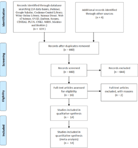
Figure 1: Prisma-chart of included-studies.
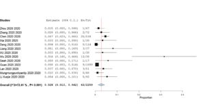
Figure 2: Forest-plot showing pooled incidence/prevalence of conjunctivitis/red-eye among patients with COVID 19.
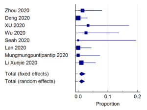
Figure 3: Forest-plot showing pooled-proportion of patients with first presentation as conjunctivitis/red-eye among patients with COVID19.
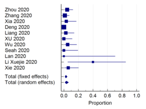
Figure 4: Forest-plot showing pooled-proportion of PCR positivity in conjunctival/tear samples in COVID19 patients.

Figure 5: Forest-plot showing association of ocular manifestation with disease severity of patient mild to moderate vs. severe to critical.

Figure 6: Forest-plot showing association of conjunctival sample PCR positivity with disease severity of patient mild to moderate vs. severe to critical.
Study, Author | Type of Study | Location | Patient Population | Ophthalmic Manifestation | Ophthalmic Interventions | Other Findings | Remarks | |
1.Zhou et al, 202044 | Retrospective cohort | Wuhan, china | N=67 (63 laboratory confirmed+ 4 suspected NCP patients) | Conjunctivitis =1. | No | Among positive conjunctival samples, one didn’t use protective eye covering while dealing with patients. | Doctors should use masks, goggles, protective clothing and gloves. | |
2.Zhang et al, 202035 | Cross-sectional study | Wuhan, China | 102 clinically diagnosed and 72 laboratory confirmed COVID-19 | Conjunctivitis= 2 | 1st case: Gancyclovir ointment. | – | Conjunctivitis may represent as an initial sign of the disease | |
3.Chen et al, 202024 | Cross-sectional study | Wuhan-china | 534 cases of COVID-19 | Conjunctival congestion= 25 conjunctival secretion=52 Ophthammalgia=22 foreign body Sensation=63 Photophobia=15 Blurred vision= 68 Tearing=55 Itching=53 Dry eye=112 | Topical ofloxacin, tobramycin, Gancyclovir and artificial tears | – | Provided in-depth results of all the ocular symptoms. | |
4.Xia et al, 202017 | Prospective interventional case series | Zhejiang, China. | 30 Laboratory confirmed NCP cases Severe= 9 Common type = 21 | Conjunctivitis=1 | Not mentioned | N.A. | Low amount of tear or conjunctival samples may affect PCR positivity | |
5.Deng et al, 202026 | Observational study | Wuhan, China | 114 patients with COVID-19 pneumonia | Nil | Nil | N.A. | Ocular protection and protective wear required for doctors | |
6.Liang et al, 2020 34 | Prospective case series | Yichang, China | 37Consecutive cases of PCR positive SARS-CoV 2 pneumonia | Conjunctivitis =3 | Not mentioned. | N.A. | SARS-CoV-2 may be present in conjunctival secretions | |
7.Xu et al 202027 | Cross sectional study | Shenyang, China | 30 COVID-19 patients (suspected=16 confirmed= 14) | Itching =1 Macular degeneration=1 | Not mentioned | Diabetes=3 Hypertension= 4 Hepatitis=1 Pneumonia=8 | Conjunctiva may be a transmission route of COVID-19 | |
8.Wu et al 202028 | Retrospective case series | Hubei province,China | 38 COVID-19 patients | Conjunctivitis=12 | Not mentioned. | Raised WBC, neutrophil, procalcitonin, C-reactive protein and LDH may be associated with ocular manifestations. | Nil | |
9.Seah et al 202029 | Prospective study | Singapore | 17 COVID-19 patients | Conjunctivitis =1 | Not mentioned. | N.A. | Nil | |
10.Guan et al 202031 | Observational | Wuhan, China | 552 Laboratory confirmed COVID-10 patient | Conjunctivitis= 9 | Not mentioned. | Diabetes, hypertension, CAD, hepatitis B | – | |
11.Lan et al 202030 | Prospective case series | China | 81 cases of confirmed COVID-19 patients | Ocular discomfort=3 Dry eye = 2 Itching= 2 Bulging eye = 2 | NA | Lymph adenopathy =2 | No new coronavirus-associated conjunctivitis has been seen in patients with COVID-19 and its prevalence is low | |
12.Mungmungpuntipantip et al 202032 | Prospective cross | Thailand | 48 COVID-19 patients | Nil | No | Not mentioned | Ocular protection should not be ignored by ophthalmologists during patient examination | |
13.Li Xuejie et al 202033 | Case series report | Wuhan, china | 92 NCP patients (Ophthalmic medical worker) | Conjunctivitis=5 | Topical Gancyclovir 4 times daily and Sodium hyaluronate eye drop. Relieved 3-5 days after treatment | All are health worker 1 patient first developed conjunctivitis and later developed systemic feature | Eye transmission may be a method of NCP transmission. | |
14. Xie 202036 | Retrospective study | Wuhan, china | 33 COVID-19 cases | No | No | – | SARS-CoV 2 might spread from normal conjunctiva of COVID-19 patients | |
NCP: Novel Corona Virus Pneumonia; WBC: White Blood Count; LDH: Lactate Dehydrogenase; CAD: Coronary Artery Disease | ||||||||
Table 1: Details of the included-studies.
Study, Author | Selection | Comparability | Outcome |
Zhou 202044 | *** | * | ** |
Zhang 202035 | *** | * | *** |
Chen 202024 | *** | * | *** |
Xia 202017 | ** | * | *** |
Deng 202026 | *** | * | *** |
Liang 202034 | *** | * | ** |
Xu 202027 | *** | * | ** |
Wu 202028 | *** | * | *** |
Seah 202029 | *** | * | *** |
Guan 202031 | **** | * | *** |
Lan 202030 | ** | * | *** |
Mungmungpuntipantip 202032 | ** | – | ** |
Li Xuejie 202033 | ** | – | ** |
Xie 202036 | *** | * | *** |
Table 2: Risk-of-bias of the included-studies.
Discussion
In our study, prevalence of red-eye/conjunctivitis in COVID-19 was 2.8% (95% CI 1.3% to 4.2%, I2=72.41%). Similar lower incidence of ocular symptoms was also noted with SARS-CoV-120. In our study, conjunctivitis/red eye was the first symptom of COVID-19 in 1.08% of patients (95% CI 0.37% to 2.44%, I2=0%). The occurrence of ocular manifestations may represent two different domains of manifestation of the problem, one is ocular tropism of the virus and the second is the adverse effects of the medications used in the management of COVID-19 [38,39].
In our study, tear sample PCR positivity rate in COVID-19 was 2.6% (1.3% to 4.5%, I2=47%). However, among COVID-19 patients with conjunctivitis/red eye, the PCR positivity rate in tear sample was 20.6% (6/29 cases). Again, among patients who were positive for the virus in tear sample by PCR, the proportion of conjunctivitis/red eye was 33.3% (4/12 cases). In our study, PCR positivity among COVID-19 cases was low (2.6%). Chan, et al., SARS-1 epidemic also reported zero percent PCR positivity out of twenty probable cases, which were confirmed with paired convalescent sera [40]. On the contrary, Loon, et al., in their study in 2003 SARS-1 epidemic, reported 37.5 % (3 out of 8 cases) PCR positive cases in tear sample in probable SARS-1cases in their study cohort (N=36) [20].
Although RT-PCR testing of viral-culture is very-specific but it lacks sensitivity [41]. To improve sensitivity multiple specimens can be tested. It can also be the case that virus and its genetic-material are present for short-period of disease and sample collection is not done at appropriate time [41]. RTPCR used in the included studies may not be sensitive enough to the detection of the small quantity of 19-CoV RNA. The time of sample collection considering the virulence of the disease and missing the significant no of cases is possible. Recently as reported by Doan, et al., to overcome the false negative error of PCR, newer technique like next generation sequencing can be applied for rapid detection of virus in tear sample as they are using in influenza and rubella virus will give us newer direction in these group of patients where the viral load in the initial period is low [42,43].
According to Xia, et al., the low amount of collected tear and conjunctival secretions may be an important determinant of PCR negativity and the simplest explanation is that sample amount/concentration might be below the detection limit of RT-PCR [17]. The window period of virus shedding may be missed. Moreover, the included studies are not mentioning the exact time of sample collection. Follow up samples also are not taken, only one time sample collection is done.
We also evaluated the association between occurrence of ocular manifestations and disease severity (mild and moderate vs. severe and critical). The odds were 0.28 (95% CI 0.12-0.67, I2=0%). This highlights that the mild to moderate severity disease had significantly lower occurrence of the ocular symptoms compared to the severe and critical group. However, we couldn’t find any association between tear sample PCR positivity and disease severity [odds ratio 0.46 (95% CI 0.06-3.45, T2=0%)].
Limitation of Our Study
One of the possible limitations is as the nature of COVID-19 is quite serious in nature, with predominantly respiratory or sepsis being areas of interest, ocular symptoms may go unnoticed or unattended, which may be one possible cause of low incidence of ocular symptoms and none of the studies have reported posterior chamber complications, however in animal model, posterior chamber complications were present. This indicates that there may less posterior chamber investigation during the disease course. Highly contagious nature of the disease is also a barrier for detailed ocular investigation.
Importance of Our Study
Our meta-analysis study was the first systematic review and metaanalysis evaluating the details of ocular manifestations associate with COVID-19 and its association with disease severity [43]. Targeted strategies to cut down the viral transmission from ocular surface by using different strategies e.g., use of PPEs (Personal Protective Equipments) e.g., goggles, N95 masks, goggles, use of transparent barrier during clinical examinations and use of agents e.g., povidone iodine eye drops may be helpful [8]. However, the findings may be considered to incorporate in day-to-day clinical practice taking into care of the level of evidence of safety and efficacy. This study also highlights the needs of long-term ocular follow up among COVID-19 patients and especially among those with ocular manifestations.
Conclusion
In patients with COVID-19, 2.8% of patients show ocular manifestation. However, among patients with ocular manifestation, only 20.6% cases showed tear/conjunctival swab RT-PCR positivity. So, ocular symptoms may warrant a COVID-19 screening test during the epidemic of COVID-19.
Acknowledgement
Acknowledgement
Authors acknowledge Mr. Siris Kr Bhattacharyya, Mrs. Anima Bhattacharyya, Dr. Linda Cottler and the FOGARTY team, INDO-US program in chronic non-communicable diseases (CNCDs) #1D43TW009120 (M Hazarika, Fellow) for their support. A pre-print version of this article is available in of the SSRN pre-print platform (http://dx.doi.org/10.2139/ssrn.3566161).
Source of Support
India-US Fogarty Training in Chronic Non-Communicable Diseases (CNCDs) across lifespan grant # 1D43TW009120.
Conflict of Interest
The authors have no conflict of interest to declare.
References
- Belser JA, Rota PA, Tumpey TM. Ocular tropism of respiratory viruses. Microbiol Mol Biol Rev 2013;77:144-56.
- Seah I, Agrawal R. Can the Coronavirus Disease 2019 (COVID-19) Affect the Eyes? A Review of Coronaviruses and Ocular Implications in Humans and Animals. Ocul Immunol Inflamm. 2020;15.
- Bhattacharyya A, Sarma P, Sarma B. Bacteriological pattern and their correlation with complications in culture positive cases of acute bacterial conjunctivitis in a tertiary care hospital of upper Assam. Medicine (Baltimore). 2020;99.
- Wang K, Chen W, Zhou YS. SARS-CoV-2 invades host cells via a novel route: CD147-spike protein. Microbiol. 2020.
- Prajapat M, Sarma P, Shekhar N, et al. Drug targets for corona virus: A systematic review. Ind J Pharmacol. 2020;52:56.
- Sarma P, Prajapat M, Avti P, Kaur H, Kumar S, Medhi B. Therapeutic options for the treatment of 2019-novel coronavirus: An evidence-based approach. Ind J Pharmacol. 2020;52:1.
- Wagner J, Jan Danser AH, Derkx FH. Demonstration of renin mRNA, angiotensinogen mRNA and angiotensin converting enzyme mRNA expression in the human eye: evidence for an intraocular renin-angiotensin system. Br J Ophthalmol. 1996;80:15963.
- Sarma P, Kaur H, Medhi B, Bhattacharyya A. Possible prophylactic or preventive role of topical povidone iodine during accidental ocular exposure to 2019-nCoV. Graefes Arch Clin Exp Ophthalmol. 2020.
- Savaskan E, Löffler KU, Meier F, Müller-Spahn F, Flammer J, Meyer P. Immunohistochemical localization of angiotensin-converting enzyme, angiotensin II and AT1 receptor in human ocular tissues. Ophthalmic Res. 2004;36:31220.
- Holappa M, Valjakka J, Vaajanen A. Angiotensin(1-7) and ACE2, “The Hot Spots” of renin-angiotensin system, detected in the human aqueous humor. Open Ophthalmol J. 2015;9:2832.
- Määttä M, Tervahartiala T, Kaarniranta K. Immunolocalization of EMMPRIN (Cd147) in the human eye and detection of soluble form of EMMPRIN in ocular fluids. Current Eye Research. 2006;31:91724.
- Nguyen TT, Sathe S, Stallone M. Expresssion of CD147 and Cyclophilin A in dry eye disease. Invest Ophthalmol Vis Sci. 2016; 57:411411.
- Li J-PO, Lam DSC, Chen Y, Ting DSW. Novel Coronavirus disease 2019 (COVID-19): The importance of recognising possible early ocular manifestation and using protective eyewear. Br J Ophthalmol. 2020;104:2978.
- In L. COVID-19 and Ophthalmology. 2020:23.
- Lai THT. Stepping up infection control measures in ophthalmology during the novel coronavirus outbreak: an experience from Hong Kong. 2020.
- Pneumonia NC. 新型冠状病毒肺炎疫情防控“一问一答” (第 3 版) 2020:02.
- Xia J, Tong J, Liu M, Shen Y, Guo D. Evaluation of coronavirus in tears and conjunctival secretions of patients with SARS-CoV-2 infection. J Medical Virol.
- Guan WJ, Ni ZY, Hu Y. Clinical characteristics of coronavirus disease 2019 in China. The New Eng J Med. 2020;113.
- Vabret A, Mourez T, Dina J. Human coronavirus NL63, France. Emerg Infect Dis. 2005;11:12259.
- Loon S-C, Teoh SCB, Oon LLE. The severe acute respiratory syndrome coronavirus in tears. Br J Ophthalmol. 2004;88:8613.
- Schoonjans F. MedCalc statistical software. MedCalc. [Last accessed on: April 12, 2023] https://www.medcalc.org/
- Funnel plots. [Last accessed on: April 12, 2023] https://handbook-5-1.cochrane.org/chapter_10/10_4_1_funnel_plots.htm
- Modesti PA, Reboldi G, Cappuccio FP. Panethnic differences in blood pressure in europe: a systematic review and meta-analysis. PLoS ONE. 2016;11:e0147601.
- Chen L, Deng C, Chen X. Ocular manifestations and clinical characteristics of 534 cases of COVID-19 in China: A cross-sectional study. MedRxiv. 2020;2020.03.12.20034678.
- Zhou Y, Zeng Y, Tong Y, Chen C. Ophthalmologic evidence against the interpersonal transmission of 2019 novel coronavirus through conjunctiva. 2020.
- Deng C, Yang Y, Chen H. Ocular Dectection of SARS-CoV-2 in 114 Cases of COVID-19 Pneumonia in Wuhan, China: An Observational Study. Rochester, NY: Social Science Research Network. 2020.
- Xu L, Zhang X, Song W. Conjunctival Polymerase Chain Reaction-Tests of 2019 Novel Coronavirus in Patients in Shenyang, China. Rochester, NY: Social Science Research Network. 2020.
- Wu P, Duan F, Luo C. Characteristics of ocular findings of patients with Coronavirus Disease 2019 (COVID-19) in Hubei Province, China. JAMA Ophthalmol. 2020.
- Seah IYJ, Anderson DE, Kang AEZ. Assessing viral shedding and infectivity of tears in Coronavirus Disease 2019 (COVID-19) Patients. Ophthalmol. 2020.
- Lan QQ, Zeng SM, Liao X, Xu F, Qi H, Li M. [Screening for novel coronavirus related conjunctivitis among the patients with corona virus disease-19]. Zhonghua Yan Ke Za Zhi. 2020;56:E009.
- Guan W, Ni Z, Hu Y. Clinical characteristics of Coronavirus Disease 2019 in China. New Eng J Med. 2020.
- Mungmungpuntipantip R, Wiwanitkit V. Ocular manifestation, eye protection and COVID-19. Graefes Arch Clin Exp Ophthalmol. 2020;1.
- Prevention and control strategies of ophthalmologists with new coronavirus infection associated with or with first-onset toxic conjunctivitis. Chinese J Experimental Ophthalmol. 2020;38:E002E002.
- Liang L, Wu P. There may be virus in conjunctival secretion of patients with COVID-19. Acta Ophthalmol. 2020.
- Sun X, Zhang X, Chen X. The infection evidence of SARS-COV-2 in ocular surface: a single-center cross-sectional study. MedRxiv. 2020;20027938.
- Xie HT, Jiang SY, Xu KK. SARS-CoV-2 in the ocular surface of COVID-19 patients. Eye Vis (Lond). 2020;7.
- Scalinci SZ, Trovato Battagliola E. Conjunctivitis can be the only presenting sign and symptom of COVID-19. IDCases 2020;20:e00774.
- Hydroxychloroquine-Induced retinal toxicity – American Academy of Ophthalmology. [Last accessed on: April 12, 2023] https://www.aao.org/eyenet/article/hydroxychloroquine-induced-retinal-toxicity
- Stokkermans TJ, Trichonas G. Chloroquine and hydroxychloroquine toxicity. In: StatPearls. Treasure Island (FL): StatPearls Publishing. 2020. [Last accessed on: April 12, 2023] http://www.ncbi.nlm.nih.gov/books/NBK537086/
- Chan WM, Yuen KSC, Fan DSP, Lam DSC, Chan PKS, Sung JJY. Tears and conjunctival scrapings for coronavirus in patients with SARS. Br J Ophthalmol. 2004;88:9689.
- Doan T, Acharya NR, Pinsky BA. Metagenomic DNA sequencing for the diagnosis of intraocular infections. Ophthalmol. 2017;124:12478.
- Sarma P, Kaur H, Kaur H. Ocular manifestations and tear or conjunctival swab PCR positivity for 2019-nCoV in patients with COVID-19: A Systematic Review and Meta-Analysis. 2020.
- Zhou Y, Zeng Y, Tong Y, Chen C. Ophthalmologic evidence against the interpersonal transmission of 2019 novel coronavirus through conjunctiva. Ophthalmol. 2020.
Supplementary File
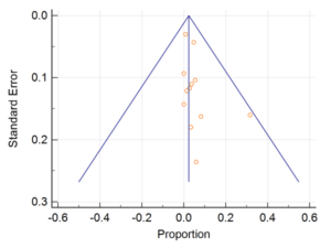
Supplementary Figure 1: Funnel plot showing publication bias among studies evaluating ocular complications of COVID-19/ conjunctivitis/red eye.
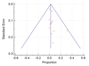
Supplementary Figure 2: Funnel plot showing publication bias among studies reporting conjunctivitis as first symptom in patients with COVID-19.
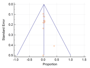
Supplementary Figure 3: Funnel plot showing publication bias among studies evaluating PCR positivity for 2019-nCoV in patients with COVID-19.
Article Type
Review Article
Publication History
Received Date: 28-11-2022
Accepted Date: 12-04-2023
Published Date: 19-04-2023
Copyright© 2023 by Sarma P, et al. All rights reserved. This is an open access article distributed under the terms of the Creative Commons Attribution License, which permits unrestricted use, distribution, and reproduction in any medium, provided the original author and source are credited.
Citation: Bhattacharyya A, et al. Ocular Manifestations and Tear or Conjunctival Swab PCR Positivity For SARS-CoV-2 In Patients With COVID-19: A Systematic Review and Meta-Analysis. J Ophthalmol Adv Res. 2023;4(1):1-14.

Figure 1: Prisma-chart of included-studies.

Figure 2: Forest-plot showing pooled incidence/prevalence of conjunctivitis/red-eye among patients with COVID 19.

Figure 3: Forest-plot showing pooled-proportion of patients with first presentation as conjunctivitis/red-eye among patients with COVID19.

Figure 4: Forest-plot showing pooled-proportion of PCR positivity in conjunctival/tear samples in COVID19 patients.

Figure 5: Forest-plot showing association of ocular manifestation with disease severity of patient mild to moderate vs. severe to critical.

Figure 6: Forest-plot showing association of conjunctival sample PCR positivity with disease severity of patient mild to moderate vs. severe to critical.
Study, Author | Type of Study | Location | Patient Population | Ophthalmic Manifestation | Ophthalmic Interventions | Other Findings | Remarks |
|
1.Zhou et al, 202044 | Retrospective cohort | Wuhan, china | N=67 (63 laboratory confirmed+ 4 suspected NCP patients)
| Conjunctivitis =1.
| No | Among positive conjunctival samples, one didn’t use protective eye covering while dealing with patients. | Doctors should use masks, goggles, protective clothing and gloves.
|
|
2.Zhang et al, 202035 | Cross-sectional study | Wuhan, China | 102 clinically diagnosed and 72 laboratory confirmed COVID-19 | Conjunctivitis= 2
| 1st case: Gancyclovir ointment. | – | Conjunctivitis may represent as an initial sign of the disease |
|
3.Chen et al, 202024 | Cross-sectional study | Wuhan-china | 534 cases of COVID-19 | Conjunctival congestion= 25 conjunctival secretion=52 Ophthammalgia=22 foreign body Sensation=63 Photophobia=15 Blurred vision= 68 Tearing=55 Itching=53 Dry eye=112 | Topical ofloxacin, tobramycin, Gancyclovir and artificial tears | – | Provided in-depth results of all the ocular symptoms. |
|
4.Xia et al, 202017 | Prospective interventional case series | Zhejiang, China. | 30 Laboratory confirmed NCP cases Severe= 9 Common type = 21
| Conjunctivitis=1
| Not mentioned | N.A. | Low amount of tear or conjunctival samples may affect PCR positivity |
|
5.Deng et al, 202026 | Observational study | Wuhan, China | 114 patients with COVID-19 pneumonia | Nil | Nil | N.A. | Ocular protection and protective wear required for doctors |
|
6.Liang et al, 2020 34 | Prospective case series | Yichang, China
| 37Consecutive cases of PCR positive SARS-CoV 2 pneumonia
| Conjunctivitis =3
| Not mentioned. | N.A.
| SARS-CoV-2 may be present in conjunctival secretions
|
|
7.Xu et al 202027 | Cross sectional study | Shenyang, China | 30 COVID-19 patients (suspected=16 confirmed= 14) | Itching =1 Macular degeneration=1
| Not mentioned | Diabetes=3 Hypertension= 4 Hepatitis=1 Pneumonia=8 | Conjunctiva may be a transmission route of COVID-19 |
|
8.Wu et al 202028 | Retrospective case series | Hubei province,China | 38 COVID-19 patients
| Conjunctivitis=12
| Not mentioned. | Raised WBC, neutrophil, procalcitonin, C-reactive protein and LDH may be associated with ocular manifestations.
| Nil
|
|
9.Seah et al 202029 | Prospective study | Singapore | 17 COVID-19 patients
| Conjunctivitis =1 | Not mentioned. | N.A.
| Nil |
|
10.Guan et al 202031 | Observational | Wuhan, China | 552 Laboratory confirmed COVID-10 patient | Conjunctivitis= 9 | Not mentioned. | Diabetes, hypertension, CAD, hepatitis B | – |
|
11.Lan et al 202030 | Prospective case series | China | 81 cases of confirmed COVID-19 patients | Ocular discomfort=3 Dry eye = 2 Itching= 2 Bulging eye = 2 | NA | Lymph adenopathy =2 | No new coronavirus-associated conjunctivitis has been seen in patients with COVID-19 and its prevalence is low |
|
12.Mungmungpuntipantip et al 202032 | Prospective cross | Thailand | 48 COVID-19 patients | Nil | No | Not mentioned | Ocular protection should not be ignored by ophthalmologists during patient examination |
|
13.Li Xuejie et al 202033
| Case series report | Wuhan, china | 92 NCP patients (Ophthalmic medical worker)
| Conjunctivitis=5 | Topical Gancyclovir 4 times daily and Sodium hyaluronate eye drop.
Relieved 3-5 days after treatment | All are health worker
1 patient first developed conjunctivitis and later developed systemic feature
| Eye transmission may be a method of NCP transmission.
|
|
14. Xie 202036 | Retrospective study | Wuhan, china | 33 COVID-19 cases | No | No | – | SARS-CoV 2 might spread from normal conjunctiva of COVID-19 patients |
|
NCP: Novel Corona Virus Pneumonia; WBC: White Blood Count; LDH: Lactate Dehydrogenase; CAD: Coronary Artery Disease | ||||||||
Table 1: Details of the included-studies.
Study, Author | Selection | Comparability | Outcome |
Zhou 202044 | *** | * | ** |
Zhang 202035 | *** | * | *** |
Chen 202024 | *** | * | *** |
Xia 202017 | ** | * | *** |
Deng 202026 | *** | * | *** |
Liang 202034 | *** | * | ** |
Xu 202027 | *** | * | ** |
Wu 202028 | *** | * | *** |
Seah 202029 | *** | * | *** |
Guan 202031 | **** | * | *** |
Lan 202030 | ** | * | *** |
Mungmungpuntipantip 202032 | ** | – | ** |
Li Xuejie 202033 | ** | – | ** |
Xie 202036 | *** | * | *** |
Table 2: Risk-of-bias of the included-studies.

Supplementary Figure 1: Funnel plot showing publication bias among studies evaluating ocular complications of COVID-19/ conjunctivitis/red eye.

Supplementary Figure 2: Funnel plot showing publication bias among studies reporting conjunctivitis as first symptom in patients with COVID-19.

Supplementary Figure 3: Funnel plot showing publication bias among studies evaluating PCR positivity for 2019-nCoV in patients with COVID-19.


