Moyseyenko NM1*
1Head of the Department of Ophthalmology, Ivano-Frankivsk National Medical University, Ivano-Frankivsk, Ukraine
*Corresponding Author: Moyseyenko NM, Head of the Department of Ophthalmology, Ivano-Frankivsk National Medical University, Ivano-Frankivsk, Ukraine; Email: [email protected]
Published Date: 06-07-2022
Copyright© 2022 by Moyseyenko NM. All rights reserved. This is an open access article distributed under the terms of the Creative Commons Attribution License, which permits unrestricted use, distribution, and reproduction in any medium, provided the original author and source are credited.
Abstract
Optic nerve neuropathy due to dental lesions is rare but causes complex persistent functional disorders that are difficult to diagnose. The aim of this case report is to show odontogenic optic neuropathy through treatment tactics and observation of structural dynamics and functional changes of the optic nerve with the help of a clinical case. The consultation of the patient was done as per the Department of Ophthalmology, Ivano-Frankivsk National University, Ukraine. Tests such as computed perimetry, Optically Coherent Tomography (OCT) of the optic nerve, Magnetic Resonance Imaging (MRI) of the brain, Computed Tomography (CT) of the paranasal sinuses and immunological blood tests were performed. The patient gave informed consent to access personal data and use it for scientific purposes. In the case of patient S., the combination of neuritis with lesions of the anterior part of the vascular tract is attributed to the background of multiple caries and prolapse of the teeth of the upper jaw in the sinus. Treatment includes a regimen by a dentist for caries and pulse therapy with corticosteroids. From the result, it was possible to reduce the swelling in the nerve fibers of the right eye by 35% and the left eye by 19%. The average sensitivity threshold in the right eye increased by 79% and in the left eye by 90%. In conclusion, we confirm that odontogenic optic neuropathy is a rare form of optic nerve damage. Two-stage treatment (dental and corticosteroids) reduces the nerve fibers swelling and increases retinal ganglion cells sensitivity.
Keywords
Odontogenic Optic Neuropathy; Optic Neuritis; Optically Coherent Tomography; Perimetry
Introduction
Optic nerve neuropathy due to dental lesions is rare but cause complex persistent functional disorders that are difficult to diagnose. According to the published scientific literature, acute dental infection causes optic nerve inflammation. At the same time, there is a sudden decrease in visual acuity, visual field defects, and oedema in Optic Disc (OD). The treatment begins with managing the dental infection, then combining it with corticosteroids [1]. In children and young people, recorded cases of retrobulbar neuritis are characterized by slow visual loss (up to 6 weeks) and complete recovers. The combination of antibiotics and corticosteroids is quite effective [2].
Ischemic components of the optic neuropathy development due to tooth extraction are also possible. The sectoral pallor of Optic Disc (OD) two months after the procedure have been studied in previous published studies [3]. This phenomenon is explained by the migration of micro embolus of dental material during extraction through the alveolar-arterial network to the short posterior ciliary vessels [3]. Thus, studying the pathogenetic mechanisms of odontogenic optic nerve damage and searching for optimal diagnosis and treatment methods are relevant because they contribute to better recovery of functional and structural damage. This case report aims to show a clinical case of odontogenic optic neuropathy with treatment tactics and observation of dynamics during structural and functional changes of the optic nerve.
Materials and Methods
The patient’s consultation was based on the Department of Ophthalmology, Ivano-Frankivsk National University, Ukraine. Tests such as computed perimetry, Optically Coherent Tomography (OCT) of the optic nerve, Magnetic Resonance Imaging (MRI) of the brain, and Computed Tomography (CT) of the paranasal sinuses were done. In addition to these tests, immunological blood tests were also performed to exclude the demyelinating nature of neuropathy. The patient gave informed consent to process the personal data for scientific purposes. The treatment included two stages of dental treatment of caries and pulse therapy with corticosteroids.
Results and Discussion
Patient S turned 20 years old in October 2021 and sought advice from a neuro-ophthalmologist after a history of iridocyclitis treatment and posterior synechiae of the right eye. The consultation was for preliminary perimeter examination and OST as preoperative preparation. The neuro-ophthalmologist found a pronounced bilateral oedema OD (Fig. 1). At the time of first consultation dated (10/2021), visual acuity of the right eye with correction was 0.5, left 1.0. The right eye was calm, and pupil was round. The posterior synechiae was at 18th year. There was a limited direct light reaction in the right eye and a concomitant light reaction in the left eye. The Perimetry at the initial examination found bilateral scotomas on the central part of the field-of-view and had different intensities (Fig. 2). OCT’s dates showed bilateral optic nerve fiber layer oedema (Fig. 3). It was this moment that led the patient to a neuro-ophthalmological consultation. The previously performed MRI of the brain found no volumetric damage.
Later the patient consulted an ENT and a dentist. The diagnosis revealed prolapse of maxillary sinus in tooth roots and multiple carious cavities in the upper and lower jaw. Eventually, the dental treatment started, which lasted from October 10, (2021) to January 1, (2022). The treatment partially reduced the oedema on the fundus to improve the visibility (Fig. 4). The intensity of cattle according to the perimeter data, also decreased (Fig. 5). The next stage of treatment lasted from 01/(2022) to 03/(2022). This treatment included pulse therapy with methylprednisolone (the first three days at a dose of 1000 mg/day, then a gradual reduction to five mg/day). At the same time significant reduction of hypostasis within the layer of nerve fibers was noted (Fig. 6). The average thickness of the nerve fiber layer in the right eye decreased from 331 mm to 215 mm (116 mm, 35%). While, the thickness for the left eye turned 182 mm from 224 mm (42 mm, 19%). The cup volume of the right eye decreased from 9.35 mm 2 to 4.27 mm 2 (by 5.08 mm 2, 54%), and the left increased from 2.97 mm 2 to 4.31 (by 1.34 mm 2, 45%), apparently by reducing parapapillary oedema. The single scotomas remained in the field-of-view for both eyes (Fig. 7). The average threshold of retinal ganglion cells in the right eye increased from -3.13 dB to -0.67 dB (2.26 dB, 79%), and in the left eye from -1.27 dB to -0.13 dB (at 1.14 dB, 90%).
Optic neuritis is a heterogeneous group of diseases. Inflammatory neuropathy can play an important role in the development of modern neuro-ophthalmology. The international classification requires consideration of many equivocal details. Typical or not, neuritis is a question that arises during the diagnosis of condition among young people [4].
The combination of neuritis with lesions in the anterior part of the vascular tract is the case of patient S. This puts the case of patient S in the category of atypical neuro uveitis, a place occupied by bacterial odontogenic or rhino-sinusogenic inflammation [5]. This diagnosis prompted the patient for consultation with an ENT and a dentist. Also, the patient opt for Computed Tomograhy (CT) scan to confirm paranasal sinuses. The result detected caries and prolapse of teeth tips in the maxillary sinus to determine the first stage of treatment-dental.
We recommend that bilateral disc oedema should be differentiated from other optic neuropathies where oedema is possible [6]. Careful examination of visual functions (field-of-view) and structural damage to the optic nerve (OCT) helped in this case study.
The second stage of treatment eliminated autoimmune inflammatory factors through pulse therapy with corticosteroids [7]. As a result, it was possible to reduce the swelling of the nerve fibers in the right eye by 35% and the left eye by 19%. The average sensitivity threshold of retinal ganglion cells in the right eye increased by 79% and in the left eye by 90%.
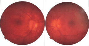
Figure 1: The state of the eye day of patient S at the initial examination.
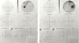
Figure 2: Perimetry of patient S at the initial examination.
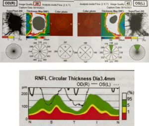
Figure 3: OCT of patient S at the initial examination.

Figure 4: The condition of the fundus after dental treatment.
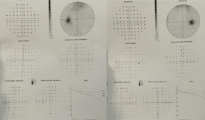
Figure 5: Perimeter of patient S after dental treatment.
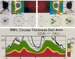
Figure 6: Dynamics of structural changes of the optic nerve of patient S after pulse therapy.
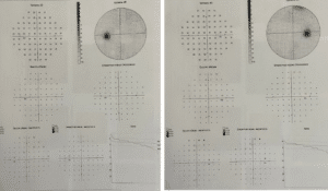
Figure 7: Perimeter of patient S after pulse therapy.
Conclusion
Odontogenic optic neuropathy is a rare form of optic nerve damage. Two-stage treatment (dental and corticosteroids) can reduce nerve fibers swelling and increase the retinal ganglion cells sensitivity.
Conflict of Interest
The authors declare that there is no conflict of interest.
References
- Chaabouni M, Hachicha L, Chebihi S. Optic neuropathy of dental origin. Apropos of 4 cases. Revue de Stomatologie et de Chirurgie Maxillo-faciale. 1991;92(4):259-61.
- Chanzy S, Routon MC, Bursztyn J, Maitrepierre E, Cheminée M, Msélati JC. Maxillary and sphenoid sinusitis complicated by acute inflammatory optic neuritis in a 12-year-old patient. Archives de Pediatrie: Organe Officiel de la Societe Francaise de Pediatrie. 2005;12(1):46-8.
- Kravitz E, Foroozan R. Anterior ischemic optic neuropathy after dental extraction. J Neuro-Ophthalmol. 2019;39(1):14-7.
- Abel A, McClelland C, Lee MS. Critical review: Typical and atypical optical neuritis. Surv Ophthalmol. 2019;64(6):770-9.
- Malik A, Ahmed M, Golnik K. Treatment options for atypical optical neuritis. Indian J Ophthalmol. 2014;62(10):982-4.
- Margolin E. The swollen optical nerve: an approach to diagnosis and management. Pract Neurol. 2019;19(4):302-9.
- Williams KJ, Allen RC. An atypical case of optic neuropathy. JAMA Ophthalmol. 2022;140(1):88-9.
Article Type
Case Report
Publication History
Received Date: 08-06-2022
Accepted Date: 28-06-2022
Published Date: 06-07-2022
Copyright© 2022 by Moyseyenko NM. All rights reserved. This is an open access article distributed under the terms of the Creative Commons Attribution License, which permits unrestricted use, distribution, and reproduction in any medium, provided the original author and source are credited.
Citation: Moyseyenko NM. Odontogenic Optic Neuropathy, Clinic, Treatment, Dynamics: A Case Report. J Ophthalmol Adv Res. 2022;3(2):1-8.

Figure 1: The state of the eye day of patient S at the initial examination.

Figure 2: Perimetry of patient S at the initial examination.

Figure 3: OCT of patient S at the initial examination.

Figure 4: The condition of the fundus after dental treatment.

Figure 5: Perimeter of patient S after dental treatment.

Figure 6: Dynamics of structural changes of the optic nerve of patient S after pulse therapy.

Figure 7: Perimeter of patient S after pulse therapy.


