Mohamed Hosny1*, Paula Sameh2, Sarah Azzam3
1Professor of Ophthalmology, Cairo University, Egypt
2Resident of Ophthalmology, Cairo University, Egypt
3Assistant Professor of Ophthalmology, Cairo University, Egypt
*Correspondence author: Mohamed Hosny, MD, FRCSEd, Professor of Ophthalmology, Cairo University, Egypt; Email: [email protected]
Published Date: 05-04-2023
Copyright© 2023 by Hosny M, et al. All rights reserved. This is an open access article distributed under the terms of the Creative Commons Attribution License, which permits unrestricted use, distribution, and reproduction in any medium, provided the original author and source are credited.
Abstract
Purpose: To determine the contribution of spherical aberration and coma to the total ocular high order aberration before and after femto laser-assisted in-situ keratomileusis using an aspheric ablation profile.
Methods: This is a prospective interventional study that was conducted on 34 eyes of 17 patients in the interval between January 2021 and June 2021. Patients did a preoperative aberrometry and corneal tomography. They underwent FS-LASIK surgery using an aspheric ablation profile. The total ocular aberrometry and corneal tomography were repeated one month postoperatively.
Results: The mean preoperative contribution of coma to total ocular HOA was 52.16 % with SD of ± 25.59% which declined to 49.48 % with SD of ± 22.53 % postoperatively. The mean preoperative contribution of SA to total ocular HOA was 38.91 % with SD of ± 15.35 % which increased significantly to 51.25 % with SD of ± 16.55 % postoperatively.
Conclusion: Post-operative contribution of corneal spherical aberrations to total ocular HOA showed a statistically significant increase compared to pre-operative. However, there was no significant change in coma contribution.
Keywords: Spherical Aberration; Coma; High Order Aberration; Femtosecond Laser-Assisted in Situ Keratomileusis; Aspheric Ablation Profile
Introduction
Laser-assisted In-Situ Eratomileu sis (LASIK) is the most frequently performed refractive surgery procedure for the correction of myopia, hyperopia, and astigmatism [1]. Improved understanding and classification of the aberrations induced by laser refractive surgery is an important requirement to design new algorithms and approaches for customized laser procedures. It has been argued that standard non wavefront nor topography guided ablations can induce higher order aberration [2]. As the most relevant clinical higher order aberrations that directly affect vision, Coma and Spherical aberrations have been found to cause most of the higher order related visual complaints after refractive surgery [3]. The contribution of each one of them to the higher order wavefront before and after refractive surgery is the purpose of this work.
Methods
This was a prospective interventional study that was conducted on 34 eyes of 17 patients in the interval between January 2021 and June 2021. The study was approved by the Cairo University Hospitals Ethical Committee in December 2020 and was registered in ClinicalTrials.gov with a UID (NCT04966806). None of the authors have any financial interests in any product used in this research. No Funding was allocated for this study from any research fund. There is no conflict of interests regarding any of the collaborating authors with this study. All patients read and signed an informed consent and the study abided by the declaration of Helsinki declaration of 1975, as revised in 1983. Patients did a preoperative aberrometry using the CSO Peramis and corneal Tomography Using the CSO Sirrius. The procedure was started by using the Ziemer LDV Z8 (ZOS Port, Switzerland) for creation of a 110 micron thickness flap with a diameter of 9 mm and a side cut angle of 90 degrees. Then LASIK was completed using an aspheric ablation profile (Aberration Free) mode on the Schwind Amaris 1050 excimer laser (SCHWIND eye-tech Kleinostheim, Germany). The total ocular aberrometry and corneal tomography were repeated one month postoperatively.
Results
This study included 34 eyes of 17 patients with mean age 25 (± 5.4 years), 35% of patients were males and 6% were females. The mean preoperative Spherical equivalent was -4.0D (SD+/- 1.6). The UCDVA of all eyes 1 month after the procedure was 0.0 LogMar. All eyes were within 0.5D Spherical equivalent after one month. No intraoperative or postoperative complications were recorded. All measured ocular aberrations were recorded in diopteric equivalent (Eq.D) instead of microns for elimination of plus (+) or (-) signs to easily calculate and interpret Spherical aberration results.
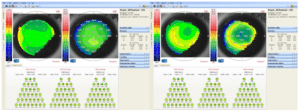
Preoperative Data (Fig. 1, Table 1):
- The mean preoperative total ocular aberrations for 34 eyes were 4.04 Eq.D with standard deviation (SD) : ± 66. Range: 1.77 to 7.13
- The mean preoperative total ocular HOA for 34 eyes were 0.33 Eq.D with standard deviation (SD) : ± 11. Range: 0.19 to 0.64
- The mean preoperative Total coma for 34 eyes were 0.16 Eq.D with standard deviation (SD) : ± 07. Range: 0.03 to 0.30
- The mean preoperative Total Spherical Aberration for 34 eyes were 0.12 Eq.D with standard deviation (SD) : ± 04. Range: 0.05 to 0.23

Figure 1: Preoperative total OA (Left), total ocular HOA, total coma and total SA (right).
|
Mean |
SD |
Median |
Minimum |
Maximum |
|
|
Preoperative Total Ocular aberrations |
4.04 |
1.66 |
3.41 |
1.77 |
7.13 |
|
Preoperative Total Ocular HOA |
0.33 |
0.11 |
0.32 |
0.19 |
0.64 |
|
Preoperative Corneal Coma |
0.16 |
0.07 |
0.18 |
0.03 |
0.30 |
|
Preoperative Corneal SA |
0.12 |
0.04 |
0.12 |
0.05 |
0.23 |
Table 1: Preoperative total OA, total ocular HOA, total coma and total SA.
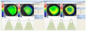
Post Operative Data (Fig. 2, Table 2):
- The mean postoperative total ocular HOA for 34 eyes were 0.41 Eq.D with standard deviation (SD) : ± 16. Range: 0.189 to 0.89
- The mean postoperative Total coma for 34 eyes were 0.19 Eq.D with standard deviation (SD) : ± 19. Range: 0.01 to 0.41
- The mean postoperative Total Spherical Aberration for 34 eyes were 0.20 Eq.D with standard deviation (SD) : ± 06. Range: 0.08 to 0.34

Figure 2: Comparison between pre-operative (left) and post-operative data (right).
|
Mean |
SD |
Median |
Minimum |
Maximum |
|
|
Postoperative Total Ocular aberrations |
1.04 |
0.46 |
1.07 |
0.37 |
2.18 |
|
Postoperative Total Ocular HOA |
0.41 |
0.16 |
0.36 |
0.18 |
0.89 |
|
Postoperative Total Coma |
0.19 |
0.09 |
0.19 |
0.01 |
0.41 |
|
Postoperative Total SA |
0.20 |
0.06 |
0.19 |
0.08 |
0.34 |
Table 2: Post-operative total OA, total ocular HOA, total coma and total SA.
Comparison Between Pre-Operative and Post-Operative Data (Table 3-5):
The mean preoperative total ocular HOA for 34 eyes increased from 0.33 Eq.D with standard deviation (SD) : ± 0.11 preoperative to 0.41 Eq.D with Standard Deviation (SD) : ± 0.16 post-operative but this increase was not statistically significant (p = 0.071) (Table 3).
|
|
Preoperative |
Postoperative |
|
|||||||||||
|
Mean |
SD |
Median |
Min. |
Max. |
Mean |
SD |
Median |
Min. |
Max. |
P-value |
||||
|
Total Ocular HOA |
0.33 |
0.11 |
0.32 |
0.19 |
0.64 |
0.41 |
0.16 |
0.36 |
0.18 |
0.89 |
0.071 |
|||
Table 3: Comparison between pre-operative and post-operative total ocular HOAs.
The mean preoperative total coma for 34 eyes increased from 0.16 Eq.D with standard deviation (SD) : ± 0.07 preoperative to 0.19 Eq.D with Standard Deviation (SD) : ± 0.19 post-operative. Again, this increase was not statistically significant (p = 0.166) (Table 4).
|
Preoperative |
Postoperative |
|
|||||||||
|
Mean |
SD |
Median |
Min. |
Max. |
Mean |
SD |
Median |
Min. |
Max. |
P-value |
|
|
Corneal Coma |
0.16 |
0.07 |
0.18 |
0.03 |
0.30 |
0.19 |
0.09 |
0.19 |
0.01 |
0.41 |
0.166 |
Table 4: Comparison between preoperative and post-operative total coma.
The mean preoperative total SA for 34 eyes increased significantly from 0.12 Eq.D with Standard Deviation (SD) : ± 0.04 preoperative to 0.20 Eq.D with standard deviation (SD) : ± 0.06 post-operative. This increase was statistically significant (p less than 0.001) (Table 5).
|
|
Preoperative |
Postoperative |
|
||||||||
|
Mean |
SD |
Median |
Min. |
Max. |
Mean |
SD |
Median |
Min. |
Max. |
P-value |
|
|
Total SA |
0.12 |
0.04 |
0.12 |
0.05 |
0.23 |
0.20 |
0.06 |
0.19 |
0.08 |
0.34 |
< 0.001 |
Table 5: Comparison between preoperative and post-operative total SA.
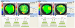
Contribution of Coma and SA to Total Ocular HOA (Fig. 3, Table 6)
- The mean preoperative contribution of coma to total ocular HOA was 52.16 % with SD of ± 25.59%, which declined to 49.48 % with SD of ± 22.53 % postoperative but this decline was not statistically significant (p = 0.844)
- The mean preoperative contribution of SA to total ocular HOA was 38.91 % with SD of ± 35 %, which showed a statistically significant increase to 51.25 % with SD of ± 16.55 % postoperative (p less than 0.001) (Table 6)

Figure 3: Comparison between pre-operative (left) and post-operative data (right).
|
Preoperative |
Postoperative |
||||||||||
|
Mean |
SD |
Median |
Min. |
Max. |
Mean |
SD |
Median |
Min. |
Max. |
P -value |
|
|
Coma to total ocular HOA (%) |
52.16 |
25.59 |
46.09 |
13.64 |
100.00 |
49.48 |
22.53 |
48.48 |
2.33 |
106.67 |
0.844 |
|
SA to total ocular HOA (%) |
38.91 |
15.35 |
40.54 |
14.00 |
74.19 |
51.52 |
16.55 |
51.14 |
15.73 |
100.00 |
< 0.001 |
Table 6: Contribution of coma and SA to total ocular HOAs.
Discussion
It is established in several studies that corneal refractive surgery causes a significant increase in the HOAs of the eye despite a good spherocylindrical refractive outcome and this causes a decline in the visual performance such as low contrast sensitivity and production of visual problems as haloes, starburst and glare that is more evident at dim light and therefore reduced patient satisfaction [4,5].
Thomas Kohnen showed in a study that the total HOA Root Mean Square (RMS) changed in the group of 50 eyes that underwent LASIK operation using conventional ablation profile by 0.167±0.180μm (factor 1.53). The mean induction of coma RMS was 0.092 ± 0.195μm; For spherical aberration (Z 4,0), the study showed a significant increase (0.130±0.120 μm; factor 1.6; P <0.001) [6]. Imene Salah-Mabed showed in his study of 50 myopic eyes underwent corneal refractive surgery using the WaveLight® Refractive Suite (Alcon® Laboratories Inc., USA) that there is a very slight but significant increase in total, corneal, and internal ocular aberrations after LASIK surgery. The most important increase in corneal and total HOAs seems to be attributed to the increase of corneal coma. The total spherical aberration increased very slightly but significantly (0.034 ± 0.063; P<0.001) [7].
Majid Moshirfar in a study on 44 eyes (22 patients), with one eye randomized to WaveLight Allegretto, and the fellow eye receiving VISX CustomVue. In the WF optimized group, total HOA increased 4% (P = 0.012), coma increased 11% (P = 0.065), and spherical aberration increased 19% (P = 0.214). In the WF guided group, total HOA RMS decreased 9% (P = 0.126), coma decreased 18% (P = 0.144), spherical aberration decreased 27% (P = 0.713) [8].
In our study performed on 34 eyes of 17 patients. The mean preoperative total ocular HOA increased from 0.33 Eq.D with Standard Deviation (SD) : ± 0.11 preoperative to 0.41 Eq.D with Standard Deviation (SD) : ± 0.16 postoperative. The mean preoperative total coma increased non significantly from 0.16 Eq.D preoperative to 0.19 Eq.D post-operative. The mean preoperative total spherical aberration increased significantly from 0.12 Eq.D to 0.20 Eq.D post-operative (Table 7).
|
Preoperative |
Postoperative |
||||||||
|
Case Number |
Eye |
Total Ocular Aberations |
Total Ocular HOA |
Corneal Coma |
Corneal SA |
Total Ocular Aberations |
Total Ocular HOA |
Corneal Coma |
Corneal SA |
|
1 |
OD |
3.1 |
0.43 |
0.26 |
0.08 |
0.54 |
0.34 |
0.25 |
0.09 |
|
OS |
3.64 |
0.64 |
0.23 |
0.09 |
0.91 |
0.44 |
0.16 |
0.13 |
|
|
2 |
OD |
4.35 |
0.25 |
0.09 |
0.13 |
0.59 |
0.29 |
0.18 |
0.2 |
|
OS |
4.36 |
0.37 |
0.18 |
0.15 |
0.61 |
0.37 |
0.36 |
0.21 |
|
|
3 |
OD |
6.76 |
0.32 |
0.1 |
0.14 |
0.82 |
0.65 |
0.36 |
0.33 |
|
OS |
6.91 |
0.36 |
0.06 |
0.18 |
0.69 |
0.54 |
0.26 |
0.34 |
|
|
4 |
OD |
5.25 |
0.29 |
0.14 |
0.09 |
1.27 |
0.68 |
0.26 |
0.32 |
|
OS |
5.7 |
0.26 |
0.19 |
0.12 |
1.88 |
0.69 |
0.41 |
0.31 |
|
|
5 |
OD |
3.12 |
0.19 |
0.15 |
0.06 |
1.21 |
0.56 |
0.27 |
0.18 |
|
OS |
3.53 |
0.31 |
0.12 |
0.05 |
0.37 |
0.3 |
0.32 |
0.08 |
|
|
6 |
OD |
1.77 |
0.37 |
0.26 |
0.15 |
1.24 |
0.33 |
0.1 |
0.15 |
|
OS |
4.24 |
0.2 |
0.17 |
0.12 |
0.59 |
0.22 |
0.03 |
0.11 |
|
|
7 |
OD |
2.27 |
0.26 |
0.06 |
0.12 |
1.52 |
0.43 |
0.01 |
0.2 |
|
OS |
2.38 |
0.26 |
0.11 |
0.12 |
1.43 |
0.31 |
0.15 |
0.18 |
|
|
8 |
OD |
3.33 |
0.45 |
0.19 |
0.1 |
1.25 |
0.29 |
0.1 |
0.17 |
|
OS |
3 |
0.44 |
0.16 |
0.12 |
1.38 |
0.43 |
0.16 |
0.16 |
|
|
9 |
OD |
2.28 |
0.33 |
0.09 |
0.08 |
0.49 |
0.21 |
0.1 |
0.15 |
|
OS |
2.7 |
0.19 |
0.03 |
0.11 |
0.64 |
0.24 |
0.11 |
0.17 |
|
|
10 |
OD |
7.12 |
0.31 |
0.3 |
0.1 |
0.46 |
0.35 |
0.23 |
0.2 |
|
OS |
7.13 |
0.22 |
0.21 |
0.08 |
0.71 |
0.49 |
0.24 |
0.29 |
|
|
11 |
OD |
2.67 |
0.44 |
0.06 |
0.11 |
1.77 |
0.5 |
0.05 |
0.21 |
|
OS |
2.34 |
0.19 |
0.11 |
0.09 |
1.55 |
0.35 |
0.17 |
0.14 |
|
|
12 |
OD |
5.85 |
0.28 |
0.23 |
0.09 |
1.27 |
0.89 |
0.19 |
0.14 |
|
OS |
5.58 |
0.21 |
0.19 |
0.11 |
2.18 |
0.53 |
0.26 |
0.18 |
|
|
13 |
OD |
3.36 |
0.39 |
0.08 |
0.1 |
0.49 |
0.3 |
0.15 |
0.2 |
|
OS |
4.12 |
0.38 |
0.13 |
0.12 |
1.06 |
0.52 |
0.28 |
0.23 |
|
|
14 |
OD |
2.71 |
0.2 |
0.2 |
0.12 |
1.15 |
0.34 |
0.14 |
0.23 |
|
OS |
3.27 |
0.21 |
0.07 |
0.13 |
1.08 |
0.43 |
0.11 |
0.27 |
|
|
15 |
OD |
2.39 |
0.31 |
0.25 |
0.23 |
1.11 |
0.18 |
0.16 |
0.18 |
|
OS |
2.44 |
0.37 |
0.27 |
0.19 |
1 |
0.31 |
0.23 |
0.2 |
|
|
16 |
OD |
3.45 |
0.47 |
0.21 |
0.2 |
1.32 |
0.3 |
0.18 |
0.19 |
|
OS |
3.15 |
0.4 |
0.19 |
0.19 |
1.55 |
0.33 |
0.21 |
0.17 |
|
|
17 |
OD |
6.5 |
0.44 |
0.25 |
0.09 |
0.52 |
0.4 |
0.2 |
0.18 |
|
OS |
6.65 |
0.5 |
0.18 |
0.07 |
0.75 |
0.41 |
0.19 |
0.22 |
|
Table 7: Comparison between preoperative and post-operative values.
Conclusion
The mean preoperative contribution of coma to total ocular HOA was 52.16% preoperative which declined to 49.48% postoperative, while the mean preoperative contribution of SA to total ocular HOA was 38.91% preoperative which increased significantly to 51.25% postoperative.
Conflict of Interest
The authors have no conflict of interest to declare.
References
- Gatinel D, Haouat M, Hoang-Xuan T. A review of mathematical descriptors of corneal asphericity. J Fr Ophtalmol. 2002;25(1):81-90.
- Dorronsoro C, Remon L, Merayo-Lloves J, Marcos S. Experimental evaluation of optimized ablation patterns for laser refractive surgery. Opt Express. 2009;17(17):15292-307.
- Kohnen T, Mahmoud K, Bühren J. Comparison of corneal higher-order aberrations induced by myopic and hyperopic LASIK. Ophthalmology. 2005;112(10):1692-e1.
- Amigo A, Bonaque-Gonzalez S. Q factor Presbylasik. Fundamentals and therapeutic approach. J Emmetropia. 2012;3:167-71
- Calossi A. Corneal asphericity and spherical aberration. J Refract Surg. 2007;23(5):505-14
- Varley, Gary A. LASIK for hyperopia, hyperopic astigmatism, and mixed astigmatism: a report by the American Academy of Ophthalmology. Ophthalmol. 2014;111(8):1604-17.
- Mrochen, Michael. Increased higher-order optical aberrations after laser refractive surgery: a problem of subclinical decentration. J Cataract Refractive Surg. 2001;27(3):362-9.
- Seiler T, Kaemmerer M, Mierdel P, Krinke HE. Ocular optical aberrations after photorefractive keratectomy for myopia and myopic astigmatism. Arch Ophthalmol. 2000;118(1):17-21.
Article Type
Research Article
Publication History
Received Date: 09-03-2023
Accepted Date: 28-03-2023
Published Date: 05-04-2023
Copyright© 2023 by Hosny M, et al. All rights reserved. This is an open access article distributed under the terms of the Creative Commons Attribution License, which permits unrestricted use, distribution, and reproduction in any medium, provided the original author and source are credited.
Citation: Hosny M, et al. Pattern of increase in Spherical Aberration and Coma After Femtosecond Laser Assisted In-Situ Keratomileusis Using an Aspheric Ablation Profile. J Ophthalmol Adv Res. 2023;4(1):1-6.

Figure 1: Preoperative total OA (Left), total ocular HOA, total coma and total SA (right).

Figure 2: Comparison between pre-operative (left) and post-operative data (right).

Figure 3: Comparison between pre-operative (left) and post-operative data (right).
| Mean | SD | Median | Minimum | Maximum |
Preoperative Total Ocular aberrations | 4.04 | 1.66 | 3.41 | 1.77 | 7.13 |
Preoperative Total Ocular HOA | 0.33 | 0.11 | 0.32 | 0.19 | 0.64 |
Preoperative Corneal Coma | 0.16 | 0.07 | 0.18 | 0.03 | 0.30 |
Preoperative Corneal SA | 0.12 | 0.04 | 0.12 | 0.05 | 0.23 |
Table 1: Preoperative total OA, total ocular HOA, total coma and total SA.
| Mean | SD | Median | Minimum | Maximum |
Postoperative Total Ocular aberrations | 1.04 | 0.46 | 1.07 | 0.37 | 2.18 |
Postoperative Total Ocular HOA | 0.41 | 0.16 | 0.36 | 0.18 | 0.89 |
Postoperative Total Coma | 0.19 | 0.09 | 0.19 | 0.01 | 0.41 |
Postoperative Total SA | 0.20 | 0.06 | 0.19 | 0.08 | 0.34 |
Table 2: Post-operative total OA, total ocular HOA, total coma and total SA.
Preoperative | Postoperative |
| ||||||||||||
Mean | SD | Median | Min. | Max. | Mean | SD | Median | Min. | Max. | P-value |
| |||
Total Ocular HOA | 0.33 | 0.11 | 0.32 | 0.19 | 0.64 | 0.41 | 0.16 | 0.36 | 0.18 | 0.89 | 0.071 |
| ||
Table 3: Comparison between pre-operative and post-operative total ocular HOAs.
Preoperative | Postoperative |
| |||||||||
Mean | SD | Median | Min. | Max. | Mean | SD | Median | Min. | Max. | P-value | |
Corneal Coma | 0.16 | 0.07 | 0.18 | 0.03 | 0.30 | 0.19 | 0.09 | 0.19 | 0.01 | 0.41 | 0.166 |
Table 4: Comparison between preoperative and post-operative total coma.
Preoperative | Postoperative |
| |||||||||
Mean | SD | Median | Min. | Max. | Mean | SD | Median | Min. | Max. | P-value | |
Total SA | 0.12 | 0.04 | 0.12 | 0.05 | 0.23 | 0.20 | 0.06 | 0.19 | 0.08 | 0.34 | < 0.001 |
Table 5: Comparison between preoperative and post-operative total SA.
Preoperative | Postoperative |
| |||||||||
Mean | SD | Median | Min. | Max. | Mean | SD | Median | Min. | Max. | P -value | |
Coma to total ocular HOA (%) | 52.16 | 25.59 | 46.09 | 13.64 | 100.00 | 49.48 | 22.53 | 48.48 | 2.33 | 106.67 | 0.844 |
SA to total ocular HOA (%) | 38.91 | 15.35 | 40.54 | 14.00 | 74.19 | 51.52 | 16.55 | 51.14 | 15.73 | 100.00 | < 0.001 |
Table 6: Contribution of coma and SA to total ocular HOAs.
Preoperative | Postoperative | ||||||||
Case Number | Eye | Total Ocular Aberations | Total Ocular HOA | Corneal Coma | Corneal SA | Total Ocular Aberations | Total Ocular HOA | Corneal Coma | Corneal SA |
1 | OD | 3.1 | 0.43 | 0.26 | 0.08 | 0.54 | 0.34 | 0.25 | 0.09 |
OS | 3.64 | 0.64 | 0.23 | 0.09 | 0.91 | 0.44 | 0.16 | 0.13 | |
2 | OD | 4.35 | 0.25 | 0.09 | 0.13 | 0.59 | 0.29 | 0.18 | 0.2 |
OS | 4.36 | 0.37 | 0.18 | 0.15 | 0.61 | 0.37 | 0.36 | 0.21 | |
3 | OD | 6.76 | 0.32 | 0.1 | 0.14 | 0.82 | 0.65 | 0.36 | 0.33 |
OS | 6.91 | 0.36 | 0.06 | 0.18 | 0.69 | 0.54 | 0.26 | 0.34 | |
4 | OD | 5.25 | 0.29 | 0.14 | 0.09 | 1.27 | 0.68 | 0.26 | 0.32 |
OS | 5.7 | 0.26 | 0.19 | 0.12 | 1.88 | 0.69 | 0.41 | 0.31 | |
5 | OD | 3.12 | 0.19 | 0.15 | 0.06 | 1.21 | 0.56 | 0.27 | 0.18 |
OS | 3.53 | 0.31 | 0.12 | 0.05 | 0.37 | 0.3 | 0.32 | 0.08 | |
6 | OD | 1.77 | 0.37 | 0.26 | 0.15 | 1.24 | 0.33 | 0.1 | 0.15 |
OS | 4.24 | 0.2 | 0.17 | 0.12 | 0.59 | 0.22 | 0.03 | 0.11 | |
7 | OD | 2.27 | 0.26 | 0.06 | 0.12 | 1.52 | 0.43 | 0.01 | 0.2 |
OS | 2.38 | 0.26 | 0.11 | 0.12 | 1.43 | 0.31 | 0.15 | 0.18 | |
8 | OD | 3.33 | 0.45 | 0.19 | 0.1 | 1.25 | 0.29 | 0.1 | 0.17 |
OS | 3 | 0.44 | 0.16 | 0.12 | 1.38 | 0.43 | 0.16 | 0.16 | |
9 | OD | 2.28 | 0.33 | 0.09 | 0.08 | 0.49 | 0.21 | 0.1 | 0.15 |
OS | 2.7 | 0.19 | 0.03 | 0.11 | 0.64 | 0.24 | 0.11 | 0.17 | |
10 | OD | 7.12 | 0.31 | 0.3 | 0.1 | 0.46 | 0.35 | 0.23 | 0.2 |
OS | 7.13 | 0.22 | 0.21 | 0.08 | 0.71 | 0.49 | 0.24 | 0.29 | |
11 | OD | 2.67 | 0.44 | 0.06 | 0.11 | 1.77 | 0.5 | 0.05 | 0.21 |
OS | 2.34 | 0.19 | 0.11 | 0.09 | 1.55 | 0.35 | 0.17 | 0.14 | |
12 | OD | 5.85 | 0.28 | 0.23 | 0.09 | 1.27 | 0.89 | 0.19 | 0.14 |
OS | 5.58 | 0.21 | 0.19 | 0.11 | 2.18 | 0.53 | 0.26 | 0.18 | |
13 | OD | 3.36 | 0.39 | 0.08 | 0.1 | 0.49 | 0.3 | 0.15 | 0.2 |
OS | 4.12 | 0.38 | 0.13 | 0.12 | 1.06 | 0.52 | 0.28 | 0.23 | |
14 | OD | 2.71 | 0.2 | 0.2 | 0.12 | 1.15 | 0.34 | 0.14 | 0.23 |
OS | 3.27 | 0.21 | 0.07 | 0.13 | 1.08 | 0.43 | 0.11 | 0.27 | |
15 | OD | 2.39 | 0.31 | 0.25 | 0.23 | 1.11 | 0.18 | 0.16 | 0.18 |
OS | 2.44 | 0.37 | 0.27 | 0.19 | 1 | 0.31 | 0.23 | 0.2 | |
16 | OD | 3.45 | 0.47 | 0.21 | 0.2 | 1.32 | 0.3 | 0.18 | 0.19 |
OS | 3.15 | 0.4 | 0.19 | 0.19 | 1.55 | 0.33 | 0.21 | 0.17 | |
17 | OD | 6.5 | 0.44 | 0.25 | 0.09 | 0.52 | 0.4 | 0.2 | 0.18 |
OS | 6.65 | 0.5 | 0.18 | 0.07 | 0.75 | 0.41 | 0.19 | 0.22 | |
Table 7: Comparison between preoperative and post-operative values.


