Radiological and Functional Outcome of Internal Fixation of Monteggia Fracture Dislocation by DCP in Adult Patient
Mushfique Manjur1*, Md Atiqul Rahaman Khan2, Md Al Mahmud Mallick3, Md Mahbub Ali4, Md Tareq Imam5
1Assistant Professor, Orthopaedics, Monno Medical College, Manikganj, Bangladesh
2Assistant Professor, Orthopaedics, Monno Medical College, Manikganj, Bangladesh
3Assistant Professor, Orthopaedics, Monno Medical College, Manikganj, Bangladesh
4Classified Specialist, Orthopaedics, Combined Military Hospital (CMH), Bogura, Bangladesh
5Medical Officer, Orthopaedics, Chattogram Medical College, Chattogram, Bangladesh
*Correspondence author: Mushfique Manjur, Assistant Professor, Orthopaedics, Monno Medical College, Manikganj, Bangladesh;
Email: mushfiquemanjurortho@gmail.com
Citation: Manjur M, et al. Radiological and Functional Outcome of Internal Fixation of Monteggia Fracture Dislocation by DCP in Adult Patients. Jour Clin Med Res. 2023;4(1):1-9.
Copyright© 2023 by Manjur M, et al. All rights reserved. This is an open access article distributed under the terms of the Creative Commons Attribution License, which permits unrestricted use, distribution, and reproduction in any medium, provided the original author and source are credited.
| Received 06 Apr, 2023 | Accepted 19 Apr, 2023 | Published 26 Apr, 2023 |
Abstract
Background: Surgical reduction and internal fixation are the mainstays for the treatment of the Monteggia fracture-dislocation in adult and there are various surgical modalities for internal fixation. Various types of implants used in fixation of Monteggia fracture dislocation in adult and the outcome also differ. Small DCP is one of the important implants. The fracture of the proximal third of the ulna with dislocation of the head of the radius was commonly known as Monteggia fracture dislocation.
Objective: To assess the radiological and functional outcome of internal fixation of monteggia fracture dislocation by (DCP) in adult patients.
Methods: This prospective observational study had been conducted in Monno Medical College and Hospital, Manikganj, to evaluate the results of open reduction and internal fixation of the ulna with small DCP and anatomical reduction of radial head in early cases of Monteggia fracture dislocation in adult from January 2022 to December 2022. Total 40 patients with radiologically proven closed Monteggia fracture-dislocation that were enrolled in this study by purposive sampling method. Radiological and functional outcome were assessed and followed up for 24 weeks.
Results: Results were evaluated by Quick DASH score, VAS scale, ROM of flexion-extension and supination-pronation. Final functional outcome was done with Anderson criteria. Results: Total 40 patients were included. Among 15 (37.5%) patients were from 20-29 years age group, 8 (20.0%) were from 30-39 years age group, 11 (27.5%) were from 40-49 years age group and 6 (15.0%) were from 50-55 years age group. The mean age of the patients was 35.96±11.48 years where minimum age was 20 years and maximum age was 55 years. 32 (80.0%) patients were male and 8 (20.0%) patients were female. Out of the 40 patients, 35 (87.5%) presented with Bado type I fracture and 5 (12.5%) presented with Bado type II fracture. The mean time interval between injury and surgery was 10.13±3.86 days. Post-operative complications (tourniquet palsy and wound infection) developed in 5 (12.5%) patients. According to Anderson criteria nine (22.5%) patients had excellent, 25 (62.5%) patients had good, 3 (7.5%) had fair and 7 (7.5%) patients had poor outcome. Final outcome was satisfactory (excellent and good) in 35 (87.5%) patients and unsatisfactory (fair and poor) in 5 (12.5%) patients. Patients who had Bado type II fracture had less satisfactory outcome (p=0.004). Again, patients who had more time interval between injury and surgery also had less satisfactory outcome (p=0.012).
Conclusion: Monteggia fractures are uncommon injuries. The commonest type of monteggia fracture dislocation in adults according to Bado’s classification is type-1. Operative treatment of Monteggia fracture-dislocation by the selected implant, leads to excellent to good radiological and functional result with uncomplicated recovery in majority of the cases.
Keywords: Functional Outcome; Monteggia Fracture; Radial Head Dislocation; Ulna Fracture
Introduction
Surgical reduction and internal fixation are the mainstays for the treatment of the Monteggia fracture dislocation in adult and there are various surgical modalities for internal fixation. Various types of implants utilized in fixation of Monteggia fracture-dislocation in person and the outcome additionally differs. Small DCP is one of the crucial implants. The fracture of the proximal 1/3 of the ulna with dislocation of the top of the radius turned into normally referred to as Monteggia fracture dislocation. The top of the radius was dislocated each from the proximal radio-ulnar joint and radio-capitellar joint. Extra recently the definition has been extended to embrace nearly any fracture of the ulna related to dislocation of the radio-capitellar joint consisting of transolecranon fracture wherein the proximal radio-ulnar joint remains intact [1]. The eponymous term “Monteggia fracture” is most exactly used to refer to dislocation of the proximal radio-ulnar joint in association with a forearm fracture. Monteggia fractures in adults progressed dramatically after the development of present-day strategies of plate and screw fixation, which facilitate early mobilization by ensuring anatomic reduction. The extraordinarily suitable outcomes associated with non-operative remedy of paediatric Monteggia injuries mirror the prevalence of solid (incomplete) fracturs in kids. Volatile (complete) ulnar fractures are at risk of residual or recurrent displacement and can require operative fixation. Overdue reconstruction of continual Monteggia lesions in kids can be complex and unpredictable. The important thing to an awesome outcome after a Monteggia-kind fracture-dislocation of the forearm stays early reputation of proximal radioulnar dissociation [2]. The Advent of Modern Methods of Internal Fixation (AO/ASIF) has had a dramatic effect on the results of operative treatment of Monteggia injuries in adults. Internal fixation with a 3.5 mm DCP or a LC-DCP plate is required. If the fracture is comminuted, purchase with at least three screws on each side of the fracture should be obtained if possible. As with other forearm fractures, autogenous cancellous bone grafting (typically from the iliac crest) is recommended for comminuted fractures (i.e., most Monteggia fracture in adults) [2-5]. Regarding the treatment of Monteggia leisons, it is essential to provide anatomic reduction and stable fixation of the ulnar fractures with 3.5 mm DCP, LC-DCP or eventually 3.5 mm reconstruction plates [6]. Restoration of anatomic length, rotation and alignment of the ulna can prove difficult in the presence of extensive comminution. The use of a distractor or a plate tensioning device for indirect reduction is preferable to extensive stripping of the periosteum and surrounding musculature from the bone and the use of circumferentially applied clamps, which risk violation of the interosseous membrane (and may increase the risk of radio-ulnar synostosis). After provisional plate fixation anatomic reduction of the ulna and radio capitellar alignment in all planes through a full arc of flexion and extension should be verified with the use of intraoperative image intensification and radiography before placement of the remaining screws. The plate should be placed in the dorsal (tension) side of the ulna. It should be contoured to the normal curvature of the ulna before fixation so that anatomic reduction of the fracture is achieved after fixation [2]. Treated by intramedullary fixation of the ulna and excision of the radial head or the detached fragment, all patients (100%) made good function and some residual disability or limitation of elbow and radio-ulnar movements were usual. Using 3.5 mm DCP provides a more stable fixation, making the need for a second surgery less likely, therefore improving chances of better functional prognosis by avoiding reoperations. Other options are LC-DCP, K-wire, Rush nail and tubular plates. Previously we used K-wire, Rush nail and tubular plates, but they provide less stable fixation, making the need for a second surgery. Moreover, internal fixation by K-wire, Rush nail and tubular plates requires long term immobilization which causes stiffness of the elbow joint. But the aim of treatment was to give a good functional limb as early as possible with sound bony union which facilitates earlier mobilization. LC-DCP is another option but it is used mainly in osteoporotic bone.
Material and Methods
This prospective observational study had been conducted from January 2022 to December 2022. Total 40 patients with radiologically proven closed Monteggia fracture-dislocation that were enrolled in this study by purposive sampling method. The results of open reduction and internal fixation of the ulna with small DCP and anatomical reduction of radial head in early cases of Monteggia fracture dislocation in adult. Radiological and functional outcome were assessed and followed up for 24 weeks. Results were evaluated by Quick DASH score, VAS scale, ROM of flexion-extension and supination-pronation. Final functional outcome was done with Anderson criteria (Fig. 1-6).
Radiological Examination
Antero-posterior and lateral view x-rays of the affected forearm including the elbow were taken. Fracture of the ulna and dislocation of the radial head were confidently assessed.
Follow-up
The patient was followed up at regular intervals (2 weeks, 6 weeks, 12 weeks and 24 weeks) for assessing the final outcome. During these follow up session, range of motion was tested, X-ray was done. VAS score for pain, quick DASH score and functional outcome according to Anderson criteria were measured during these follow ups. Assessment of any late complications was done. Improvement was noted.
Surgical Technique
Internal fixation of the ulna and anatomical reduction of radial head were done. The dislocation of the radial head was reduced first by traction on the forearm and counter- traction on the arm followed by flexion of the elbow to 120°. Then an incision was made along the subcutaneous border of the ulna to expose the fracture of this bone. The fracture ends were cleaned, apposed and fixed by a Dynamic Compression Plate (small DCP) and 3.5 mm cortical screws. A 6 or 7 whole plate was placed over the postero-medial surface of the ulna after periosteal elevation and removal of soft tissue interposition. After holding the plate with a Luman’s clamp drill holes were made, at first most distal hole, then towards the fracture site and the screws were fixed. Compression was given to the fracture site. The muscles were allowed to fall. Haemostasis was ensured and the wound was closed in layers. Long arm cast was applied at 90° flexion of the elbow with the forearm supinated.
After Care
Active movements of the fingers were started in the evening after the patient recovered from anaesthesia. The limb was elevated and checked for early signs of compartment syndrome frequently. Adequate analgesic was administered at regular intervals. After 2 weeks the cast was removed and sutures were cut maintaining the elbow in the same position of flexion and supination. The long arm back slab was reapplied. After 6 weeks the cast was removed and active exercises of the elbow and forearm were started. Radiological checkup of the elbow and forearm was advised. From then time to time check X-ray were done and prognosis noted.
Statistical Analysis
The statistical analysis was conducted using SPSS (Statistical Package for the Social Science) version 25 statistical software. The findings of the study were presented by frequency, percentage in tables. Means and standard deviations for continuous variables and frequency distributions for categorical variables were used to describe the characteristics of the total sample. Associations of categorical data were assessed using Fisher Exact test where p<0.05 was considered significant. Here, all p-values were two sided.
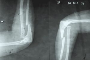
Figure 1: Pre-operative X-ray.
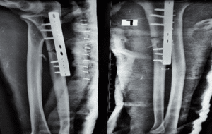
Figure 2: Post -operative X-ray.
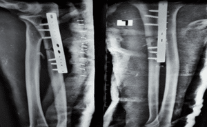
Figure 3: X-ray at 6 weeks.
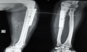
Figure 4: X-ray at 12 weeks.
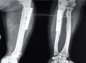
Figure 5: X-ray at final follow up (24 weeks).
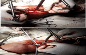
Figure 6: Per-operative photograph after operative reduction of ulna with the small DCP.
Results
Total 40 patients were included. Among 15 (37.5%) patients were from 20-29 years age group, 8 (20.0%) were from 30-39 years age group, 11 (27.5%) were from 40-49 years age group and 6 (15.0%) were from 50-55 years age group. The mean age of the patients was 35.96±11.48 years where minimum age was 20 years and maximum age was 55 years (Table 1). 32 (80.0%) patients were male and 8 (20.0%) patients were female. out of the 40 patients, 40% (n=16) presented with right sided fracture and 60% (n=24) with left sided fractures.
Age (in years) | N (%) | Sex | N (%) | Side Involvement | N (%) |
20-30 | 15 (37.5) | Male | 32 (80) | Right | 16 (40) |
31-40 | 08 (20.0) | Female | 08 (20) | Left | 24 (60) |
41-50 | 11 (27.5) | Mean ± SD | 35.96±11.48 | ||
51-55 | 06 (15.0) | Range(min-max) | 20-55 |
Table 1: Distribution of patients by age, Sex and affected limb (n=40).
Table 2 shows that out of the 40 patients, 87.5% (n=35) presented with Bado type I fracture and 12.5% (n=5) presented with Bado type II fracture. 15 (37.5%) of injuries were caused by physical assault, 13 (32.5%) were caused by RTA and 12 (30.0%) were due to fall from high. Among the 40 patients, 25 (62.5%) had surgery within 10 days while 15 (37.5%) had surgery within 11-21 days. The mean time interval between injury and surgery was 10.13±3.86 days where minimum time interval between injury and surgery was 5 days and maximum time interval between injury and surgery was 20 days.
Bado Classification | N (%) | Cause of Injury | N (%) | Time Interval (in days) | N (%) |
Type I | 35 (87.5) | Assault | 15(37.5) | Up to 10 | 25(62.5) |
Type II | 05 (12.5) | RTA | 13(32.5) | 11-21 | 15 (37.5) |
Fall | 12 (30.0) | Mean ± SD | 10.13±3.86 |
Table 2: Distribution of patients by Bado classification of Monteggia fracture, cause of injury and surgery time interval (n=40).
Table 3 shows that 35 (87.5%) patients had no complication post-operatively while 5(12.5%) patients developed post-operative complications like tourniquet palsy (7.5%, f=3) and wound infection (7.5%, f=3). Out of 40 cases, 37 (92.5%) was united but only 3 (7.5%) case neither unite nor found any sign of union up to 24 weeks of follow up.
Post-Operative Complication | Union Status at Last Follow Up | ||
Absent | 35(87.5) | United | 37(92.5) |
Present | 05 (12.5) | Nonunion | 03(7.5) |
Table 3: Post-operative complication and Union status at last follow up of patients (n=40).
Six weeks after surgery, the mean Quick DASH score of the cases were 54.2%±5.11%. At 12 weeks, it has decreased to 34.4%±7.71%. Furthermore, at last follow up, it has significantly decreased to 16.6%±10.08% (Fig. 7).
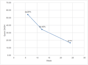
Figure 7: Distribution of patients according to quick DASH score (n=40).
The pain statuses of the cases were measured with VAS scale (Fig. 8). At first follow, the mean VAS score was 43.83±8.57. At second follow up, it has decreased to 18.50±10.09. Again, at last follow up, it has significantly decreased to 1.66±5.14.
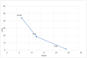
Figure 8: Distribution of patients according to pain status (VAS scale) (n=40).
Final outcome was measured according to Anderson criteria (Fig. 9). Among the 40 cases, 9 (22.5%) patients had excellent, 25 (62.5%) patients had good, 3 (7.5%) had fair and 3 (7.5%) patients had poor outcome.
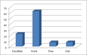
Figure 9: Final outcome according to Anderson criteria (n=40).
Table 4 shows that the final outcome was satisfactory (excellent and good) in 35 (87.5%) patients and unsatisfactory (fair and poor) in 5 (12.5%) patients.
Outcome | N | % |
Satisfactory (Excellent and Good) | 35 | 87.5 |
Unsatisfactory (Fair and Poor) | 5 | 12.5 |
Table 4: Distribution of the patients by final outcome (n=40).
Discussion
Monteggia fracture dislocations are rare injuries that comprise less than five percent of all forearm fractures. Good results in monteggia fractures depend on early and accurate diagnosis, rigid fixation of ulna, accurate reduction of radial head and post-operative immobilization to allow ligamentous healing about the dislocated radial head. Among the 40 patients, 15 (37.5%) patients were from 20-29 years age group, 8 (20.0%) were from 30-39 years age group, 11 (27.5%) were from 40-49 years age group and 6 (15.0%) were from 50-55 years age group. The mean age of the patients was 35.96±11.48 years where minimum age was 20 years and maximum age was 55 years. Retrospective study conducted in India among adult patients showed similar result [7]. Eighty percent patients of this study were male which was consistent with other studies [6-8]. This fracture often occurs after a highway accident or aggression, which explains the male predominance of age. Monteggia fractures are part of a spectrum of forearm injuries and commonly result either from a fall on the outstretched arm with forced pronation or from a direct injury [9]. Physical assault, RTA and fall from high were the causes of injury found in the present study. One third of the patients had surgery within 11-21 days. The mean time interval between injury and surgery was 10.13±3.86 days. This long duration of time had several factors. So, patients were given back slab until swelling subsides and then surgery was done. The most frequent complications of surgery after Monteggia lesion are implant loosening, misalignment of the ulna, radio ulnar dislocation, nonunion of the ulna, necrosis of the radial head, postoperative infection, heterotopic ossification, loosening of osteosynthesis of radial head and neck, delayed consolidation of the neck of the radius fracture, radio-ulnar synostosis, deficiency neuropathy, and posterolateral rotatory instability [6]. Majority of the patients did not face any kind of post-operative complications while two patients (6.7%) had tourniquet palsy. The retrospective study of Bruce and Wilson, et al., reported the incidence of nerve palsies 14% after treatment [10]. The dissimilarity of result might be due to the fact that 17% patients of their study had nerve palsies on admission while no patient of the present study had this. Two patients suffered from wound infection. These patients were treated according to the C/S report and with regular dressing. No patient needed revision of surgery. Regarding the treatment of Monteggia lesions, it is essential to provide anatomic reduction and stable fixation of the ulnar fractures with 3.5 mm DCP. Using 3.5 mm plates provides a more stable fixation, making the need for a second surgery less likely, therefore improving chances of better functional prognosis by avoiding reoperations [6]. The mean arc of motion (Supination-Pronation) at first follow up was 790±6.990. The mean arc of motion at 2nd follow up was 94.660±11.210. Finally, at last follow up it has significantly improved to 120.830±15.260. From first follow up to last follow up, arc of motion has significantly improved. In the study of Gill, et al., the mean ROM of supination-pronation was 137.90 ± 17.70 which was similar to present study [11]. Final outcome was measured according to Anderson criteria [12]. Majority of the patients had good functional outcome and one fourth had excellent outcome. Two patients had poor outcome. The poor outcome was due to nonunion of that case. In the present study the excellent and good functional outcome were considered as satisfactory and others were considered as unsatisfactory. Majority of the patients had satisfactory outcome. Suarez, et al., also classified functional outcome as satisfactory and unsatisfactory and reported 84.0% patients had satisfactory outcome [6]. The patients of Suarez, et al., had no associated injuries which helped them to achieve more satisfactory results which was true for the present study also [6]. The study of Reddy and Prasad found 80.5% patients had satisfactory outcome [7]. The proportion of satisfactory results was a bit lower in the study of Reddy and Prasad as they had 16.1% patients having Bado type III fracture [7]. But the present study had patients only having Bado type I and II fractures. No significant association was found between age of the patients and functional outcome. Konrad, et al., [8] also reported that age of the patient did not influence the outcome. However, patients who had more time interval between injury and surgery had less satisfactory outcome. In case of delayed surgery there is chance of stiffness and fibrosis of joint due to soft tissue interposition. Again, early surgery can relief pain early which will help early mobilization of elbow.
Summary and Conclusion
Monteggia fractures are uncommon injuries. The commonest type of monteggia fracture dislocation in adults according to Bado’s classification is type-1. Operative treatment of Monteggia fracture-dislocation by the selected implant, leads to excellent to good radiological and functional result with uncomplicated recovery in majority of the cases. The technique of early closed reduction of radial head and open reduction and internal fixation of ulna using compression plate, is a simple and effective means of treating monteggia fracture dislocation in adults with excellent functional outcome.
Conflict of Interest
The authors have no conflict of interest to declare.
References
- Blom A, Warwick D, Whitehouse M. Apley and solomon’s system of orthopaedics and trauma. CRC Press. 2017.
- Ring D, Jupiter JB, Waters PM. Monteggia fractures in children and adults. JAAOS-J Am Acad Ortho Surg. 1998;6(4)L215-24.
- Scott J, Huskisson EC. Graphic representation of pain. Pain. 1976;2(2):175-84.
- Stein F, Grabias SL, Deffer PA. Nerve injuries complicating Monteggia lesions. J Bone and Joint Surg. 1971;53(7):1432-6.
- Stoll TM, Willis RB, Paterson DC. Treatment of the missed Monteggia fracture in the child. J Bone and Joint Surg. 1992;74(3):436-40.
- Suarez R, Barquet A, Fresco R. Epidemiology and treatment of Monteggia lesion in adults: series of 44 cases. Acta Ortopedica Brasileira. 2016;24:48-51.
- Reddy GR, Prasad PN. A study to assess epidemiological, clinical profile and outcome of Monteggia fracture dislocation in adults: a retrospective study. Int J Res Ortho. 2017;3(3):472.
- Konrad GG, Kundel K, Kreuz PC, Oberst M, Sudkamp NP. Monteggia fractures in adults: long-term results and prognostic factors. J Bone and Joint Surg. British volume. 2007;89(3):354-60.
- Ramisetty NM, Revell M, Porter KM, Greaves I. Monteggia fractures in adults. Trauma. 2004;6(1):13-21.
- Bruce HE, Harvey Jr JP, Wilson Jr JC. Monteggia fractures. J Bone and Joint Surg. 1974;56(8):1563-76.
- Gill SP, Mittal A, Raj M, Singh P, Kumar S, Kumar D. Stabilisation of diaphyseal fractures of both bones forearm with limited contact dynamic compression or locked compression plate: comparison of clinical outcomes. Int J Res Orthop. 2017;3(3):623-31.
- Anderson LD, Sisk D, Tooms RE, Park 3rd Compression-plate fixation in acute diaphyseal fractures of the radius and ulna. J Bone and Joint Surg. 1975;57(3):287.
This work is licensed under a Creative Commons Attribution 2.0 International License.


