Anastasios John Kanellopoulos1*
1Laservision GR Clinical and Research Eye Institute, Athens, Greece and the Department of Ophthalmology, New York University Medical School, New York, USA
*Correspondence author: Anastasios John Kanellopoulos, MD, Laservision GR Clinical and Research Eye Institute, Athens, Greece and the Department of Ophthalmology, New York University Medical School, New York, USA; Email: [email protected]
Published Date: 29-11-2023
Copyright© 2023 by Kanellopoulos AJ. All rights reserved. This is an open access article distributed under the terms of the Creative Commons Attribution License, which permits unrestricted use, distribution, and reproduction in any medium, provided the original author and source are credited.
Abstract
Purpose: The purpose of this retrospective study was to evaluate and analyze visual outcomes by recording pre and postoperative trefoil, coma and refractive astigmatism in wavefront optimized myopic LASIK.
Methods: In this retrospective case review 200 eyes (one hundred patients) that had undergone myopic (with corresponding astigmatism) wavefront-optimized LASIK using the FS200 femtosecond and EX500 excimer lasers (Alcon/Wavelight, Erlagen, Germany) were evaluated. The 12 months post-operative UDVA and CDVA, low (myopia and/or astigmatism) along with high order aberration C6 to C9 changes were compared to the pre-operative values. Pre-operative topography data were available and used to generate for this study hypothetical treatment data (low and high order aberrations) if Topography-Guided (TG) with TMR cylinder amount and axis adjustment was used instead of the actual WFO.
Results: Mean values at 12 months: UDVA of 20/22 and CDVA of 20/20. The postoperative refractive error in Diopters was -0.20±0.46 sphere and – 0.45±0.27 cylinder. The average absolute value for the high order aberrations studied were pre-op: C6: 0.10±0.12, C7: 0.19±0.16, C8: 0.15±0.12, C9: 0.09±0.09 μm and respectively post-op, C6: 0.11±0.10, C7: 0.46±0.38, C8: 0.34±0.30, C9: 0.11±0.13 μm. If topography-guided customization with TMR was originally employed an addition mean -0.36D of astigmatism would have been attempted.
Conclusion: Wavefront optimized ablations do not address HOA, pre-existing trefoil (C6, C9) in this group essentially did not change while coma (C7 and C8) increased despite the essential achievement of emmetropia. In theory topography-guided customization with TMR may had offered improved C7 and -C8 outcomes, along with superior cylindrical correction.
Keywords: Topography-Modified Refraction; Topography-Guided; Laser Vision Correction; LASIK; Coma, Higher Order Aberrations
Introduction
Clinical refraction has been the gold standard in refractive surgery for decades. Most clinical studies for excimer laser safety and efficacy have been based on a combination or either a dry manifest or cycloplegic refraction [1,2].
It has been through the study of topography guided excimer LASIK treatments internationally and especially in highly irregular eyes, that often the clinical refraction astigmatism does not agree with the topographic astigmatism in amount and/or axis [3]. The subjective measurements present usually less cylindrical power and of different axis [4,5]. Our clinical experience reported in over 1000 cases so far in peer-reviewed literature, has shown that modifying the topographic astigmatism in amount and axis in the refractive (and adjusting accordingly the sphere, so not to affect the spherical equivalent) has produced superior refractive outcomes in irregular corneas, but most interestingly in normal eyes as well with shift in gain of 1 line of vision from 30% to almost 70% and 2 lines from 10% to over 30% [6].
These findings are compelling. Following introduction of this concept at our previous study comparing contralateral eye topography-guided LASIK to Smile, we have studied routine myopic cases very carefully and found documentation in the EX500 treatment software, following importation of Placido topography (Vario Topolyzer, Alcon/Wavelight, Erlagen Germany) and clinical data and following the conversion of the treatment refraction to 0 sphere and 0 cylinder, that many eyes present significant angle kappa (we consider significant: deviation of the pupillary center from the cornea vertex equal or more than 0.1 mm (100µm) in the x, y or both axes [7]. Cases with “significant” coma, even if they have eluded topographic clinical assessment, can be identified on this treatment software setting, as the platform presents an obvious pattern, to normalize it.
The compelling finding in these cases is that all standard refraction modalities, measuring the total functional eye refraction “need”, excluding the wavefront suggested refraction and coma (C7 and C8) and trefoil (C6 and C9) explained in the next paragraph, show very little evidence of this. It is therefore in our clinical experience studied and reported that the actual manifest refraction includes an adjustment chosen by each subject to compensate for coma and trefoil induced by regular corneal astigmatism due to angle kappa.
Accommodative contribution by the crystalline lens may also contribute to dynamic manifest refraction shifts [8-10].
This cylinder measurement bias may “shift” the accurate topographic axis and power to what is measured subjectively by clinicians and later may present as a sphero-cylindrical “regression” of the treatment [11-13]. The significance of this potential finding is that-if this holds true-a large percentage of routine myopic and myopic astigmatic LASIK corrections are not up to now treated to the most accurate extent and at some point, following the initial LASIK intervention they may produce a refractive error (usually small amounts of myopic astigmatism that relate to de-compensation of the subjective astigmatic power measured as described above). If the pre-operative topographic data is recorded at the time of WFO LASIK treatment based on the clinical subjective refraction (myopic sphere and astigmatism), the theoretical difference of the design of a topographic-guided with Topography Modified Refraction (TMR) treatment can be calculated in retrospect with low and high order aberration data. These data can be compared to the actual low and high order postoperative outcomes of the WFO treatments. This retrospective, consecutive case study was designed to compare the above data.
Patients and Methods
Study Design
In this retrospective data review, 200 eyes of 100 patients with bilateral myopia or myopic astigmatism, were included. The study received approval by the Ethics Committee of Laservision Clinical and Research Eye Institute and adhered to the tenets of the Declaration of Helsinki. Informed consent was provided and documented in writing from each patient prior to the time of the interventions that specifically data from their procedures and postoperative care maybe used for research and quality control purposes.
Inclusion-Exclusion Criteria
All patients enrolled in the study had no other previous ocular surgery prior to the LASIK procedure studied in retrospect, had at the time of their procedure documented refractive stability for at least 3 year and had discontinued contact lens use for at least 2 weeks. Additional inclusion criteria for this study: Patients of 18-65 years of age with refractive spherical error up to -12.00 diopters (D), astigmatism of 0.00 to -6.00 (D) and central corneal thickness of at least 500μm. Last, preoperative corrected distance visual acuity was least 20/30 in all subjects studied.
Exclusion criteria included: history of corneal dystrophy and/or herpetic eye disease, topographic evidence of ectatic corneal disorder /keratoconus (as evidenced by Placido topography or Scheimpflug based tomography) and epithelial warpage from contact lens use, corneal scarring, glaucoma, severe dry eye and collagen vascular disease. Any other abnormalities that in the surgeon’s opinion would have negatively affected the potential for optimal visual outcomes were also included.
Virtual Refractive Procedure Design
A retrospective data review was performed on 200 myopic eyes of 100 patients treated with wavefront-optimized LASIK. The correction target was based on the manifest subjective refraction, with emmetropia being the target in all patients. The FS200 Femtosecond laser was used for flap creation and the EX500 Excimer Laser (Alcon Laboratories, Ft Worth, TX, USA) for ablation in all eyes. All cases had planned flap thickness of 110 µm and planned flap diameter of 8.5 mm. The 110-µm-flap thickness was chosen in all LASIK cases because this has been optimal in our practice and has been used as the clinical standard during the past 10 years. Topographic data were captured by the Vario topolyzer (WaveLight, Erlagen, Germany) and corneal pachymetric data were captured and imported for the treatments by the Oculyzer II (WaveLight), a Scheimpflug-based tomography device associated with the Refractive Suite, a diagnostic device that is essentially based on the Pentacam HD (Oculus Optikgeräte GmbH, Wetzlar, Germany). The Vario topographic data were used in this study to produce in retrospect a hypothetical topography-guided with Topography Modified Refraction (TMR) simulated treatment planning.
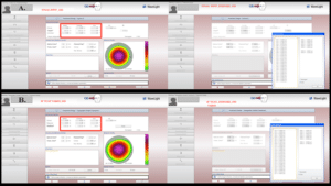
The Root-Mean-Square (RMS) of HOAs from the second to third Zernike aberration orders were measured with the vario topolyzer (WaveLight, Erlagen, Germany) preoperatively and postoperatively representing mainly C7 and C8. For the purpose of the virtual design in retrospect in these cases, the mean of eight consecutive scan measurements were taken without pupil dilation in all eyes and the best map was chosen to put into final fit software when designing the hypothetical topography-guided ablation treatment profile, where the Zernike polynomials were noted (Fig. 1). The cylindrical refraction was adjusted by the surgeon to match the amount and axis of the topographically measured cylinder and appropriate sphere adjustments were made to keep the same spherical equivalent in our accordance to our recently introduced technique of TMR (Topography-Modified Refraction) [3].
Past Clinical Examinations
All 100 patients enrolled in this study underwent a wavefront-optimized LASIK for the correction of myopia and/or astigmatism, performed by the same surgeon (AJK) in, Athens, Greece). All eyes were evaluated preoperatively and on each postoperative visit for CORRECTED DISTANCE VISUAL ACUITY (CDVA), Uncorrected Distance Visual Acuity (UDVA) and manifest spherical equivalent, slit-lamp biomicroscopy, fundus examination including retinal periphery and applanation tonometry.
Data Analysis
All statistical analyses were calculated on a computer using IBM SPSS version 20 (IBM Corporation, New York, NY). A P-value of 0.05 or less was considered to be significant at a 95% confidence interval.
Results
Of the 100 patients enrolled in this study, 64 were female and 36 were male. Mean age at the time of the operation was 30±8 years (range: 18 to 60 years). Preoperative patient demographics are listed in Table 1. The mean uncorrected distance visual acuity was 0.05±0.09 (decimal), (range: 0.01 to 0.63) and the mean best-corrected distance visual acuity was 0.98±0.12 (decimal), (range: 0.32 to 1.25). The mean preoperative spherical equivalent was -5.89±2.76 (range: -14.00 to -0.50) D and the mean preoperative manifest cylinder was -0.74±0.74 (range: -3.50 to 1.75) D.
Table 2 compares the intraoperative parameters corrected between the wavefront-optimized LASIK treatment plan that was actually used and was based on the clinical manifest refraction (myopic sphere and astigmatism) to the-in retrospect-calculated for the purpose of this study, hypothetical topography-adjusted refraction treatment plan. There was statistically significant difference between treatment plans regarding astigmatism (P ≤ .05) and corneal asphericity (Q value) (P = 0.037). The Wavefront-Optimized (WFO group) and hypothetical topography-modified refraction intraoperative parameters (topography-guided group) were well matched in preoperative spherical equivalent (SE) of refractive error and optical zone of treatment.
Refractive Outcome
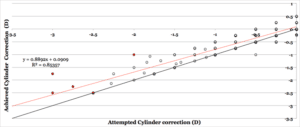
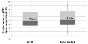
The corrected visual acuity (distance monocular) outcome at 12 months shows that 69.2% of the eyes had postoperatively CDVA better than 1.0 (decimal). Forty-eight percent of eyes gained at least one line of CDVA and 0.3% lost ≥2 lines of CDVA. There was no statistically significant difference between preoperative CDVA and postoperative UDVA. 12 months postoperatively, 74.2% of eyes had UDVA of 1.0 (decimal) and 95.2% of eyes had 0.65 (decimal). The mean of wavefront-optimized attempted cylindrical correction was -0.76±0.27 D and the mean achieved cylindrical correction was -0.20±0.23 D at 12 months (Fig. 2). The Mean Manifest Refraction Spherical Equivalent (MRSE) 12 months postoperatively was -0.16±0.32 D (range: -1.87 to 1.00 D). Fig. 3 shows a graphical depiction of prediction error SEQ for wavefront optimized LASIK and hypothetical topography-guided LASIK. The definition of Prediction Error (PE) is given by the following equation: Prediction error = Desired postoperative SEQ-Actual postoperative SEQ. There was no significant difference (P = 0.517) in the percentage of eyes achieving emmetropia.
Changes in High Order Aberrations
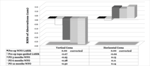
Table 3 illustrates the pre-operative to 12-month postoperative differences in the HOA studied: specifically, C6-C9. Only the coma values both C7 and C8 demonstrated statistically significant increase. No significant difference was found in either C6 (vertical trefoil) or C9 (Oblique Trefoil). Specifically, the average absolute value for the high order aberrations studied were pre-op: C6: 0.10±0.12, C7: 0.19±0.16, C8: 0.15±0.12, C9: 0.09±0.09μm and respectively post-op, C6: 0.11±0.10, C7: 0.46±0.38, C8: 0.34±0.30, C9: 0.11±0.13μm (Fig. 4,5 and Table 4).

Figure 1: A: The left image illustrates the actual wavefront-optimized treatment plan and ablation profile of one of the cases in this study. The dry manifest refraction was sph: -5.25 cyl: -0.50 @ 35 degrees, UCVA and BDVA were 20/20. The right image illustrates the Zernike coefficients and the RMS values for the current treatment parameters; B: The left image illustrates the hypothetical topography-guided treatment plan and ablation profile of the same case, after the refraction has been adjusted by the user to the desired sphere and cylinder and includes the changes noted in image. The right image illustrates the Zernike coefficients and the RMS values for the current treatment parameters.

Figure 2: Equivalency plot of 3 months postoperatively achieved and WFO desired cylinder data in minus notation (statistically significant, P <0.05). Fifty percent of the cases were under-corrected by at least 0.25D, 42% were corrected within±0.24D and 8% were overcorrected by at least 0.25D. The regression line is shown in solid red (Achieved correction = 0.88*Desired correction + 0.09D) with R2 = 0.85 and the standard error = 0.23D.

Figure 3: Comparison of the Predictive error (in dioptric spherical equivalent) at 3 months postoperatively. No statistically significant difference, P = .517. WFO = wavefront optimized; Topo-guided = topography-guided customized; Mean values of differences.

Figure 4: Graphical representation of vertical and oblique trefoil (in microns) pre- and 3, 6 and 12 months postoperatively. Each is based on the mean of the absolute values of the Zernike polynomials.

Figure 5: Graphical representation of vertical and horizontal coma (in microns) pre- and 3, 6 and 12 months postoperatively. Each is based on the mean of the absolute values of the Zernike polynomials.
|
Demographic Data of the Included Patients |
|
|
Variable |
|
|
Age |
|
|
Mean±SD |
30±8 |
|
Range |
18 to 60 |
|
Preop SE (D) |
|
|
Mean±SD |
-5.89±2.76 |
|
Range |
-14 to -0.5 |
|
Preop cylinder |
|
|
Mean±SD |
-0.74±0.74 |
|
Range |
-3.50 to 1.75 |
|
Preop UCVA (decimal) |
|
|
Mean±SD |
0.05±0.09 |
|
Range |
0.01 to 0.63 |
|
Preop BCVA (decimal) |
|
|
Mean±SD |
0.98±0.12 |
|
Range |
0.32 to 1.25 |
Table 1: Preoperative patient demographics. Preop=preoperative; SE=spherical equivalent; SD=standard deviation; D=diopters; UCVA=uncorrected visual acuity; BCVA=best-corrected visual acuity.
|
Between Group Comparison of Intraoperative Parameters |
|||
|
|
WFO Treatment Plan |
Hypothetical Topo-guided Treatment Plan |
P-value |
|
SE (D) |
|||
|
Mean±SD |
-5.60±2.46 |
-5.80±2.42 |
.410 |
|
Range |
-14.12 to -0.87 |
-14.12 to -1.12 |
|
|
Cylinder (D) |
|||
|
Mean±SD |
-0.75±0.70 |
-1.09±0.72 |
.000* |
|
Range |
-3.50 to 0.00 |
-3.75 to 2.00 |
|
|
Sphere (D) |
|||
|
Mean±SD |
-5.22±2.42 |
-5.25±2.43 |
.892 |
|
Range |
-13.75 to -0.50 |
-14.00 to -0.75 |
|
|
Q value |
|||
|
Mean±SD |
-0.50±0.00 |
-0.48±0.06 |
.037* |
|
Range |
-0.50 to -0.50 |
-0.52 to -0.16 |
|
|
OZ (mm) |
|||
|
Mean±SD |
6.51±0.45 |
6.50±0.45 |
.956 |
|
Range |
5.00 to 7.20 |
5.00 to 7.00 |
|
|
Trans Zone (mm) |
|||
|
Mean±SD |
1.16±0.13 |
1.17±0.14 |
.943 |
|
Range |
0.90 to 2.00 |
1.00 to 2.00 |
|
Table 2: Wavefront optimized LASIK treatment plan based on the clinically derived refraction (myopic sphere and astigmatism) versus hypothetical Topographic adjusted refraction treatment plan. WFO = wavefront optimized; Topo-guided = topography-guided customized; SE = spherical equivalent; SD = standard deviation; OZ = optical zone; Trans Zone = transition zone *Statistically significant, P < .05.
|
Comparison of Preoperative RMS of Total, Lower (Second) Order Aberrations and Higher (Third) Order Aberrations between WFO LASIK and topography – guided LASIK |
|||
|
RMS (μm) |
WFO |
Topo-guided |
P value |
|
RMS (Total) |
0.3176 |
0.5009 |
.000* |
|
RMS2 |
8.1277 |
8.1229 |
.985 |
|
Oblique Primary Astigmatism Z (2, -2) |
-0.0456 |
-0.0088 |
.431 |
|
Vertical Primary Astigmatism Z (2, 2) |
-0.3639 |
-0.8621 |
.000* |
|
Defocus Z (2, 0) |
8.0650 |
8.0134 |
.844 |
|
RMS3 |
0.0107 |
0.3278 |
.000* |
|
Vertical Trefoil Z (3, -3) – C6 |
0 |
-0.0462 |
– |
|
Oblique Trefoil Z (3, 3) – C9 |
0 |
0.0009 |
– |
|
Vertical Coma Z (3, -1) – C7 |
0 |
-0.0739 |
– |
|
Horizontal Coma Z (3, 1) – C8 |
0 |
-0.0191 |
– |
Table 3: Comparison of Preoperative RMS of Total, Lower (Second) Order Aberrations and Higher (Third) Order Aberrations between WFO LASIK and topography – guided LASIK, WFO = wavefront optimized; Topo-guided = topography-guided customized; RMS = root-mean-square; *Statistically significant, P < .05.
|
Comparison of the Differences between Preoperative WFO Minus Postoperative WFO Higher (Third) Order Aberrations (HOAs) to Preoperative Topo-guided Minus Postoperative WFO HOAs |
|||
|
Zernike Mode (μm) |
WFO |
Topo-guided |
P-value |
|
3 months post-op |
|||
|
Vertical Trefoil Z (3, -3) |
-0.0178 |
-0.0717 |
.009* |
|
Oblique Trefoil Z (3, 3) |
0.0137 |
0.0113 |
.885 |
|
Vertical Coma Z (3, -1) |
0.1663 |
0.0137 |
.000* |
|
Horizontal Coma Z (3, 1) |
-0.1265 |
-0.1460 |
.676 |
|
6 months post-op |
|||
|
Vertical Trefoil Z (3, -3) |
-0.0168 |
-0.0604 |
.014* |
|
Oblique Trefoil Z (3, 3) |
0.0065 |
0.0069 |
.981 |
|
Vertical Coma Z (3, -1) |
0.2782 |
0.2014 |
.041* |
|
Horizontal Coma Z (3, 1) |
-0.1136 |
-0.1342 |
.626 |
|
12 months post-op |
|||
|
Vertical Trefoil Z (3, -3) |
-0.0167 |
-0.0596 |
.001* |
|
Oblique Trefoil Z (3, 3) |
0.0007 |
0.0010 |
.984 |
|
Vertical Coma Z (3, -1) |
0.2891 |
0.2121 |
.038* |
|
Horizontal Coma Z (3, 1) |
-0.1263 |
-0.1459 |
.504 |
Table 4: Comparison of the Differences between Preoperative WFO Minus Postoperative WFO Higher (Third) Order Aberrations (HOAs) to Preoperative Topo-guided Minus Postoperative WFO HOAs; WFO = wavefront optimized; Topo-guided = topography-guided customized; *Statistically significant, P < .05.
Discussion
The documentation of these data appears to support our study theory: Instead of using wavefront-optimized LASIK as the initial procedure as was performed in these cases, if topography-guided with TMR was used instead, the majority of coma (horizontal and and vertical) may had been addressed resulting in lower postoperative high order aberrations and possibly better quality of vision. It appears from the relatively large database evaluated, that horizontal and oblique trefoil were not found to be significant pre-operatively and did not seem to have been affected either by increase or decrease by the WFO procedure that was actually performed. Our speculation that specials attention should be given to horizontal and vertical coma that do persist and actually appear increased in absolute value post-operatively along with residual refractive astigmatism-that could had been addressed by TMR at the time of the original procedure, appear to be further supported by the data presented herein [3,7].
These data theoretically support that if, instead of the WFO ablation used in these cases, topography-guided with employment of TMR (topography-modified refraction) was used, coma and astigmatic correction may have been superior, as presented and reported by our team 3,7 as well as by other investigators [14,15]. This study appears to justify our paradigm shift these last 1o years from wavefront-optimized myopic LASIK to customization with topography-guided and TMR. Prospective randomized studies comparing topography-guided with the use of the standard clinical manifest refraction versus topography-guided with TMR, may further aid in elucidating these findings.
Conclusion
This retrospective study suggests that in myopic LASIK, coma and astigmatic correction may be better addressed by a topography-guided with adjunct TMR ablation instead of a standard wavefront optimized treatment utilizing the manifest clinical refraction.
Conflict of Interest
The author has no conflict of interest to declare.
References
- Nayak BK, Ghose SU, Singh JP. A comparison of cycloplegic and manifest refractions on the NR-1000F (an objective Auto Refractometer). British J Ophthalmol. 1987;71(1):73-5.
- Hofmeister EM, Kaupp SE, Schallhorn SC. Comparison of tropicamide and cyclopentolate for cycloplegic refractions in myopic adult refractive surgery patients. J Cataract and Refractive Surg. 2005;31(4):694-700.
- Kanellopoulos AJ. Topography-Modified Refraction (TMR): adjustment of treated cylinder amount and axis to the topography versus standard clinical refraction in myopic topography-guided LASIK. Clin Ophthalmol. 2016;10:2213-21.
- Kanellopoulos AJ, Asimellis G. Distribution and repeatability of corneal astigmatism measurements (magnitude and axis) evaluated with color light emitting diode reflection topography. Cornea. 2015;34(8):937-44.
- Alpins N. Topography-modified refraction: adjustment of treated cylinder amount and axis to the topography versus standard clinical refraction in myopic topography-guided LASIK. Clin Ophthalmol. 2017;11:1203-4.
- Kanellopoulos AJ. Reporting acuity outcomes and refractive accuracy after LASIK. J Refract Surg. 2014;30(12):798-9.
- Kanellopoulos AJ. Topography-guided LASIK versus small incision lenticule extraction (smile) for myopia and myopic astigmatism: a randomized, retrospective, contralateral eye study. J Refract Surg. 2017;33(5):306-12.
- Li Wang, MD, Douglas DK. Ocular higher-order aberrations in individuals screened for refractive surgery. J Cataract Refract Surg. 2003;29(10):1896-903.
- Artal P, Benito A, Tabernero J. The human eye is an example of robust optical design. J Vision. 2006;6(1):1.
- Sun M, Birkenfeld J, de Castro A, Ortiz S, Marcos S. OCT 3-D surface topography of isolated human crystalline lenses. Biomed Optics Express. 2014;5(10):3547-61.
- Shetty R, Shroff R, Deshpande K, Gowda R, Lahane S, Jayadev C. A prospective study to compare visual outcomes between wavefront-optimized and topography-guided ablation profiles in contralateral eyes with myopia. J Refract Surg. 2017;33(1):6-10.
- Toda I, Ide T, Fukumoto T, Tsubota K. Visual outcomes after LASIK using topography-guided vs wavefront-guided customized ablation systems. J Refract Surg. 2016;32(11):727-32.
- Jain AK, Malhotra C, Pasari A, Kumar P, Moshirfar M. Outcomes of topography-guided versus wavefront-optimized laser in situ keratomileusis for myopia in virgin eyes. J Cataract Refract Surg. 2016;42(9):1302-11.
- Galvis V, Tello A, Carreño NI, Berrospi RD, Niño CA. Wavefront-Guided LASIK and preoperative higher order aberrations. J Refract Surg. 2016;32(12):862.
- Goyal JL, Garg A, Arora R, Jain P, Goel Y. Comparative evaluation of higher-order aberrations and corneal asphericity between wavefront-guided and aspheric LASIK for myopia. J Refract Surg. 2014;30(11):777-84.
Article Type
Research Article
Publication History
Received Date: 29-10-2023
Accepted Date: 23-11-2023
Published Date: 29-11-2023
Copyright© 2023 by Kanellopoulos AJ. All rights reserved. This is an open access article distributed under the terms of the Creative Commons Attribution License, which permits unrestricted use, distribution, and reproduction in any medium, provided the original author and source are credited.
Citation: Kanellopoulos AJ. Retrospective Analysis of Wavefront-Optimized Myopic LASIK: Comparison of Preoperative to Postoperative Astigmatism and High Order Aberrations: Trefoil and Coma Specifically: Could Topography-Guided Original Customization Had Addressed the Above? J Ophthalmol Adv Res. 2023;4(3):1-9.

Figure 1: A: The left image illustrates the actual wavefront-optimized treatment plan and ablation profile of one of the cases in this study. The dry manifest refraction was sph: -5.25 cyl: -0.50 @ 35 degrees, UCVA and BDVA were 20/20. The right image illustrates the Zernike coefficients and the RMS values for the current treatment parameters; B: The left image illustrates the hypothetical topography-guided treatment plan and ablation profile of the same case, after the refraction has been adjusted by the user to the desired sphere and cylinder and includes the changes noted in image. The right image illustrates the Zernike coefficients and the RMS values for the current treatment parameters.

Figure 2: Equivalency plot of 3 months postoperatively achieved and WFO desired cylinder data in minus notation (statistically significant, P <0.05). Fifty percent of the cases were under-corrected by at least 0.25D, 42% were corrected within±0.24D and 8% were overcorrected by at least 0.25D. The regression line is shown in solid red (Achieved correction = 0.88*Desired correction + 0.09D) with R2 = 0.85 and the standard error = 0.23D.

Figure 3: Comparison of the Predictive error (in dioptric spherical equivalent) at 3 months postoperatively. No statistically significant difference, P = .517. WFO = wavefront optimized; Topo-guided = topography-guided customized; Mean values of differences.

Figure 4: Graphical representation of vertical and oblique trefoil (in microns) pre- and 3, 6 and 12 months postoperatively. Each is based on the mean of the absolute values of the Zernike polynomials.

Figure 5: Graphical representation of vertical and horizontal coma (in microns) pre- and 3, 6 and 12 months postoperatively. Each is based on the mean of the absolute values of the Zernike polynomials.
Demographic Data of the Included Patients | |
Variable | |
Age | |
Mean±SD | 30±8 |
Range | 18 to 60 |
Preop SE (D) | |
Mean±SD | -5.89±2.76 |
Range | -14 to -0.5 |
Preop cylinder | |
Mean±SD | -0.74±0.74 |
Range | -3.50 to 1.75 |
Preop UCVA (decimal) | |
Mean±SD | 0.05±0.09 |
Range | 0.01 to 0.63 |
Preop BCVA (decimal) | |
Mean±SD | 0.98±0.12 |
Range | 0.32 to 1.25 |
Table 1: Preoperative patient demographics. Preop=preoperative; SE=spherical equivalent; SD=standard deviation; D=diopters; UCVA=uncorrected visual acuity; BCVA=best-corrected visual acuity.
Between Group Comparison of Intraoperative Parameters | |||
| WFO Treatment Plan | Hypothetical Topo-guided Treatment Plan | P-value |
SE (D) |
|
|
|
Mean±SD | -5.60±2.46 | -5.80±2.42 | .410 |
Range | -14.12 to -0.87 | -14.12 to -1.12 |
|
Cylinder (D) |
|
|
|
Mean±SD | -0.75±0.70 | -1.09±0.72 | .000* |
Range | -3.50 to 0.00 | -3.75 to 2.00 |
|
Sphere (D) |
|
|
|
Mean±SD | -5.22±2.42 | -5.25±2.43 | .892 |
Range | -13.75 to -0.50 | -14.00 to -0.75 |
|
Q value |
|
|
|
Mean±SD | -0.50±0.00 | -0.48±0.06 | .037* |
Range | -0.50 to -0.50 | -0.52 to -0.16 |
|
OZ (mm) |
|
|
|
Mean±SD | 6.51±0.45 | 6.50±0.45 | .956 |
Range | 5.00 to 7.20 | 5.00 to 7.00 |
|
Trans Zone (mm) |
|
|
|
Mean±SD | 1.16±0.13 | 1.17±0.14 | .943 |
Range | 0.90 to 2.00 | 1.00 to 2.00 |
|
Table 2: Wavefront optimized LASIK treatment plan based on the clinically derived refraction (myopic sphere and astigmatism) versus hypothetical Topographic adjusted refraction treatment plan. WFO = wavefront optimized; Topo-guided = topography-guided customized; SE = spherical equivalent; SD = standard deviation; OZ = optical zone; Trans Zone = transition zone *Statistically significant, P < .05.
Comparison of Preoperative RMS of Total, Lower (Second) Order Aberrations and Higher (Third) Order Aberrations between WFO LASIK and topography – guided LASIK | |||
RMS (μm) | WFO | Topo-guided | P value |
RMS (Total) | 0.3176 | 0.5009 | .000* |
RMS2 | 8.1277 | 8.1229 | .985 |
Oblique Primary Astigmatism Z (2, -2) | -0.0456 | -0.0088 | .431 |
Vertical Primary Astigmatism Z (2, 2) | -0.3639 | -0.8621 | .000* |
Defocus Z (2, 0) | 8.0650 | 8.0134 | .844 |
RMS3 | 0.0107 | 0.3278 | .000* |
Vertical Trefoil Z (3, -3) – C6 | 0 | -0.0462 | – |
Oblique Trefoil Z (3, 3) – C9 | 0 | 0.0009 | – |
Vertical Coma Z (3, -1) – C7 | 0 | -0.0739 | – |
Horizontal Coma Z (3, 1) – C8 | 0 | -0.0191 | – |
Table 3: Comparison of Preoperative RMS of Total, Lower (Second) Order Aberrations and Higher (Third) Order Aberrations between WFO LASIK and topography – guided LASIK, WFO = wavefront optimized; Topo-guided = topography-guided customized; RMS = root-mean-square; *Statistically significant, P < .05.
Comparison of the Differences between Preoperative WFO Minus Postoperative WFO Higher (Third) Order Aberrations (HOAs) to Preoperative Topo-guided Minus Postoperative WFO HOAs | |||
Zernike Mode (μm) | WFO | Topo-guided | P-value |
3 months post-op |
|
|
|
Vertical Trefoil Z (3, -3) | -0.0178 | -0.0717 | .009* |
Oblique Trefoil Z (3, 3) | 0.0137 | 0.0113 | .885 |
Vertical Coma Z (3, -1) | 0.1663 | 0.0137 | .000* |
Horizontal Coma Z (3, 1) | -0.1265 | -0.1460 | .676 |
6 months post-op |
|
|
|
Vertical Trefoil Z (3, -3) | -0.0168 | -0.0604 | .014* |
Oblique Trefoil Z (3, 3) | 0.0065 | 0.0069 | .981 |
Vertical Coma Z (3, -1) | 0.2782 | 0.2014 | .041* |
Horizontal Coma Z (3, 1) | -0.1136 | -0.1342 | .626 |
12 months post-op |
|
|
|
Vertical Trefoil Z (3, -3) | -0.0167 | -0.0596 | .001* |
Oblique Trefoil Z (3, 3) | 0.0007 | 0.0010 | .984 |
Vertical Coma Z (3, -1) | 0.2891 | 0.2121 | .038* |
Horizontal Coma Z (3, 1) | -0.1263 | -0.1459 | .504 |
Table 4: Comparison of the Differences between Preoperative WFO Minus Postoperative WFO Higher (Third) Order Aberrations (HOAs) to Preoperative Topo-guided Minus Postoperative WFO HOAs; WFO = wavefront optimized; Topo-guided = topography-guided customized; *Statistically significant, P < .05.


