Shreya Thatte1*, Komal Jaiswal2
1Professor and Head of Department of Ophthalmology, Sri Aurobindo Medical College and PG Institute, Indore, Madhya Pradesh, India
2Junior Resident 2nd year of Department of Ophthalmology, Sri Aurobindo Medical College and PG Institute, Indore, Madhya Pradesh, India
*Corresponding Author: Shreya Thatte, Prof and Head of Department of Ophthalmology, Sri Aurobindo Medical College and PG Institute, Indore, Madhya Pradesh, India; Email: [email protected]
Published Date: 22-07-2022
Copyright© 2022 by Thatte S, et al. All rights reserved. This is an open access article distributed under the terms of the Creative Commons Attribution License, which permits unrestricted use, distribution and reproduction in any medium, provided the original author and source are credited.
Abstract
Background: Penetrating Keratoplasty with cataract extraction and Intraocular Lens (IOL) implantation (Triple procedure) is very challenging procedure because in cases of Opaque cornea, status of anterior segment is difficult to predict that can lead to intraoperative surprises. If not managed properly, these can severely affect visual outcome. To avoid this, we performed Ultrasound Biomicroscopy (UBM) preoperatively for detailed analysis of anterior segment structures and associated pathologies and later compared it with intraoperative findings to judge predictive accuracy of UBM in guiding surgical strategies, decision making and modifications intraoperatively.
Methods and findings: 20 eyes of 20 patients with different grades of corneal opacities like simple corneal opacities (6 eyes) or adherent leucoma (7 eyes) or anterior staphyloma (7 eyes) that underwent Triple procedure (Penetrating Keratoplasty+ cataract extraction+ IOL implantation) were evaluated preoperatively by UBM and the findings were compared intraoperatively to find predictive accuracy of UBM. Extent and depth of corneal lesions, corneal thickness, anterior chamber depth, type, position and extent of synechiae, pupillary membrane and status of lens and capsule were assessed. Scan was performed to visualize any posterior segment pathology and to find Axial Length (AL). Keratometry readings of other eye were taken to calculate IOL power for implantation. Preoperative UBM findings when co-related intraoperatively was found to be accurate in 71.42%- 100% parameters and accordingly these cases underwent penetrating Keratoplasty with other modifications like Iridectomy, Membranectomy, Synechiolysis, iris reconstruction, Trabeculectomy, cataract extraction and IOL implantation.
Conclusion: In cataract coexisting with corneal pathologies, UBM is Best and reliable guide in predicting and preplanning of surgical strategies for best visual outcomes.
Keywords
UBM (Ultrasound Biomicroscopy); Triple Procedure; Cataract, Intraocular Lens (IOL); Keratoplasty; Keratometry; Opaque Media
Introduction
Globally, blindness due to cataract is the second leading cause whereas corneal diseases rank fifth. Many a times, cataract can co-exist with corneal pathologies as a result of which, visualizing the anterior segment becomes very difficult [1]. In such cases, Cataract surgery can be very challenging and these cases may require Triple procedure (Keratoplasty+ cataract extraction + IOL implantation). Hence, proper preoperative assessment of corneal diseases and other associated ocular pathologies like synechiae, adherent leucoma, pupillary membrane, calcified capsule etc. can aid in optimal treatment, surgical planning and visual outcomes [2]. UBM (Ultrasound Biomicroscopy) plays a very important role, as unlike other modalities (anterior segment OCT and Pentacam) using light waves for visualizing anterior segment structures, UBM works on the ultrasound waves which can penetrate through opaque media and helps to visualize anterior segment structures [3-6]. It uses high frequency ultrasound transducer, usually performed with a 50 MHz probe. It is helpful in studying the position of the lens, iris, and ciliary body, and the configuration of the anterior chamber angle with dense corneal pathologies. Frequently, surgeries may also encompass surprises regarding the integrity of the posterior capsule and Zonular structure, presence of synechiae or pupillary membrane. If statuses of these structures are known pre- operatively, the decision in modification of intraoperative surgical steps can be planned. The study was conducted to estimate the predictive value of pre-operative UBM in corneal pathologies undergoing triple procedures, in predicting the status of anterior segment, assessment of finer pre-operative details, intraoperative complications and better surgical outcome.
Materials and Method
Our study was conducted as per Helsinki law after taking approval from Institutional Ethical Committee (IEC). This observational study was performed on 20 eyes of 20 patients with various grades of corneal opacities with cataract that required triple procedure. Patients were evaluated preoperatively with the help of UBM for detailed assessment of anterior segment.
Inclusion Criteria
- Patients with full thickness, healed corneal pathologies (non-infective)
- Healthy posterior segment
- Patients those are willing to participate in the study by giving written consents
Exclusion Criteria
- Infective corneal pathologies
- active inflammation of cornea
After taking informed written consents for the study, case history regarding the course of disease, previous treatment taken (if any) and systemic comorbidities was recorded in detail. A complete ocular examination for both eyes was carried out including visual acuity, IOP (Intraocular Pressure), slit-lamp bio microscopy for visualizing anterior segment. Posterior segment pathologies were ruled out by performing B-Scan. It also gave us a measure of Axial Length (AL). Keratometry readings of other eye was taken to calculate IOL power for implantation.
Optos OTI scan 3000 Ultrasound bio microscope with a 50 MHz transducer probe was used to analyze the anterior segment, after taking consent from the patient. Patient being in supine position, after instilling topical anesthetic drops, an appropriate sized silicon rubber eye cup is placed between the lids and distilled water or normal saline is filled in it which acts as coupling medium. UBM Scanning was done by an ophthalmologist. The pathology was described in detail for various parameters such as extent and depth of corneal pathology, edema and thickness of cornea, anterior chamber depth, angle position, extent of anterior and posterior synechiae, iris details, lens, posterior capsule and Zonular integrity. The operating surgeon used these details to plan the surgical strategies for that particular patient and similar findings were then observed intra operatively and compared with those of the preoperative UBM predictions to note the accuracy of UBM in different parameters.
Result
We conducted UBM examination for those eyes with opaque cornea who were planned for penetrating Keratoplasty, to view anterior segment details. We examined total 62 such patients and 20 patients were found to have opaque cornea with dense cataract as per UBM findings as shown in Table 1. These patients underwent penetrating Keratoplasty along with cataract extraction and certain modifications as required like iris reconstruction, Iridectomy, Membranectomy, Synechiolysis and Trabeculectomy. For few cases IOL implantation was done simultaneously whereas few were left Aphakic or planned for secondary IOL implantation in subsequent surgeries.
As per types of corneal pathologies, our 20 patients were divided into 3 groups
- Simple corneal opacities (6 eyes)
- Adherent leucoma (7 eyes)
- Anterior staphyloma (7 eyes)
Taking into account the various corneal parameters as shown in Table 2, full thickness corneal involvement was seen in all three groups except 1 case of corneal opacity where half stroma with endothelial scarring was seen.
On UBM, we could analyze extent and thickness of corneal pathology which helped us to determine appropriate graft size and trephination depth preoperatively and showed 100% positive predictive value in all the three categories as shown in Table 3.
The UBM findings of anterior chamber are described in Table 4. On Anterior Chamber Depth (ACD) evaluation, shallow anterior chamber was found in 1 case (16.66%) of simple corneal opacity and 4 cases (57.14 %) of anterior staphyloma. ACD was variable in 1 case (16.66%) of simple corneal opacity, all 7 cases (100%) of adherent leucoma due to anterior adherence and 3 (42.85%) cases of anterior staphyloma. 1 case (16.66%) of simple corneal opacity and 2 cases (28.57% ) each of anterior staphyloma and adherent leucoma had closed angles due to membrane formation. Rest of the 5 cases in each of the three categories had normal angle position.
As per Table 5. The anterior chamber entry was not achieved as per prediction (under predicted) in 1 case of adherent leucoma and 2 cases of anterior staphyloma due to iris incarceration. Hence positive predictive value in these cases is 71.42% and 85.71% respectively. In cases of corneal opacity, the positive predictive value of anterior chamber entry was found to be 100% accurate.
Anterior synechaie was found in only 1 case of corneal opacity and in all cases (100%) of both, adherent leucoma (membranous in1, sectoral in 5 and central in 1) and anterior staphyloma (membranous in 2, sectoral in 5) (Table 3).
Posterior synechaie was found in only 2 cases (33.33%) of corneal opacity (1 annular and 1 sectoral), 4 cases (57.14%) of adherent leucoma (1 annular and 3 sectoral) and 2 cases (28.57%) of anterior staphyloma (both sectoral) (Table 3).
Lens and Zonular status is described in Table 4. In all cases of simple corneal opacities, adherent leucoma and anterior staphyloma, Cataractous lens changes were noted. Zonules were found to be intact in 5 (83.33%) cases of corneal opacity whereas 1 (16.66%) case had Zonular dialysis. Zonular dehiscence was seen in only 1 (14.28%) case of adherent leucoma while 6 (85.71%) had intact zonules. Also, all the 7 cases (100%) of anterior staphyloma had intact zonules.
As per Table 6, in cases of corneal opacity, 83.33% positive predictive value was obtained regarding extent of synechiae. It was under predicted only in 1 case of corneal opacity (predicted from 2-3 O’clock but was found to be 2-5 O’clock). Likewise, a positive predictive value was seen in 85.71% cases and 71.42% cases of adherent leucoma and anterior staphyloma respectively. The extent of anterior synechiae was under predicted in 1 case of adherent leucoma (predicted from 9 O’clock to 11 O’clock but was found to be from 9 O’clock to 1 o’clock) and 2 cases of anterior staphyloma. (Predicted as 8-9 O’clock and 4-6 O’clock but was found to be 8-10 O’clock and 4-7 O’clock respectively).
Extent of posterior synechiae also showed 100% positive predictive value in all three groups.
Iris reconstruction was done in total 8 cases. Out of these 8 cases, 6 cases (75%) were predicted accurate preoperatively whereas it was under predicted in 2 cases (28.57%) of anterior staphyloma and decision was taken intraoperatively for iris reconstruction. In cases of iris incarceration with meticulous dissection, iris loss could be prevented.
Preoperative UBM findings with intraoperative comparison about lens status; posterior capsule and Zonules is shown in Table 7. All cases were found to have dense Cataractous lens. Lens position was predicted accurately (100% positive predictive value) in all cases by UBM. All cases had lens in the bag. Posterior capsule and Zonular integrity prediction was observed to be 100% accurate. This parameter was deciding factor for the type of IOL to be used and whether IOL placement can be done in primary procedure itself or secondary IOL placement is to be planned. 1 (16.66%) case of corneal opacity had dialysis whereas dehiscence was seen in only 1 (14.28%) case of adherent leucoma. Rest all cases had intact Zonules.
On the basis of UBM findings, 1 case of adherent leucoma and 2 cases of anterior staphyloma were left Aphakic. In 3 cases of adherent leucoma, anterior chamber IOL was implanted and 1 case in each of the three categories was planned for secondary IOL implantation, whereas in rest of the cases IOL implantation was done simultaneously. All these modifications could be explained to patients preoperatively itself with the help of UBM findings.
Total 9 parameters were compared intraoperatively from the preoperative UBM findings as follows and the results ranged from 71.42% to 100% as shown in Table 8.
Corneal Pathologies | Number of Cases (20) |
Corneal opacities | 6 |
Adherent Leucoma | 7 |
Anterior Staphyloma | 7 |
Table 1: Corneal pathologies grouping.
Corneal Involvement | Corneal Opacity (6) | Adherent Leucoma (7) | Anterior Staphyloma (7) |
Full thickness | 5 (83.33%) | 7 (100%) | 7 (100%) |
Half stroma with endothelial involvement with scarring | 1 (16.67%) | 0 | 0 |
Table 2: Corneal involvement on UBM in various corneal pathologies.
Anterior Chamber Depth (ACD) | Corneal Opacity (6) | Adherent Leucoma (7) | Anterior Staphyloma (7) |
Normal | 4 (66.66%) | 0 | 0 |
Shallow | 1 (16.66%) | 0 | 4 (57.14%) |
Variable | 1 (16.66%) | 7 (100%) | 3 (42.85%) |
Angle position | |||
Normal | 5 (83.33%) | 5 (71.42%) | 5 (71.42%) |
Membrane closed | 1 (16.66%) | 2 (28.57%) | 2 (28.57%) |
Receding angle | 0 | 0 | 0 |
Anterior synechaie | |||
Membrane (involving angles) | 0 | 1 (14.28%) | 2 (28.57%) |
Sectoral | 1 (16.66%) from 2-3 o’clock | 5 (71.42%) | 5 (71.42%) |
Central | 0 | 1 (14.28%) | 0 |
Absent | 5 (83.33%) | 0 | 0 |
Posterior Synechaie | |||
Annular | 1 (16.66%) | 1 (14.28%) | 0 |
Sectoral | 1 (16.66%) | 3 (42.85%) | 2 (28.57%) |
Membrane | 0 | 0 | 0 |
Absent | 4 (66.66%) | 3 (42.85%) | 5(71.42%) |
Table 3: Status of anterior chamber on UBM.
Zonules | Corneal opacity (6) | Adherent Leucoma (7) | Anterior Staphyloma (7) |
Intact | 5 (83.33%) | 6 (85.71%) | 7 (100%) |
Dehiscence | 0 | 1 (14.28%) | 0 |
Dialysis | 1 (16.66%) | 0 | 0 |
Lens | |||
Phakic | 6 (100%) | 6 (100%) | 7 (100%) |
Psuedophakic | 0 | 0 | 0 |
Aphakic | 0 | 0 | 0 |
Table 4: Status of lens and Zonules on UBM.
| Size of graft | Trephination depth | Anterior chamber entry | ||||||||
| Adequate | Not adequate | Positive Predictive | Adequate | Not adequate | Prediction value | Achieved in estimated clock hours | Not achieved as per prediction | Positive Predictive Value | ||
Under predicted | Over Predicted | Under predicted | Over predicted | ||||||||
Corneal opacity (6) | 6 | – | – | 100% + | 6 | – | – | 100% + | 6 | – | 100% + |
Adherent Leucoma (7) | 7 | – | – | 100% + | 7 | – | – | 100% + | 6 | 1 (underpredicted) | 85.71% + |
Anterior Staphyloma (7) | 7 | – | – | 100% + | 7 | – | – | 100% + | 5 | 2 (underpredicted) | 71.42% + |
Table 5: Intraoperative confirmation of preoperative predictability of corneal findings and anterior chamber on UBM.
| Extent of anterior synechiae | Extent of posterior synechiae | Iris reconstruction | |||||||||
| Over predicted | Under predicted | Same as predicted | Positive Predictive | Over predicted | Under predicted | Same as predicted | Positive Predictive Value | Same As predicted | Changes | Positive PredictiveValue |
|
Simple Corneal opacities (6) | 0 | 1 | 5 | 83.33% + | 0 | 0 | 6 | 100% + | 6 | 0 | 100% + |
|
Adherent Leucoma (7) | 0 | 1 | 6 | 85.71% + | 0 | 0 | 7 | 100% + | 7 | 0 | 100% + |
|
Anterior Staphyloma (7) | 0 | 2 | 5 | 71.42% + | 0 | 0 | 7 | 100% + | 5 | 2 (underpredicted) | 71.42% + |
|
Table 6: Intraoperative confirmation of prediction of preoperative UBM findings on status of iris.
| Lens status | Lens position | Posterior capsule status | Status of zonules | ||||||||||||||
As predicted | Not as predicted |
| As predicted | Not as predicted |
| |||||||||||||
| Cataractous | Non cataractous |
| Positive Predictive | At normal anatomical position | Else where |
| Positive Predictive | Intact | Ruptured | Positive Predictive | Intact | Dehiscence/ dialysis | Variations from prediction | Positive Predictive Value |
| ||
Corneal opacities (6) | 6 | – | – | 100% + | 6 | – | – | 100% + | 6 | 0 | 100% | 5 | 1 | – | 100% |
| ||
Adherent Leucoma (7) | 7 | – | – | 100% + | 7 | – | – | 100% + | 4 | 3 | 100% | 6 | 1 | – | 100% |
| ||
Anterior Staphyloma (7) | 7 | – | – | 100% + | 6 | – | – | 100% + | 6 | 1 | 100% | 7 | 0 | – | 100% |
| ||
Table 7: Intraoperative confirmation of lens status, posterior capsule and zonular status to preoperative UBM findings.
| Corneal Opacity | Adherent Leucoma | Anterior Staphyloma |
Size of graft | 100% | 100% | 100% |
Trephination depth | 100% | 100% | 100% |
Anterior chamber entry | 100% | 85.71% | 71.42% |
Extent of synechiae | 83.33% | 85.71% | 71.42% |
Iris reconstruction prediction | 100% | 100% | 71.42% |
Lens status (cataractous) | 100% | 100% | 100% |
Lens position | 100% | 100% | 100% |
PC status | 100% | 100% | 100% |
Status of zonules | 100% | 100% | 100% |
Table 8: Positive Prediction value for various parameters in different groups.
Discussion
Both cataract and corneal pathologies are most common causes of blindness worldwide. Many a times, cataract can co-exist with corneal pathologies as a result of which, visualizing the anterior segment becomes very difficult [1]. Though, various modalities like Ultrasound Biomicroscopy (UBM) ,anterior segment Optical Coherence Tomography (OCT), Pentacam and scanning slit-lamp systems (e.g., Orbscan) are available now a days to visualize anterior segment details but their use is limited in cases of opaque media as they work on light waves which cannot penetrate through opaque media, whereas UBM works on ultra sound waves which can easily penetrate through the opaque media.
In our study case selection itself was UBM based where we focused only at cases of “corneal pathologies with cataract” where Triple procedure (Keratoplasty+ cataract extraction + IOL implantation) was planned. Total 20 cases were selected out of which 6 cases (30%) were of simple corneal opacity, 7 cases (35%) of anterior staphyloma and 7 cases (35%) of adherent leucoma.
Various other studies where anterior segment details were evaluated using UBM for keratoplasty was focused at cases of glaucoma in post penetrating keratoplasty opaque grafts Trauma, corneal opacities, congenital corneal opacification, Penetrating Keratoplasty in children with Mucopolysaccharidoses [2,3,6-12].
On slit lamp examination, it was difficult to predict precisely the depth of corneal involvement. Here, UBM was very helpful in prediction of thickness of corneal involvement. Also, it was helpful in deciding the diameter of corneal involvement which helped us in deciding appropriate graft size, size of trephine, and to avoid inadvertent damage to iris or lens, which when compared intraoperatively was found to have 100% positive predictive value in all the 20 cases and no variability was found intraoperatively.
Study by Samantha Harding, et al., also mentioned use of UBM to guide trephination depth in children with mucopolysaccharidoses [3]. Consistent with the findings of our study, other study groups in general also suggested that UBM provides high resolution imaging for visualizing corneal extent and depth involvement and anterior segment structures in patients planned for keratoplasty but accuracy of prediction in terms of number of cases or percentage were not provided by any of these studies [2].
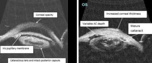
Depth of anterior chamber, entry into Anterior Chamber (AC), angle structures, iris synechiae, and iris deformity are another important findings which are difficult to be observed on slit lamp and thus UBM was done which helped in assessing the AC depth and accordingly site of entry into the anterior chamber can be planned in order to avoid injury to iris and lens (Fig. 2) which , in our study, was 100% accurately achieved in estimated clock hours for all cases of simple corneal opacity, 71.42% in cases of adherent leucoma and 85.71% in cases of anterior staphyloma, which was a high positive prediction number. We could not find any other study mentioning about entry into anterior chamber such precisely.
The anterior synechiae extensions (in terms of clock hours) were 83.33%, 85.71% and 71.42% positively predicted in cases of corneal opacity, adherent leucoma and anterior staphyloma respectively. It was under predicted only in 1 case (16.66%) of corneal opacity, 1 case (14.28%) of adherent leucoma and 2 cases (28.57%) of anterior staphyloma, whereby under prediction means that more extensive synechiae were seen intraoperatively than predicted on UBM. No over prediction was seen for this parameter, whereby over prediction means that synechiae were less extensive than predicted on UBM.
Similarly, in study by Madhavan, et al., for penetrating Keratoplasty in corneal opacity (16 cases), the anterior synechial positive prediction was 55.6% (5/9) and PAS was 73.1% (19/26) [6].
Similar to our study, only Zhang, et al., in their study highlighted clock hours and locations of lens-iris-corneal touch [13].
In the study of Penetrating Keratoplasty in children with Mucopolysaccharidoses UBM was considered useful preoperative guide [3]. But, the study emphasized only on congenital ocular abnormalities such as congenital aphakia and Aniridia but details of the extent of involvement in terms of percentage or predictive values were not mentioned [8,9].
Posterior synechaie was found in only 2 cases (33.33%) of simple corneal opacity (1 annular and 1 sectoral), 4 cases of adherent leucoma (57.14%) (1 annular and 3 sectoral) and 2 cases of anterior staphyloma (28.57%) (1 sectoral and 1 membranous), which was found 100% positively predicted when confirmed intraoperatively in all the 3 groups in our study.
Our results were consistent with the findings of Madhavan, et al., where positive predictive value for annular posterior synechiae was 23% and sectoral posterior synecheia was found in 11.2% cases [6].
The findings of anterior and posterior synechiae helped us to make decision for Membranectomy, iris reconstruction and Synechiolysis.
Iris reconstruction was done in total 8 out of 20 cases (40%). Out of these 8 cases, 6 cases (75%) were predicted accurate preoperatively whereas 2 cases (25%) of anterior staphyloma was under predicted and led to more iris loss than predicted by UBM. As predicted by UBM, in cases of extensive iris incarceration, patient could be explained for Aniridia and the need for iris reconstruction and such patients could be counseled preoperatively and managed in a better way intraoperatively.
No study has mentioned about the status of iris reconstruction in their cases.
Posterior capsule status and Zonular status predictability (Fig 1,2) which was 100% positive predicted in our study helped us to decide whether IOL placement should be done simultaneously or it should be planned for secondary IOL implantation. It also helped us in deciding the type of IOL to be placed in a particular case.
In 3 cases of adherent leucoma, anterior chamber IOL was implanted and 1 case in each of the three categories was planned for secondary IOL implantation, whereas in rest of the cases IOL implantation was done simultaneously. All these modifications and possibilities can be explained to patients preoperatively itself with the help of UBM.
Few studies also accurately predicted Zonular status using UBM in cases planned for cataract surgery with corneal pathology [6,8].
Madhavan, et al., observed 92.9% positive predictive value for Posterior Capsule (PC) compared to our study where it was 100%. Lens status and location were also observed in their study where crystalline lens was found in 100% (4/4) and cataractous changes in 50% (1/2) [6].
Other studies had not commented on posterior capsule and Zonules in such detail [7,12,13].
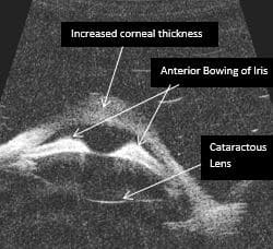
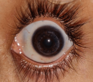
Thus, UBM plays extremely important role in detecting and overcoming surgical surprises and preventing the unforeseen complications by preplanning surgical strategies for best possible outcome (Fig. 3,4) and also for explaining the visual prognosis to patients beforehand in a best possible way.
Conclusion
Study shows that UBM is very supportive in cases planned for triple procedure as accurate predictions of maximum parameters could be achieved in all three groups. The positive prediction value ranging from 71.42% to 100% is obtained for various different parameters where lowest prediction value in our study being 71.42% which means , in all the cases, preoperative UBM findings were co-related intraoperatively and was found to be accurate in a minimum of 71.42% cases. Thus, UBM is a reliable guide in preplanning complicated surgeries like triple procedure and required intraoperative modifications for best visual outcomes.
Conflict of Interest
The authors declare that there is no conflict of interest.
References
- Bhatt DC. Ultrasound biomicroscopy: An overview. J Clin Ophthalmol Res. 2014;2(2):115.
- Dada T, Aggarwal A, Vanathi M, Gadia R, Panda A, Gupta V, et al. Ultrasound biomicroscopy in opaque grafts with post-penetrating keratoplasty glaucoma. Cornea. 2008;27(4):402-5.
- Harding SA, Nischal KK, Upponi-Patil A, Fowler DJ. Indications and outcomes of deep anterior lamellar keratoplasty in children. Ophthalmology. 2010;117(11):2191-5.
- Janssens K, Mertens M, Lauwers N, de Keizer RJ, Mathysen DG, De Groot V. To study and determine the role of anterior segment optical coherence tomography and ultrasound biomicroscopy in corneal and conjunctival tumors. J Ophthalmology. 2017;2016.
- Konstantopoulos A, Hossain P, Anderson DF. Recent advances in ophthalmic anterior segment imaging: a new era for ophthalmic diagnosi. Br J Ophthalmol. 2007;91:551-7.
- Madhavan C, Basti S, Naduvilath TJ, Sangwan VS. Use of ultrasound biomicroscopic evaluation in preoperative planning of penetrating keratoplasty. Cornea. 2000;19(1):17-21.
- Nischal KK, Naor J, Jay V, MacKeen LD, Rootman DS. Clinicopathological correlation of congenital corneal opacification using ultrasound biomicroscopy. Br J Ophthalmol. 2002; 86(1):62-9.
- Özdal MÇ, Mansour M, Deschenes J. Ultrasound biomicroscopic evaluation of the traumatized eyes. Eye. 2003;17(4):467-72.
- Pavlin CJ, Buys YM, Pathmanathan T. Imaging zonular abnormalities using ultrasound biomicroscopy. Arch Ophthalmol. 1998;116(7):854-7.
- Rutnin SS, Pavlin CJ, Slomovic AR. Preoperative ultrasound biomicroscopy to assess ease of haptic removal before penetrating keratoplasty combined with lens exchange. J Cataract and Refractive Surgeries. 1997;23:239-43.
- Thatte S, Gupta C. A study of anterior chamber morphology using UBM (Ultrasound Biomicroscopy) in cases with corneal pathology undergoing penetrating keratoplasty. Int J Ophthalmology and Optometry. 2019;1(1):18-24.
- Agraval U, Kincaid W, Mantry S, Ramaesh K. Predicting anterior segment surgical anatomy: application of Ultrasound Biomicroscopy (ubm) in surgical intervention. Invest Ophthalmol Vis Sci. 2014;55(13):4847.
- Zhang Y, Liu Y, Liang Q, Miao S, Lin Q, Zhang J, et al. Indications and outcomes of penetrating keratoplasty in infants and children of Beijing, China. Cornea. 2018;37(10):1243-8.
Article Type
Research Article
Publication History
Received Date: 13-06-2022
Accepted Date: 15-07-2022
Published Date: 22-07-2022
Copyright© 2022 by Thatte S, et al. All rights reserved. This is an open access article distributed under the terms of the Creative Commons Attribution License, which permits unrestricted use, distribution, and reproduction in any medium, provided the original author and source are credited.
Citation: Thatte S, et al. Role of Ultrasound Biomicroscopy in Predicting Intraoperative Surgical Strategies in Penetrating Keratoplasty with Cataract Extraction and IOL Implantation. J Ophthalmol Adv Res. 2022;3(2):1-14.

Figure 1: Various groups of corneal pathologies.

Figure 2: Anterior chamber and lens details on UBM in various categories of corneal pathologies.

Figure 3: UBM showing anterior staphyloma with anterior bowing of lens-iris diaphragm.

Figure 4: Post Penetrating keratoplasty photograph.
Corneal Pathologies | Number of Cases (20) |
Corneal opacities | 6 |
Adherent Leucoma | 7 |
Anterior Staphyloma | 7 |
Table 1: Corneal pathologies grouping.
Corneal Involvement | Corneal Opacity (6) | Adherent Leucoma (7) | Anterior Staphyloma (7) |
Full thickness | 5 (83.33%) | 7 (100%) | 7 (100%) |
Half stroma with endothelial involvement with scarring | 1 (16.67%) | 0 | 0 |
Table 2: Corneal involvement on UBM in various corneal pathologies.
Anterior Chamber Depth (ACD) | Corneal Opacity (6) | Adherent Leucoma (7) | Anterior Staphyloma (7) |
Normal | 4 (66.66%) | 0 | 0 |
Shallow | 1 (16.66%) | 0 | 4 (57.14%) |
Variable | 1 (16.66%) | 7 (100%) | 3 (42.85%) |
Angle position | |||
Normal | 5 (83.33%) | 5 (71.42%) | 5 (71.42%) |
Membrane closed | 1 (16.66%) | 2 (28.57%) | 2 (28.57%) |
Receding angle | 0 | 0 | 0 |
Anterior synechaie | |||
Membrane (involving angles) | 0 | 1 (14.28%) | 2 (28.57%) |
Sectoral | 1 (16.66%) from 2-3 o’clock | 5 (71.42%) | 5 (71.42%) |
Central | 0 | 1 (14.28%) | 0 |
Absent | 5 (83.33%) | 0 | 0 |
Posterior Synechaie | |||
Annular | 1 (16.66%) | 1 (14.28%) | 0 |
Sectoral | 1 (16.66%) | 3 (42.85%) | 2 (28.57%) |
Membrane | 0 | 0 | 0 |
Absent | 4 (66.66%) | 3 (42.85%) | 5(71.42%) |
Table 3: Status of anterior chamber on UBM.
Zonules | Corneal opacity (6) | Adherent Leucoma (7) | Anterior Staphyloma (7) |
Intact | 5 (83.33%) | 6 (85.71%) | 7 (100%) |
Dehiscence | 0 | 1 (14.28%) | 0 |
Dialysis | 1 (16.66%) | 0 | 0 |
Lens | |||
Phakic | 6 (100%) | 6 (100%) | 7 (100%) |
Psuedophakic | 0 | 0 | 0 |
Aphakic | 0 | 0 | 0 |
Table 4: Status of lens and Zonules on UBM.
| Size of graft | Trephination depth | Anterior chamber entry | ||||||||
| Adequate | Not adequate | Positive Predictive | Adequate | Not adequate | Prediction value | Achieved in estimated clock hours | Not achieved as per prediction | Positive Predictive Value | ||
Under predicted | Over Predicted | Under predicted | Over predicted | ||||||||
Corneal opacity (6) | 6 | – | – | 100% + | 6 | – | – | 100% + | 6 | – | 100% + |
Adherent Leucoma (7) | 7 | – | – | 100% + | 7 | – | – | 100% + | 6 | 1 (underpredicted) | 85.71% + |
Anterior Staphyloma (7) | 7 | – | – | 100% + | 7 | – | – | 100% + | 5 | 2 (underpredicted) | 71.42% + |
Table 5: Intraoperative confirmation of preoperative predictability of corneal findings and anterior chamber on UBM.
| Extent of anterior synechiae | Extent of posterior synechiae | Iris reconstruction | |||||||||
| Over predicted | Under predicted | Same as predicted | Positive Predictive | Over predicted | Under predicted | Same as predicted | Positive Predictive Value | Same As predicted | Changes | Positive PredictiveValue |
|
Simple Corneal opacities (6) | 0 | 1 | 5 | 83.33% + | 0 | 0 | 6 | 100% + | 6 | 0 | 100% + |
|
Adherent Leucoma (7) | 0 | 1 | 6 | 85.71% + | 0 | 0 | 7 | 100% + | 7 | 0 | 100% + |
|
Anterior Staphyloma (7) | 0 | 2 | 5 | 71.42% + | 0 | 0 | 7 | 100% + | 5 | 2 (underpredicted) | 71.42% + |
|
Table 6: Intraoperative confirmation of prediction of preoperative UBM findings on status of iris.
| Lens status | Lens position | Posterior capsule status | Status of zonules | ||||||||||||||
As predicted | Not as predicted |
| As predicted | Not as predicted |
| |||||||||||||
| Cataractous | Non cataractous |
| Positive Predictive | At normal anatomical position | Else where |
| Positive Predictive | Intact | Ruptured | Positive Predictive | Intact | Dehiscence/ dialysis | Variations from prediction | Positive Predictive Value |
| ||
Corneal opacities (6) | 6 | – | – | 100% + | 6 | – | – | 100% + | 6 | 0 | 100% | 5 | 1 | – | 100% |
| ||
Adherent Leucoma (7) | 7 | – | – | 100% + | 7 | – | – | 100% + | 4 | 3 | 100% | 6 | 1 | – | 100% |
| ||
Anterior Staphyloma (7) | 7 | – | – | 100% + | 6 | – | – | 100% + | 6 | 1 | 100% | 7 | 0 | – | 100% |
| ||
Table 7: Intraoperative confirmation of lens status, posterior capsule and zonular status to preoperative UBM findings.
| Corneal Opacity | Adherent Leucoma | Anterior Staphyloma |
Size of graft | 100% | 100% | 100% |
Trephination depth | 100% | 100% | 100% |
Anterior chamber entry | 100% | 85.71% | 71.42% |
Extent of synechiae | 83.33% | 85.71% | 71.42% |
Iris reconstruction prediction | 100% | 100% | 71.42% |
Lens status (cataractous) | 100% | 100% | 100% |
Lens position | 100% | 100% | 100% |
PC status | 100% | 100% | 100% |
Status of zonules | 100% | 100% | 100% |
Table 8: Positive Prediction value for various parameters in different groups.


