Noor Hanisah Rahim1, Nurul Izyani Johari1, Maria Angela Gonzalez2, Prema Sukumaran3*
1Faculty of Dentistry, University of Malaya, Kuala Lumpur, Malaysia
2College of Dentistry, National University, Manila, Philippines
3Department of Restorative Dentistry, Faculty of Dentistry, University of Malaya, Kuala Lumpur, Malaysia
*Corresponding Author: Prema Sukumaran, Department of Restorative Dentistry, Faculty of Dentistry, University of Malaya, Kuala Lumpur, Malaysia; Email: [email protected]
Published Date: 27-07-2022
Copyright© 2022 by Sukumaran P, et al. All rights reserved. This is an open access article distributed under the terms of the Creative Commons Attribution License, which permits unrestricted use, distribution, and reproduction in any medium, provided the original author and source are credited.
Abstract
Objective: This study was designed to use Optical Coherence Tomography (OCT) to quantify depth of occlusal fissures on premolar teeth and to quantitatively evaluate the penetration of pit and fissures sealants relative to fissure depth. The study aimed to evaluate the penetrability of Pit and Fissure Sealant (PFS) in covering the full-depth of different occlusal fissure depths using Swept-Source Optical Coherence Tomography (SS-OCT).
Methods: Ninety-seven investigation sites on f occlusal fissures of fifteen premolar teeth with International Caries Detection and Assessment System (ICDAS) scores of 01 or 02 were categorized using SS-OCT into four groups (smooth, shallow, intermediate, and deep fissures). After PFS placement, cross-sectional images of these fissures were observed again with SS-OCT.
Results: The result of this study shows that PFS can fully penetrate the smooth fissures (100%) followed by shallow fissures (94.2%) and intermediate fissures (47.6%). Incomplete full depth PFS penetration can be seen in all deep fissures (100%).
Conclusion: The depth of the fissure will largely affect the penetrability of PFS into the fissure.
Keywords
Dental Caries; Pit and Fissure Sealant; Optical Coherence Tomography
Introduction
Intraoral cariogenic bacteria, mainly Streptococcus mutans, interacts with dietary carbohydrates, producing acidic by products. Over time, the accumulation of these by-products interferes with the delicately balanced demineralization and remineralization process on tooth surfaces, leading to caries formation.
Studies have shown pit and fissure surfaces are most susceptible to caries formation whereas smooth, lingual and labial surfaces are less susceptible [1]. Microscopic investigations have shown that the occlusal morphology consists of pits of considerable length positioned at various angles relative to different depth of fissures [2]. Ito and colleagues classified these fissure depths into smooth, shallow, intermediate and deep fissure [3]. Due to its complex occlusal anatomy of intricate pits and fissures, it is often difficult to completely remove bacteria and food on the occlusal surfaces with tooth brushing or other mechanical debridement method [4].
Effective non-surgical approaches to caries prevention, include community water fluoridation, topical fluoride therapy, plaque control, and dietary sugar controlregulating sugar intake. These approaches may contribute to the reduction of smooth surface caries. However, researchers have reservations on the impact these measures may have on pit and fissure caries, mainly due to the difference in the anatomy of the surfaces involved [5,6].
In the 1960s, Pit and Fissure Sealants (PFS) were introduced as a preventive management to reduce the incidences of occlusal caries in posterior teeth. Flowing PFS onto the occlusal surfaces effectively seals the pits and fissures, preventing lodging of food debris or bacterial growth. This clinical procedure was also prescribed for occlusal surface with incipient caries as a preventive measure to avoid formation of more severe caries lesions [7,8]. There are a few options for occlusal sealant materials, but resin and glass ionomers comprise the main material types [9]. Placement techniques for PFS vary based on sealant type and the manufacturer or brand. PFS are usually applied as a liquid that is allowed to flow into the crevices of the occlusal surface of the tooth and harden after polymerization. PFS bonds to the tooth micromechanically, providing a physical barrier that keeps bacteria away from their source of nutrients.
Intraoral radiography is the most common tool used in clinical setting to diagnose carious lesions. Bitewing radiographs are the most recommended intraoral radiograph used to detect caries, as it provides view of the interproximal surfaces of the posterior teeth that cannot be clinically probed [10]. Although the percentage of the intraoral radiograph to detect the presence of caries in dentine is fairly high (50-70%), there are several limitations with the use of this type of radiograph [11]. Bitewing radiograph is unable to differentiate between active and arrested caries, to detect early caries lesion, as well the presence of PFS on occlusal surface [12]. Since it is difficult to visualize changes in the occlusal surface with radiographs, it is mostly used for detection of interproximal caries.
Optical Coherence Tomography (OCT) is a non-invasive imaging technique that produces high resolution, cross-sectional images of biological hard and soft tissue. OCT technology has been significantly advanced by the development of Spectral Domain (SD) techniques, which provide a substantial increase in sensitivity over earlier systems. The Swept Source Optical Coherence Tomography (SS-OCT) is one of these SD developments and uses a wavelength-tuned near-infrared laser as the light source, thus providing improved imaging resolution and scanning speed. In laboratory studies, itIt was reported that SS-OCT has ability to differentiate between carious and sound tissues in both primary and permanent teeth, estimate the lesion depth in demineralized tissues, observe gaps at tooth-restoration interface and defects in dental restorations and detect tooth cracks [13]. In a study by Holztman and colleagues, SS-OCT was used also used as a diagnostic tool for the detection of occlusal fissure depth and the completeness of PFS penetration via rudimentary visual qualitative evaluation of OCT images [14].
According to Ito and co-workers, the sensitivity and specificity of fissure depth evaluation using SS-OCT was higher than visual inspection for all fissure depths [3]. In this study, The results from their study showed that SS-OCT correlated with confocal laser screening microscopy while visual inspection did not show close association.
Material and Methods
Sample Preparation and Visual Inspection
A power calculation was performed using GPower software to determine the number of samples for this in-vitro study. Fifteen extracted maxillary and mandibular sound premolar teeth or with ICDAS 01 or 02 caries were selected for this study. Caries assessment on the occlusal surface was performed visually and using SS-OCT. The tooth samples were collected and selected based on the criteria below:
a) Inclusion criteria:
- Extracted maxillary or mandibular premolar tooth
- Sound tooth or tooth with caries (ICDAS 01 or 02)
b) Exclusion criteria:
- Tooth with crack line on the occlusal surface
- Tooth with gross caries extending depth of enamel
The selected teeth were stored in 0.05 mol/L chloramine-T solution before the start of the experiment. The roots of all fifteen premolar teeth were sectioned with a diamond cutting disc bur attached to straight handpiece. A customized jig (22 mm x 30 mm) was prepared for each tooth by mounting it in addition silicone putty (Vonflex S Putty, Vericom Co., Ltd., Chuncheon, Korea). The prepared jig allowed for repositioning of tooth samples on SS-OCT scanning platform during all scanning and measurement exercises.
OCT Observation of Fissure Depth
A Swept-source OCT Imaging System (OCS1300SS, Thorlabs Ltd., UK) with emission wavelength centered at 1325nm and axial and transverse resolution of 9μm and 11μm in air, respectively, was used to scan the investigation sites. The Thorlabs OCT capturing software (Swept Source OCT Imaging System Version 2.3.1, Thorlabs) was used to configure the SS-OCT settings, guide the light beam and capture the image [17].
Ninety-seven locations on the occlusal fissures were obtained from the fifteen premolars and marked with felt-tip pen as the inspection sites. The occlusal surfaces of these teeth were cleaned to remove any visible debris and dried for five seconds with compressed air before inspection was done.
The teeth were set at a fixed distance with the scanning beam oriented at 90º with respect to the occlusal plane of the tooth according to the location selected. The scanned location for each fissure selected was noted and recorded from the X and Y coordinates. X and Y coordinates for selected B-scans for each tooth were used to reposition the jig after PFS placement. This was to ensure B-scans from similar Regions of Interest (ROI) are selected to measure the depth of resin penetration. The OCT images were obtained as cross-sections of the investigation sites (B-scans) and these images were taken as pre-resin infiltration data to score each of the fissure locations according to fissure classification adopted from the publication by Ito and colleagues [3].
- Smooth fissure (score 0): fissure base shows no cleaving between cuspal inclines
- Shallow fissure (score 1): base of fissure cleft is less than one third of enamel thickness
- Intermediate fissure (score 2): base of fissure cleft is deeper than one third but less than two thirds of enamel thickness
- Deep fissure (score 3): fissure is deeper than two thirds of enamel thickness, near enamo-dentinal junction
Representative B-scan images of different types of fissure depth are shown in Fig. 1.

Figure 1: Representative B-scan images of different types of fissure depth. (a) smooth fissure (score 0), (b) shallow fissure (score 1), (c) intermediate fissure (score 2), and (d) deep fissure (score 3). The Enamo-Dentinal Junction (EDJ) is indicated by the yellow arrowhead and the base of fissure is indicated by the red arrow.
Pre-sealant Measurement / Depth of Fissure Measurement
Using the measuring tools in the software, a horizontal intercuspal reference line (rl) of 500 μm in length was constructed between the buccal and lingual slopes, perpendicular to the cusp section on a B-scan.
A fissure depth line (fd) was constructed perpendicular to the (rl), downwards, towards the base of fissure (Fig. 2). The pre-sealant fissure depth (fd) measurements for each tooth were recorded and tabulated accordingly.

Figure 2: Measurement of fissure pre- (a) and post-sealant (b) penetration. (rl) refers to reference line, (fd) is the fissure depth and (sp) is the PFS penetration.
Sealant Placement and OCT Imaging of Sealant Penetration
The researcher trained in applying PFS on a few occlusal surfaces of teeth prior to the study in order to achieve a standardized amount of PFS used and placement procedure. During the study, each tooth was cleaned with a slurry of pumice using prophylactic brush attached to slow speed handpiece, before it was rinsed thoroughly with water and dried with compressed air [18]. The occlusal fissure system on each tooth was etched with 35% phosphoric acid (Scotchbond Etchant 3M ESPE, St Paul, MN, USA) for 15 seconds, rinsed thoroughly with water for ten seconds and subsequently gently air-dried. The applicator nozzle of 3M™ Clinpro™ Sealant (3M ESPE) was placed against the fissure system, and the PFS was gently flowed into the pits and fissures. PFS was light-cured for 20 seconds using light-curing unit (Bluephase N, Ivoclar Vivodent). Following PFS placement, observation and measurement was performed with OCT to evaluate the PFS penetration into pit and fissure depth. The B-scan images (in the same ROI) after placement of PFS were recorded for each tooth. The before and after B-scan images for each tooth sample was compared and depth of PFS penetration into fissure was measured using measuring tools available in the software.
Post Sealant Measurement
After placement of PFS, the reference line (rl) was re-established to reproduce the exact reference location as pre-sealant measurements (Fig. 2). Once this was re-established, we could assess depth of penetration of PFS into fissure using the previously identified base of fissure as reference (Fig. 3). When there is complete penetration of PFS to the base of fissure, SS-OCT image shows a homogenous brightness extending to the base. On the contrary, when there is incomplete penetration of PFS, the resulting image shows there is some degree of homogenous brightness extending to only half or more than half of fissure depth. In addition, a dark zone could also be observed at the base of the fissure. Comparative images of complete and incomplete PFS penetration are shown in Fig. 3.

Figure 3: (a) SS-OCT image shows complete penetration of PFS into shallow fissure (score 1). Vertical lines indicate the width is at 500 μm, upper and lower yellow horizontal lines indicate the reference line and the base of fissure respectively. The complete PFS penetration into the base of the fissure was observed as homogenous brightness; (b) SS-OCT image shows incomplete penetration of PFS into intermediate fissure (score 2). Vertical lines indicate the width is at 500 μm, upper and lower yellow horizontal lines indicate the reference line and the base of fissure respectively. Red horizontal line indicates the base of PFS. Lack of complete PFS penetration at the base of fissure could be observed as a dark zone extending from the red line to the yellow bottom line.
Penetrability of PFS (P) was measured as percentage of PFS penetration (sp) to the fissure depth (fd). PFS penetration (sp) was measured from the reference line (rl) to the lowest base of the PFS that was presented.
Complete penetration of PFS was achieved when the penetrability of PFS was recorded as 100%, where the PFS reached the base of fissure. In incomplete PFS penetration, there was light scattering in the SS-OCT image adjacent to the base of the fissure, indicating PFS did not completely penetrate the fissure, leaving unfilled zones.
Stereomicroscope
After PFS penetration, each tooth sample from the four different fissure types was sectioned and subjected to stereomicroscopy (Olympus, Model SZX 7- S4, Tokyo, Japan) imaging to visually correlate with OCT B-scans. Prior to sectioning, one site was chosen from each tooth sample and marked with fine tip felt pen. The chosen site was sectioned vertically using a diamond disc. The sectioned sample was positioned and viewed under the stereomicroscope and the resulting image was visually compared with corresponding OCT B-scan.
Data Analysis
Data was tabulated and statistical analysis was performed using SPSS Version 25 to ascertain the complete or incomplete penetration of the PFS according to the fissure depth scores. Chi-Square test was performed to determine the association between fissure depth scores and PFS penetration. The strength of association between fissure depth scores and PFS penetration was analyzed using Cramer’s V. Statistical testing was performed at α = 0.05.
Results
The ninety-seven investigation sites that were obtained were classified into: 33 smooth fissures without a cleft (score 0), 35 shallow fissures (score 1), 21 intermediate fissures (score 2), and 6 deep fissures (score 3) as shown in Table 1.
The range of fissure depth for each fissure score overlapped. The ranges were between 59 μ-398 μ, 305 μ-750 μ, 451 μ-1002 μ and 809 μ-1307 μ for scores 0,1,2 and 3 respectively.
As reported in Table 1, PFS completely penetrated all of the smooth fissures (100%) and almost all of shallow fissures (94.2%). However, none of the deep fissures were completely penetrated by PFS into the base.
Fissure Depth Scores | PFS Penetration |
Total Investigation Sites | |
Complete Penetration N (%) | Incomplete Penetration N (%) | ||
Smooth fissure (score 0) | 33 (100) | 0 (0) | 33 |
Shallow fissure (score 1) | 33 (94.2) | 2 (5.7) | 35 |
Intermediate fissure (score 2) | 10 (47.6) | 11 (52.3) | 21 |
Deep fissure (score 3) | 0 (0) | 6 (100) | 6 |
Table 1: Proportion of complete and incomplete PFS penetration by fissure depth scores.
Figure 4 shows representative B-scan images of complete and incomplete penetration of PFS for each fissure type and how the measurements were made.

Figure 4: Representative B-scan images of complete and incomplete penetration of PFS in different fissure depth scores. Vertical lines indicate the width is at 500 μm, upper and lower yellow horizontal lines indicate the reference line and the base of fissure respectively. a) SS-OCT image shows complete penetration of PFS into smooth fissure (Score 0). The complete PFS penetration into the base of the fissure was observed as homogenous brightness; b) SS-OCT image shows complete penetration of PFS into shallow fissure (Score 1). The complete PFS penetration into the base of the fissure was observed as homogenous brightness; c) SS-OCT image shows incomplete penetration of PFS into shallow fissure (Score 1). Red horizontal line indicates the base of PFS. Lack of complete PFS penetration at the base of fissure could be observed as a dark zone extending from the red line to the yellow bottom line; d) SS-OCT image shows complete penetration of PFS into intermediate fissure (Score 2). The complete PFS penetration into the base of the fissure was observed as homogenous brightness; e) SS-OCT image shows incomplete penetration of PFS into intermediate fissure (Score 2). Red horizontal line indicates the base of PFS. Lack of complete PFS penetration at the base of fissure could be observed as a dark zone extending from the red line to the yellow bottom line; f) SS-OCT image shows incomplete penetration of PFS into deep fissure (Score 3). Red horizontal line indicates the base of PFS. Lack of complete PFS penetration at the base of fissure could be observed as a dark zone extending from the red line to the yellow bottom line.
Chi-square test result shows that there is an association between PFS penetration and depth of fissure depth with p-value = 0.00. The effect size was large with Cramer’s V value of 0.538.
Table 2 presents average depth of each fissure score (in µm) after application of PFS for each fissure score. In fissures with Score 1, the average depth of fissure with incomplete penetrations was lesser than fissures with complete penetration. The opposite was demonstrated for fissures with Score 2, whereby, the fissures with incomplete PFS penetration were deeper than those with complete penetration. Regardless of the quality of penetration, Table 2 shows that fissures with score 0 have the most shallow depth followed by fissures with Scores 1,2 and 3. Total average depth of the various fissures types.
Fissure Score | Average depth of fissure in μm (SD) | Total average depth in μm (SD) | |
Complete | Incomplete | ||
Score 0 | 218.0 (90.3) | – | 218.0 (90.3) |
Score 1 | 504.7 (102.7) | 460.0 (4.2) | 502.1 (100.2) |
Score 2 | 609.9 (102.7) | 778.7 (123.9) | 698.3 (141.0) |
Score 3 | – | 989.3 (186.4) | 989.3 (186.4) |
Table 2: Mean fissure depth for each type of PFS penetration.
Fig. 5 shows the comparative images of complete and incomplete PFS penetration under OCT machine and stereomicroscope. The yellow arrow shows the base of fissure, and the red arrows indicate the base of PFS. In fissures with scores 0, 1 and 2 (fissures 1 and 2 with complete penetration of PFS), the red and yellow arrows are at the same level, indicating penetration of PFS to the base on the fissure. For the rest of the fissure types, the red arrow is above the yellow arrow, demonstrating the lack of PFS penetration to the base of fissure.


Figure 5: Comparative images of complete and incomplete PFS penetration under OCT machine and stereomicroscope. The yellow arrow shows the base of fissure, and the red arrow shows the base of PFS. (E) is the enamel and (D) is the dentine.
Discussion
In this study, the depth of pit and fissure into enamel influenced the ability of PFS to completely penetrate the fissure. PFS almost completely filled the smooth and shallow fissures whilst deeper fissures were incompletely filled. The findings from this study are similar with the results reported by Ito and colleagues. The results in both studies were supported by SS-OCT images confirming that PFS flowed to the base of shallow fissures more frequently [3]. In this study, the depth of pit and fissure into enamel influenced the ability of PFS to completely penetrate the fissure. PFS almost completely filled the smooth and shallow fissures whilst deeper fissures were incompletely filled. The findings from this study are similar with the results reported by Ito and colleagues. The results in both studies were supported by SS-OCT images confirming that PFS flowed to the base of shallow fissures more frequently [4,19]. In our study, the measurement of fissure depth was based on marked reference lines utilized by Markovic and his colleagues that used a horizontal reference line of 500 μm between cusp slopes. The 500 μm of reference horizontal line was chosen based on the fact that this line embraces both fissure depth and width as equally important factors for sealant penetration [20]. We found that the reference point is more reproducible in each investigation site and fissures depth can easily be scored accordingly. However, there is no universal classification of pit and fissure with respect to their morphology and depth but in many studies, fissures are classified according to the morphology of the fissures (U, V, I and IK) [18,21].
The depth of the fissure overlapped from one fissure score group to the other. Shallower fissures were observed to have complete penetration over deeper fissures and vice versa within the same fissure score group as displayed by fissures in Score 1 group. The fissure depth score classification that we used, which is relative to enamel thickness, does not consider fissure depth alone. This was why we took into consideration the depth of each fissure. We found that the depth of fissure relative to enamel thickness has a positive correlation to PFS penetration. The complex anatomy of the fissure may be the cause of this observation.
Based on the guideline for the use of PFS for posterior permanent teeth by American Academy of Pediatric Dentistry and American Dental Association (AAPD), there are two main purposes in sealing the occlusal fissure that we need to understand. First, as the occlusal fissures are the most susceptible area for food accumulation and can promote the production of bacterial biofilm, there will be a high risk of caries formation. Thus, by effectively penetrating and sealing the occlusal fissure with PFS, we may help to prevent caries lesion. Secondly, the use of PFS also will help to impede the progression or development of non-cavitated carious lesion (AAPD) [10].
Even though we can claim from our study that the penetration of PFS is affected by the fissure depth scores, but in reality, there is no consensus on whether complete resin penetration into fissure depth is considered the key to successful bonding of the PFS. In studies by Marks, Owens and Johnson, no correlation was found between material microleakage and PFS penetration [22]. This statement was supported by a previous study claiming that overall penetration into full depth of fissure is not necessarily required for restorative success. Studies by Duangthip and Lussi and Garg and co-workers agreed that PFS effectiveness is determined by adequate bonding at the coronal part of fissure and not necessarily depending on complete penetration of the PFS into the base of fissure [18,21]. However, in a study by Garcia-Godoy and De-Araujo, it was proven that total penetration of PFS into full fissure depth will determine the success rate of PFS [23].
In this study, we cleaned the occlusal surfaces of sample teeth using slurry of pumice prior to sealant placement. According to Sheyholeslam, salivary pellicles, organic debris and handpiece lubricating oil are the most common potential causes of stagnation of the occlusal fissure and later causing impediment in proper cleaning of the tooth surface [24]. These contaminants also will contribute to marginal leakage or poor retention of the PFS to tooth surface. Thus, proper cleaning and preparing of the tooth fissure is important to achieve good retention of the PFS. In a study by Hedge and Coutinho, they found that cleaning occlusal fissure with slurry of pumice and surface conditioning prior to PFS placement showed a better result of PFS retention after being reviewed in 3, 6 and 12 months [25].
In order to achieve complete penetration of PFS, some improvement in sealant materials and techniques were established. One of the techniques that claimed to enhance PFS penetration is enameloplasty. The result of a study by Garcia-Godoy and De Araujo demonstrated that enameloplasty allowed a deeper PFS penetration and a superior PFS adaptation than the conventional PFS technique [23]. Enlarging the fissures by using Enameloplasty Sealant Technique (EST) will increase surface area that will enhance PFS retention. EST can be indicated for placing PFS on any occlusal surface with deep fissures. However, it should be especially indicated on deep and narrow discoloured fissures, suspected of being carious. There are, however, claims that PFS penetration is not an important factor in having a good success rate of PFS longevity. If PFS retention is gained from good adaptation of PFS to the enamel, routine removal of sound fissures by enameloplasty to achieve complete penetration of PFS to the bottom of fissures might not be an important step in sealant success [18]. Furthermore, enameloplasty is an invasive technique that widens the base of a fissure on a sound tooth which disturbs the equilibrium of the fissure and exposes a patient unnecessarily to the use of a handpiece or air abrasion. In our opinion, there is a need for removal of most organic substances in the fissures in order to obtain sufficient bonding to tooth structure, but removal of sound tooth tissue by the use of instruments, such as a bur, is unnecessary and undesirable.
Properties of the PFS such as surface tension and viscosity are the most important factors that affect the penetration of PFS. Most commercially used PFS are resin-based sealants (filled or unfilled) and glass ionomer sealants. In this study, 3M™ Clinpro™ Sealant (3M ESPE), an unfilled resin, was used, also used in the present authors’ clinical practice. Unfilled sealant has lower viscosity that increases the PFS penetrability into deep and narrow fissures. Whereas, filled sealant has increased resistance towards wear, it also increases the viscosity of the PFS that eventually will decrease the PFS penetrability. Resin-based materials have been proven to have high retention rates and wear resistance. However, it requires excellent moisture control as it is hydrophobic in nature and requires a dry working area. Glass ionomer sealants are alternatives with advantages such as high fluoride release and antibacterial properties. In addition, studies have also documented that newer glass ionomer sealants have improved adherence to enamel, better flow properties and easier to handle clinically [7,8].
In this study, SS-OCT machine was used as a diagnostic tool to assess the occlusal fissure depth scores and PFS penetration. This machine is capable in providing clear cross-sectional images of occlusal fissures, able to detect demineralized enamel, and can discriminate complete and incomplete PFS penetration based on contrasting brightness of PFS. Teeth with gross caries and crack line presenting on the occlusal surfaces were excluded from this study because the OCT derived B-scans of these teeth would show increase back scattering intensity extending either to the depth of enamel and/or beyond the Enamo-Dentinal Junction (EDJ). The increased back scattering intensity denotes increased porosity due to demineralization such as caries. This would later interfere with the observation of surface of the fissures and the calculation of the depth of fissures.
By using OCT imaging, we are able to eradicate ionization radiation, and patient would be comfortable as we will not be using the intraoral radiographic sensors or film as seen in conventional radiography. After PFS application, caries on radiographs may be even more difficult to detect because the radiographic appearance of sealant is indistinguishable from the tooth structure and PFS may mask early carious lesions. Thus, the ability to view a demineralized (early caries) tooth surface pre- and post-sealant placement are better in OCT imaging than clinical examination or radiographs. Compact, mobile, user-friendly clinical OCT systems are now becoming available to clinicians, offering a small, flexible fibreoptic OCT probe that can easily access the oral cavity and provide chair side in-vivo clinical imaging, and immediate, real-time OCT images, with total imaging times of less than a minute. Digital data files provide a quick and easy data storage, retrieval, and printouts as needed. The potential use of OCT for demineralization detection under PFS may result in better utilization of manpower and time, as well as reduced health care cost [14].
The limitation of this study is in terms of the capability of the SS-OCT system to capture the image of fissure depths due to technical obstacles such as light attenuation through the structure. Demineralization in the fissures typically strongly attenuates the OCT signal, preventing an accurate depth determination of the lesion. In this study, the axial orientation and distance from the probe were adjusted for each tooth during SS-OCT scan to ensure an optimal fissure depth imaging. However, due to variation of structure of deep occlusal fissures, it was difficult to obtain a clear image of the deep fissures in some cases. Accurate depth determination also failed to be recorded when there is obstruction of debris inside the fissure especially deep fissures that would limit the laser light to penetrate. In this study, opaque or colored shades of PFS were used, as it is easier to see the PFS during application and to assess retention at later time intervals when compared with a clear PFS. But opaque shades contain an optical opacifier, titanium dioxide, which strongly attenuates near-infrared light thus image of the PFS and fissures are difficult to capture accurately.
Conclusion
From our study, we can conclude that pit and fissure sealant penetration is affected by the fissure depth of teeth relative to enamel thickness. Based on the classification by Ito and colleagues, most of the smooth fissures are completely penetrated by the PFS to the base of the fissure, while in deep fissures there is difficulty for the PFS to completely penetrate the fissure.
Conflict of Interest
The authors report no conflict of interest. The authors alone are responsible for the content and writing of the manuscript.
References
- Chestnutt IG, Schäfer F, Jacobson AP, Stephen KW. Incremental susceptibility of individual tooth surfaces to dental caries in Scottish adolescents. Community Dent Oral Epidemiol. 1996;24(1):11-6.
- Juhl M. Three‐dimensional replicas of pit and fissure morphology in human teeth. Eur J Oral Sci. 1983;91(2):90-5.
- Ito S, Shimada Y, Sadr A, Nakajima Y, Miyashin M, Tagami J, et al. Assessment of occlusal fissure depth and sealant penetration using optical coherence tomography. Dent Mater J. 2016;35(3):432-9.
- Symons AL, Chu CY, Meyers IA. The effect of fissure morphology and pretreatment of the enamel surface on penetration and adhesion of fissure sealants. J Oral Rehabil. 1996;23(12):791-8.
- Demirci M, Tuncer S, Yuceokur AA. Prevalence of caries on individual tooth surfaces and its distribution by age and gender in university clinic patients. Eur J Dent. 2010;4(3):270.
- Kitchens DH. The economics of pit and fissure sealants in preventive dentistry: a review. J Contemp Dent Pract. 2005;6(3):95-103.
- Buonocore M. Adhesive sealing of pits and fissures for caries prevention, with use of ultraviolet light. J Am Dent Assoc. 1970;80(2):324-8.
- Ahovuo‐Saloranta A, Forss H, Walsh T, Nordblad A, Mäkelä M, Worthington HV. Pit and fissure sealants for preventing dental decay in permanent teeth. Cochrane Database of Systematic Reviews. 2017(7).
- Naaman R, El-Housseiny AA, Alamoudi N. The use of pit and fissure sealants-A literature review. Dent J. 2017;5(4):34.
- Wright JT, Crall JJ, Fontana M, Gillette EJ, Nový BB, Dhar V, et al. Evidence-based clinical practice guideline for the use of pit-and-fissure sealants: a report of the American Dental Association and the American Academy of Pediatric Dentistry. J Am Dent Assoc. 2016;147(8):672-82.
- Wenzel A. Current trends in radiographic caries imaging. Oral Surg Oral Med Oral Pathol Oral Radiol. 1995;80(5):527-39.
- Wenzel A. Bitewing and digital bitewing radiography for detection of caries lesions. J Dent Res. 2004;83:72-5.
- Karteva E, Manchorova-Veleva N, Stefanova V, Atanasov M, Atanasov A, Pashkouleva D, et al. Novel methods for the assessment of crack propagation in endodontically treated teeth. J IMAB–Annu Proceeding (Sci Pap). 2016;22(3):1308-13.
- Holtzman JS, Osann K, Pharar J, Lee K, Ahn YC, Tucker T, et al. Ability of optical coherence tomography to detect caries beneath commonly used dental sealants. Lasers Surg Med. 2010;42(8):752-9.
- Amaechi BT, Higham SM, Podoleanu AG, Rogers JA, Jackson DA. Use of optical coherence tomography for assessment of dental caries: quantitative procedure. J Oral Rehabil. 2001;28(12):1092-3.
- Amaechi BT, Podoleanu AG, Higham SM, Jackson DA. Correlation of quantitative light-induced fluorescence and optical coherence tomography applied for detection and quantification of early dental caries. J Biomed Opt. 2003;8(4):642-8.
- Zain E, Zakian CM, Chew HP. Influence of the loci of non-cavitated fissure caries on its detection with optical coherence tomography. J Dent. 2018;71:31-7.
- Duangthip D, Lussi A. Effects of fissure cleaning methods, drying agents, and fissure morphology on microleakage and penetration ability of sealants in-vitro. Pediatr Dent. 2003;25(6):527-33.
- Oong EM, Griffin SO, Kohn WG, Gooch BF, Caufield PW. The effect of dental sealants on bacteria levels in caries lesions: a review of the evidence. J Am Dent Assoc. 2008;139(3):271-8.
- Markovic D, Petrovic B, Peric T, Miletic I, Andjelkovic S. The impact of fissure depth and enamel conditioning protocols on glass-ionomer and resin-based fissure sealant penetration. J Adhes Dent. 2011;13(2):171.
- Garg N, Indushekar KR, Saraf BG, Sheoran N, Sardana D. Comparative evaluation of penetration ability of three pit and fissure sealants and their relationship with fissure patterns. J Dent. 2018;19(2):92.
- Marks D, Owens BM, Johnson WW. Effect of adhesive agent and fissure morphology on the in-vitro microleakage and penetrability of pit and fissure sealants. Quintessence Int. 2009;40(9):763-72.
- Garcia-Godoy F, De Araujo FB. Enhancement of fissure sealant penetration and adaptation: The enameloplasty technique. J Clin Pediatr Dent. 1994;19:13-9.
- Sheykholeslam Z, Brandt S. Some factors affecting the bonding of orthodontic attachments to tooth surface. J Clin Ortho. 1977;11(11):734-43.
- Hegde RJ, Coutinho RC. Comparison of different methods of cleaning and preparing occlusal fissure surface before placement of pit and fissure sealants: An in-vivo J Indian Soc Pedod Prev Dent. 2016;34(2):111.
Article Type
Research Article
Publication History
Received Date: 28-06-2022
Accepted Date: 20-07-2022
Published Date: 27-07-2022
Copyright© 2022 by Sukumaran P, et al. All rights reserved. This is an open access article distributed under the terms of the Creative Commons Attribution License, which permits unrestricted use, distribution, and reproduction in any medium, provided the original author and source are credited.
Citation: Sukumaran P, et al. Sealant Penetration into Occlusal Fissures: An In-Vitro Optical Coherence Tomography Study. J Dental Health Oral Res. 2022;3(2):1-17.
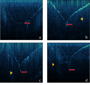
Figure 1: Representative B-scan images of different types of fissure depth. (a) smooth fissure (score 0), (b) shallow fissure (score 1), (c) intermediate fissure (score 2), and (d) deep fissure (score 3). The Enamo-Dentinal Junction (EDJ) is indicated by the yellow arrowhead and the base of fissure is indicated by the red arrow.
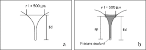
Figure 2: Measurement of fissure pre- (a) and post-sealant (b) penetration. (rl) refers to reference line, (fd) is the fissure depth and (sp) is the PFS penetration.
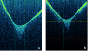
Figure 3: (a) SS-OCT image shows complete penetration of PFS into shallow fissure (score 1). Vertical lines indicate the width is at 500 μm, upper and lower yellow horizontal lines indicate the reference line and the base of fissure respectively. The complete PFS penetration into the base of the fissure was observed as homogenous brightness; (b) SS-OCT image shows incomplete penetration of PFS into intermediate fissure (score 2). Vertical lines indicate the width is at 500 μm, upper and lower yellow horizontal lines indicate the reference line and the base of fissure respectively. Red horizontal line indicates the base of PFS. Lack of complete PFS penetration at the base of fissure could be observed as a dark zone extending from the red line to the yellow bottom line.
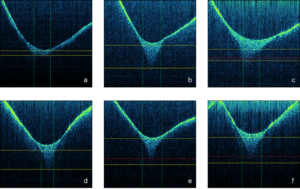
Figure 4: Representative B-scan images of complete and incomplete penetration of PFS in different fissure depth scores. Vertical lines indicate the width is at 500 μm, upper and lower yellow horizontal lines indicate the reference line and the base of fissure respectively. a) SS-OCT image shows complete penetration of PFS into smooth fissure (Score 0). The complete PFS penetration into the base of the fissure was observed as homogenous brightness; b) SS-OCT image shows complete penetration of PFS into shallow fissure (Score 1). The complete PFS penetration into the base of the fissure was observed as homogenous brightness; c) SS-OCT image shows incomplete penetration of PFS into shallow fissure (Score 1). Red horizontal line indicates the base of PFS. Lack of complete PFS penetration at the base of fissure could be observed as a dark zone extending from the red line to the yellow bottom line; d) SS-OCT image shows complete penetration of PFS into intermediate fissure (Score 2). The complete PFS penetration into the base of the fissure was observed as homogenous brightness; e) SS-OCT image shows incomplete penetration of PFS into intermediate fissure (Score 2). Red horizontal line indicates the base of PFS. Lack of complete PFS penetration at the base of fissure could be observed as a dark zone extending from the red line to the yellow bottom line; f) SS-OCT image shows incomplete penetration of PFS into deep fissure (Score 3). Red horizontal line indicates the base of PFS. Lack of complete PFS penetration at the base of fissure could be observed as a dark zone extending from the red line to the yellow bottom line.
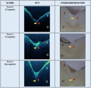

Figure 5: Comparative images of complete and incomplete PFS penetration under OCT machine and stereomicroscope. The yellow arrow shows the base of fissure, and the red arrow shows the base of PFS. (E) is the enamel and (D) is the dentine.
Fissure Depth Scores | PFS Penetration |
Total Investigation Sites | |
Complete Penetration N (%) | Incomplete Penetration N (%) | ||
Smooth fissure (score 0) | 33 (100) | 0 (0) | 33 |
Shallow fissure (score 1) | 33 (94.2) | 2 (5.7) | 35 |
Intermediate fissure (score 2) | 10 (47.6) | 11 (52.3) | 21 |
Deep fissure (score 3) | 0 (0) | 6 (100) | 6 |
Table 1: Proportion of complete and incomplete PFS penetration by fissure depth scores.
Fissure Score | Average depth of fissure in μm (SD) | Total average depth in μm (SD) | |
Complete | Incomplete | ||
Score 0 | 218.0 (90.3) | – | 218.0 (90.3) |
Score 1 | 504.7 (102.7) | 460.0 (4.2) | 502.1 (100.2) |
Score 2 | 609.9 (102.7) | 778.7 (123.9) | 698.3 (141.0) |
Score 3 | – | 989.3 (186.4) | 989.3 (186.4) |
Table 2: Mean fissure depth for each type of PFS penetration.


