Kjær I1*
1Department of Odontology, Faculty of Health and Medical Sciences, University of Copenhagen, Copenhagen, Denmark
*Correspondence author: Kjær I, Department of Odontology, Faculty of Health and Medical Sciences, University of Copenhagen, Copenhagen, Denmark; E-mail: [email protected]
Published Date: 31-12-2024
Copyright© 2024 by Kjær I, et al. All rights reserved. This is an open access article distributed under the terms of the Creative Commons Attribution License, which permits unrestricted use, distribution, and reproduction in any medium, provided the original author and source are credited.
Abstract
This overview of the human maxilla focuses on the prenatal embryological development as a basis for understanding the postnatal abnormal development.
In the normal prenatal development, the neural crest cell migration, the early bone formation, the innervation as marker of developmental fields in the bony maxilla and in the dentition are highlighted as well as maxillary growth pattern.
Five examples of maxillary malformations and dental abnormalities in the maxilla are explained.
Keywords: Human Maxilla; Maxillary Growth Pattern; Dental Abnormalities
Introduction
This present overview focusing on figures of the human maxilla gives information on the steps in normal human maxillary development in the pre- and postnatal development and exemplifies how pathological cases in the maxilla can be understood from steps in normal development. The results are based on published research and new viewpoints based on histological, anthropological and radiological methods performed on human material during a period of 35 years (1989-2024).
The studies were inspired by professor Odont Dr Arne Bjørk, who in 1970 suggested histological research on human material included in the research profile at the institute of orthodontics in Copenhagen. How prenatal tissue could be relevant for clinical orthodontics remained a question at that time. But remembering the sentence from Winston Churchill: “The further you can look back, the further you can look ahead” encouraged the project which initiated several publications on the human prenatal maxilla performed at the Anatomy medical institute A, University of Copenhagen.
Later admission was given to study other normal craniofacial regions and fetal pathology. At that time the relevance for insight in normal embryology was obvious not only for fetal pathology but also for postnatal pathology and Odontology [1]. This present presentation summarizes several out of many investigations including the normal and pathological maxilla. For improving medical diagnostics different hospitals gave assess to fetal pathological material. Furthermore, these studies gave inspiration to understand the interrelationship between bone, peripheral nerves and the central nervous system [2-5].
Embryological and Anatomical Development of the Human Maxilla
Steps in normal embryological development of the human maxilla were studied radiographically, histologically and anthropologically until about age 20 weeks of gestation [6-8]. Maxillae from human fetuses older than 20 weeks of gestation underwent anthropological examination.
These steps include the following:
- Neural crest cell migration forming the maxilla
- Formation of the palate
- Bone development of the maxilla
- Innervation and developmental fields in the maxilla
- Tooth formation and the development of the alveolar process
- Maxillar Foramina and canals
- Growth of the maxilla
Neural Crest Cell Migration Forming the Maxilla
The cells forming the maxilla have migrated from the neural crest as demonstrated schematically in Fig. 1. The cells migrating from the neural crest gave rise to vessels, nerves, ectomesenchymal tissues such as cartilage and bone.
The peripheral nerves can demonstrate the migration path from the neural crest. Fig. 1 demonstrates the maxillary nerve in a bundle of neural crest cells before the ossification starts.
Formation of the Palate
During development the soft tissue processes change position from a vertical to a horizontal position (Fig. 1) [9-10]. This movement is associated with a muscular retraction of the tongue [11]. These physiological movements occur bilaterally before development of the mandibular condyle (Fig. 1) [12-13]. Accordingly, the mandibular components suddenly changed position caused by a muscular contraction by the musculus genio-glossis and musculus genio-hyoideus (Fig. 1). This contraction moves the jaw components backward resulting in an enlarged distance between the distal parts of the mandibular components. This movement occurs exactly when the maxillary palatal shelved change from a vertical to a horizontal position, forming the palate. This backward movement of the tongue and mandible necessary for forming the palate can be observed in the S-shape of the Meckels cartilage (Fig. 1) [13].
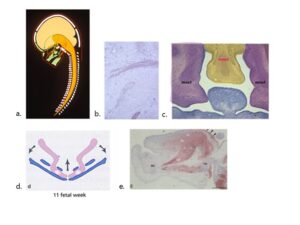
Figure 1: Overview of the maxillary development before ossification. A: Schematic drawing demonstrating cell migration (green arrows) from the neural crest of the brainstem (orange) to the maxilla. The craniofacial skeleton is marked green. The brain hemispheres are marked yellow and the red lines indicate ectoderme. The white structures indicate other bones in the body axis; B: Histological section demonstrating early neural cell migration to the maxilla. The dark red structures visualize maxillary nerves; C: Histological frontal section of the craniofacial region from a human fetus 10 weeks of Gestation colored blue for demonstrating the tongue. The yellow color in this section demonstrates the fronto-nasal neural crest tissue in the mid-axial field (fnncf) and the purple tissue illustrates the bilateral maxillary neural crest fields (mncf); D: Drawing demonstrating muscular movements (arrows) in the developing mandible during the elevation of the vertical maxillare processes (see c) resulting in palate formation in a human fetus 11 weeks of Gestation. The purple color demonstrates the Meckel cartilage and the blue color the early bone formation in the mandible. E: Histological sagittal section in which the muscles are marked red in a human fetus 12 weeks of Gestation. Anterior direction is toward left. Mc indicates the mid-axial mandibular cartilage and bone and Hy the mid-axial Hyoid cartilage and bone. Two bundles of muscular fibers are demonstrated. A horizontal bundle between Mc and HY, called musculus genio hyoideus and a bundle of muscular fibers called musculus genio-glossus from Mc towards the muscular fibers in the tongue. The contraction of these muscular bundles draws the tongue backwards, resulting in a change in tongue morphology (arrows) and creating the space necessary for the maxillary processes to elevate and form the palate.
Bone Development of the Maxilla
The bony development of the claviculae are the first bones to ossify in the human body, shortly followed by initial ossification of the mandible and the maxilla. The bony formation of the maxilla starts in the canine regions and palatine regions (Fig. 2) and develops intra-membranously [6]. Ossification of the palate occur in spiculae radiating out from the canine areas and palatine areas (Fig. 2). Postnatal maxillae are visualized anthropologically in Fig. 3. A scanning image of a body plane from a patient 12 years of age demonstrates the maxilla with one central incisor (Fig 3). This condition called SMMCI (Single Median Maxillary Central Incisor) is described in the pathological section of this article.
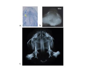
Figure 2: Overview of stages in early bone formation of the human fetal maxilla. A: Horizontal section of a human prenatal maxilla about 12 ½ weeks of Gestation The blue line in the center of the figure indicates where the maxillary processes have fused during palate formation. The blue arrow indicates early bone formation in the later canine region close to the maxillary nerve marked with a yellow arrow. The yellow circles mark the location of the two mid-axial naso-palatine nerves anteriorly in the section. Posteriorly in the section the bilateral palatal nerves are marked. Red arrow indicates the future growth direct of the maxilla; B: Radiograph taken by a faxitron radiographic apparatus visualizes the first osseous contours in the human maxilla and palatal bone at the Gestation age 12 ½ observed in a horizontal view. A dark midline shadow indicates that the posterior part of the palate (lower part in the figure) has not completely closed. In the upper part of the figure the white bony contours of the bilateral maxillary bone components appear and in the lower part of the figure the white bony contours indicate the ossification of the early palatal bones; C: Radiograph taken by a faxitron radiographic apparatus visualizing the maxillary bony appearance at 16 weeks of Gestation observed in a horizontal view. The upper part of the figure is the anterior direction. Note that the mid-palatal suture and the transverse palatal transversal suture have formed. Also, bony structures are seen in the palate radiating out from the bilateral canine areas.
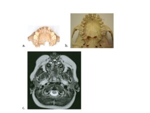
Figure 3: Postnatal human maxillae. A: Occlusal view of an anthropological maxilla from a child approximal 4 years old. A square part of the palatal bone forming the posterior part of the osseous palate is missing; B: Occlusal view of an anthropological maxilla from an adult individual; C: Scanning image at the body plane where the maxilla can be studied. In the figure the maxilla appears in the upper third of the image. The indication for scanning this patient 12 years of age with one single maxillary central incisor (arrow) was to examine the patient for a possible brain malformation.
Innervation and Developmental fields in the Maxilla
Cell migration from different regions on the neural crest were spread in different craniofacial locations so-called fields illustrated in Fig. 4. The neural crest field forming the maxilla are named the fronto-nasal field, the maxillary field and the palatal field (Fig. 4) [1]. Each field has a different main nerve supply (Fig. 4). The fields extend from the surface of the head to the pituitary gland/sella turcica (Fig. 4). Within each field different calcified structures can be analyzed as exemplified in the right figure belong to Fig. 4.
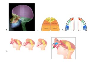
Figure 4: Overview of the three maxillary fields, the fronto-nasal fields, the maxillary field and the palatal field observed in three dimensions. A: In a profile radiograph from an individual 13 years of age different developmental regions are marked by colors. The three maxillary fields inserted are: the naso-frontal field, yellow, the maxillary field, red and the mandibular fields, orange; B: A drawing of the maxilla demonstrates the tree maxillary fields observed in the occlusal view. Note the borderlines between the fields and the individual teeth belonging to each field; C: Innervation of the maxilla is marked on this drawing. The incisors are innervated from the bilateral naso-frontal nerves (red). The canines and premolars are innervated from the bilateral maxillary nerves ( green) and the molars from the bilateral palatal nerves (blue); D: The developmental fields extend from the surface three-dimensionally to the sella turcica/ pituitary gland. This is demonstrated in 3 small figures. The figure to the right is a magnification of the fronto-nasal field exemplifying the hard tissue structures which can be observed within this field. These are the crista gally the nasal bone the anterior part of the maxilla the incisors and the anterior wall of the sella turcica.
Tooth Formation and Development of the Alveolar Process
The primary dentition develops early from the dental lamina (Fig. 5). The tooth buds consist before gestional age 20 weeks of early dentine and enamel in the coronal part of the surrounding dental follicle which is highly innervated in the later apically region (Fig. 5) [14,15]. The alveolar processes develop after the tooth buds have been formed [16]. In the region of the primary canine and in the first primary molar region the facial alveolar process is late in development. The permanent tooth buds develop from the dental lamina after gestional age 20 weeks. Tooth eruption depends on the crown follicle and then the periroot sheet and the apical innervation [17-20].
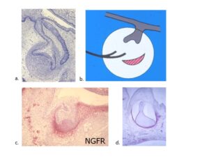
Figure 5: Tissue types involved in early human tooth formation. A: Histological section from a human fetus 10 weeks of Gestation demonstrating that the oral mucosa forms the shape of the early primary tooth bud surrounded by mesenchymal tissue. At a later stage the oral mucosa also forms the permanent tooth bud; B: Schematic drawing of the three tissue types involved in early tooth formation. These tissues are: Oral mucosa from the ectoderm marked grey, ectomesenchyme marked red from the neuroectoderm and the peripheral nerves marked black; C: Histological section colored with NGFR (nerve-growth factor receptor.) demonstrates positive red reaction for nerve tissue in the apical region of a tooth bud; D: This section demonstrates a tooth germ from a human fetus 2 weeks of Gestation colored with NGFR. The red color indicates the apical part of the tooth bud before dentine formation the odontoblasts also react positively for NGFR.
Maxillar Foramina and Canal
Foramina
The incisive foramen. This foramen encloses the bilateral naso-palatine/incisor nerves in the naso-frontal field (Fig. 6) [1]. The infra-orbital foramen. This foramen develops initially between the tooth buds of the primary canine and the first primary molar. The bony tissue gradually encircling the infra-orbital. The distance between the infra orbital foramen close to the primary teeth enlarges during vertical growth of the maxilla (Fig. 6) [15-18].
The palatal foramen. The palatal foramen is formed between the horizontal part of the palatal bone and the posterior part of the maxillary alveolar process (Fig 6). During growth the maxillary alveolar process moves in the anterior direction resulting in a change in the position of the palatal foramen from a location close to the first permanent molar and later close to the second permanent maxillary molar (Fig. 6) [1].
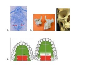
Figure 6: Maxillary foramina and canals, important for understanding maxillary growth patterns. A: The incisive foramen has developed around two bilateral naso-palatine nerves. The nerves marked with red arrows are always present before the foramen develops. By bone apposition an incisive canal develops in a vertical direction; B: The bilateral infra-orbital foramen at age 4 and 17 (left) have developed early in the canine regions by bone encircling the infra-orbital nerves. From these foramina the infra-orbital canals develop by bone apposition. The anterior and vertical maxillary growth can be visualized by a pin inserted in the canal (right); C: The palatine foraminae have developed by bone encircling the palatine nerves. The foraminae are located at the edges of the horizontal process of the palatine bone, which ensures a sliding anterior growth movement, when the maxillary bone marked green moves forward. The entrance to the pterygo-maxillary canal stays stable in the palatine bone marked red. The activity in the Trans-Palatal Suture (TPS) is larger in the maxillary bone compared to the palatal bone, At the bilateral Tuber Maxillae (TM) the alveolar process enlarges by bone apposition (white area).
Canals
Appositional growth changes the holes to channels during growth. This results in the formation of the incisive, infra orbital canals and the palatine canals. The direction of the canals reflects the direction of the maxillary growth Fig. 6 right.
Growth of the Maxilla
Growth of the maxilla occurs in the sutures and by bone apposition [21-30].
Sutures
Intermaxillary suture is located midaxially, not only in the area of the fused midpalatal soft tissue processes (maxillary field) and between the palatal soft tissue process of the palatal bone (palatal field) but also anteriorly in the frontonasal region between the central incisors (Fig 7).
Transpalatal suture is developed between the maxillary and palatal soft tissue processes after the palate has formed Fig. 6 and Fig. 7.
The sutural growth in the transversal direction occurs in the midpalatal suture. Growth in the anterior and downward direction occurs in the transpalatal suture Fig. 7.
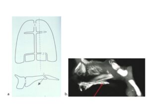
Figure 7: The palatal sutures and incisive fissure. A: Two drawings illustrating the mid-palatal suture, marked 1 and the transpalatal suture marked 2 in the horizontal plane (upper drawing) and the transpalatal suture is also demonstrated in the vertical plane. The Incisive fissure is marked 3. The alveolar process is only interrupted in the region of the central incisors, where the term maxillary interincisive suture is used at a later developmental stage; B: The lower figure illustrates a radiograph of a mid-sagittal section through the cranial part of the craniofacial skeleton. Note the palate where the red arrow illustrates the transpalatal suture between the maxillary and palatal part of the palate. Growth in this suture explains the downwards and forwards growth of the maxilla. This growth direction is illustrated by an arrow in the lover part of the upper figure a.
Fissure
Incisive fissure stretches from the midpalatal suture in a 90-degree angle to the primary canines Fig 7. The fissure is not a suture because this bony structure ends blind og does not cross over the alveolar process. The fissure is not a borderline structure between the frontonasal field and the maxillary field. The fissure arises in connection with the growth of the incisors. The fissure and the intermaxillary suture create the local space necessary for the enlarging permanent incisors.
Bone Apposition
Growth by apposition occur at the tuber maxillae and in the alveolar processes.
Pathological development of the human maxilla.
The phenotypic variability of the pathological maxilla is exemplified in five pathological cases.
Example 1. Cleft lip and palate occur in three phenotypes [32-36].
Cleft Lip: Unilateral or bilaterally cleft occur between the frontonasal field and the maxillary field (Fig. 8). These clefts are associated with short nasal bones indicating that the neural crest cells in the frontonasal fields are malformed or delate during development. The frontonasal field including the nasal bones is demonstrated on an anthropological cranium in Fig 8. Cleft Palate: This cleft arises between the soft tissue palatal shelves which during development did not fuse (Fig. 8). The nasal bones are seemingly normal in this condition which indicate abnormal development of the maxillary and palatal fields. Combined cleft lip and palate. This type of cleft is the most severe type which is associated with retrognatia of the mandible and late closure of the synchondrosis spheno-basilaris Fig. 8 center.
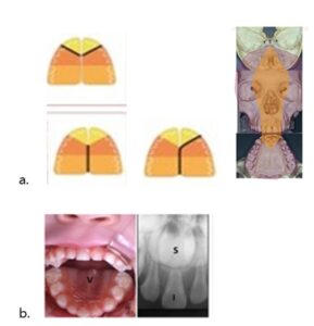
Figure 8: Overview of different malformations in the human maxilla. A: In the left half of this figure drawings of the palatal view of the developmental fields in three maxillae are demonstrated. The black lines drawn between fields in the upper left figure demonstrate that the frontonasal fields are involved in bilateral cleft lip and in the lower left figure that the maxillary and palatal fields are involved in isolated cleft palate. In the center figure all fields are involved in unilateral cleft lip and palate. In the right half of this figure the extension of the frontonasal field in palatal, facial and intracranial regions is marked yellow on a human, anthropological cranium; B: To the left is an intraoral photo, supplemented by a palatal view observed in a mirror from a child 5 years of age. Note the single primary central incisor and palatal ridge marked v. To the right an intra oral radiograph of the same child demonstrates that not only the primary incisor marked I but also the permanent Incisor S appear in the midline. An interincisal suture is not seen. The patient demonstrated in b is a case diagnosed with SMMCI, (Single Median Maxillary Central Incisor). In this condition the median part of the nasofrontal field is missing. This malformation is associated with a short interoccular distance, absence of the interincisal suture, abnormal lip contour and of absence of the suo-prior frenulum labii superior and of the incisive papilla, lip contour; C: Frontal view of an anthropological cranium from a newborn child only the mid-axial nasofrontal part (field) appears. (The yellow part of the cranium in Fig. 4); D: Frontal view of an anthropological cranium from a newborn child absence of the lower part of the fronto-nasal field is missing. (The yellow part of the cranium in Fig. 4); E: Lateral view of a narrow and long human cranium from a newborn child (orange part of the cranium seen in Figure 4a. The cranium demonstrates absence of the zygomatic bone, the lateral part of the maxillary bone (red area in Fig. 4) and also the posterior part of the palatal bone.
Example 2. Single medial maxillary central incisor in the malformed fronto-nasal field [37-45].
Single central incisor. This malformation is associated with lip and palatal malformation but often first diagnosed when the single central primary incisor erupts Fig. 8. This malformation is caused by a reduction or narrowness in the fronto-nasal field. The mid-axially permanent incisor is malformed seemingly by fusion (or not separation) of the two distal parts of the permanent central incisors Fig. 8. The incisive papilla and the inter incisive suture are missing in this condition and a tissue ridge appears in the center of the palate Fig. 8.
Example 3. Absence of maxillary developmental field [46].
This condition is observed in a frontal view where only the nasopalatine field appears in a human cranium of a newborn child Fig. 8.
Example 4. Median cleft in the fronto-nasal process [46].
This abnormality is part of the holoprosencephalic spectrum, often associated with abnormal development of the frontal lope of the brain. The incisors are not present. The condition has different genotypes. An anthropological case is demonstrated Fig. 8d. In this condition the fronto nasal field has not developed normally.
Example 5. Malformation or absence of the maxillary and palatine processes [46].
This maxillary malformation has only been observed in an anthropological cranium demonstrated in Fig. 8.
Dental abnormalities in the human maxilla. Several publications on tooth abnormalities in the maxilla have been published [47-65]. Of these 4 examples are highlighted in Fig. 9 the text, illustrating tooth agenesis, tooth malformation, ectopic eruption and pathological root resorption.
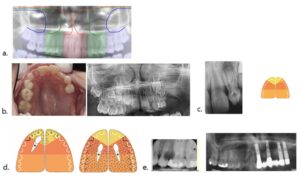
Figure 9: A: Demonstrates the maxillary part of an orthopantomogram from a normally developed young adult. In this radiograph the peripheral nerves to the different maxillary developmental fields are schematically marked. Red, incisor field, green premolar/canine field and blue molar field; B: demonstrates a clinical photo with absence of molars and one second premolar in the left side of the maxilla from a 9-year-old child. The radiograph next to this photo proves that absence of teeth is inborn predominantly in the molar field; C: demonstrates a malformed lateral incisor Fig. 9 left, which might be associated with malformation at the borderline between the fronto-nasal and the maxillary fields Fig. 9 right; D: These drawings illustrate unilateral and bilateral canine ectopia and the association between tooth malformation and abnormal tooth eruption; E: These radiographs demonstrate pathological resorption seemingly due to neurogen virus infection. This resorption gradually extended along the nerves to the left half of the maxilla, but did not pass over the maxillary midline. All teeth in the left of the maxilla were extracted and replaced by dental implants.
Conclusion
This overview illustrates the importance of analyzing prenatal human material for understanding normal and abnormal maxillary and dental development based on neural crest fields. Furthermore, the studies focus on how the peripheral nervous system influences bone growth, tooth formation and tooth eruption, which creates a new scientific approach to future craniofacial analysis, described before [66-69].
Conflict of Interest
There are no potential conflicts of interest to declare in this paper.
Acknowledgement
Thanks to all colleagues mentioned in the list of references for collaboration through many years on scientific craniofacial projects and thanks to the departmental secretary Linda Michelsen for preparation of this manuscript.
Funding
The author reported that there is no funding associated with publication of this article.
Author Contributions
All authors contributed equally for this paper.
References
- Kjær I. Etiology-based dental and craniofacial diagnostics. Wiley Blackwell. 2017.
- Kjær I. Neuroosteologi – a discipline of importance for evaluation of human craniofacial development. EC Neurology. 2018;10.6.
- Kjær I. Human prenatal craniofacial development related to brain development under normal and pathologic conditions. Acta Odontol Scand. 1995;53:135-43.
- Kjær I. Orthodontics and foetal pathology: a personal view on craniofacial patterning. Eur J Orthod. 2010;32:140-7.
- Kjær I. Neuro-Orthodontics – a sub-speciality in orthodontics, important for diagnostics and treatment planning. E.C. Dental Science. 2020;19.10.
- Kjær I. Prenatal skeletal maturation of the human maxilla. J Craniofac Genet Dev Biol. 1989;9:257-64.
- Kjær I. Human prenatal palatal closure related to skeletal maturity of the jaws. J Craniofac Genet Dev Biol. 1989;9:265-70.
- Kjær I. Correlated appearance of ossification and nerve tissue in human fetal jaws. J Craniofac Genet Dev Biol. 1990;10:329-36.
- Kjær I. Human prenatal palatal shelf elevation related to craniofacial skeletal maturation. Eur J Orthod. 1992;14:26-30.
- Kjær I, Bach Petersen S, Græm N, Kjær T. Changes in human palatine bone location and tongue position during prenatal palatal closure. J Craniofac Genet Dev Biol. 1993;13:18-23.
- Takashi O, Hansen BF, Nolting D, Kjær I. Nerve growth factor receptor immunolocalization during human palate and tongue development. Cleft Palate-Craniofacial J. 2003;40:116-25.
- Kjær I. Mandibular movements during elevation and fusion of palatal shelves evaluated from the course of Meckel´s cartilage. J Craniofac Genet Dev Biol. 1997;17:80-5.
- Kjær I. The human mandible – embryological anatomical and pathological aspects. Ann Otolaryngol Rhinol. 2024;11:1335.
- Christensen LR, Møllgård K, Kjær I, Janas MS. Immunocytochemical demonstration of Nerve Growth Factor Receptor (NGF-R) in developing human fetal teeth. Anat Embryol. 1993;188:247-55.
- Christensen LR, Janas MS, Møllgård K, Kjær I. An immunocytochemical study of the innervation of developing human fetal teeth using protein gene product 9.5 (PGP 9.5). Arch Oral Biol. 1993;38:1113-20.
- Kjær I, Bagheri A. Prenatal development of the alveolar bone of human deciduous incisors and canines. J Dent Res. 1999;78:667-72.
- Kjær I, Nolting D. The human periodontal membrane – focusing on the spatial interrelation between the epithelial layer of Malassez, fibers and innervation. Acta Odontol Scand. 2009;67:134-8.
- Kjær I. Mechanism of Human Tooth Eruption: Review Article Including a New Theory for Future Studies on the Eruption Process. Scientifica. 2014.
- Kjær I, Kocsis G, Nodal M, Christensen LR. Aetiological aspects of mandibular tooth agenesis – focusing on the role of nerve, oral mucosa and supporting tissues. Eur J Orthod. 1994;16:371-5.
- Kock M, Nolting D, Kjaer KW, Hansen BF, Kjær I. Immunohistochemical expression of p63 in human prenatal tooth primordia. Acta Odont Scand. 2005;63:253-7.
- Caspersen LM, Christensen IJ, Kjær I. Inclination of the infraorbital canal studied on dry skulls expresses the maxillary growth pattern: a new contribution to the understanding of change in inclination of ectopic canines during puberty. Acta Odontol Scand. 2009;67:341-5.
- Kjær I Arvedsen K, Danielsen J. Can overlap of dermatome-like fields in the maxillary canine region explain canine transposition and canine agenesis? Dental Hypotheses. 2018;9:64-7.
- Kjær I. Can the location of tooth agenesis and the location of initial bone loss seen in juvenile periodontitis be explained by neural developmental fields in the jaws? Acta Odontol Scand. 1997;55:70-2.
- Kjær I. Neuro-osteology. Crit Rev Oral Biol Med. 1998;9:224-44.
- Caspersen LM, Christensen IJ, Kjær I. Inclination of the infraorbital canal studied on dry skulls expresses the maxillary growth pattern: a new contribution to the understanding of change in inclination of ectopic canines during puberty. Acta Odontol Scand. 2009;67:341-5.
- Njio B, Kjær I. The development and morphology of the incisive fissure and the transverse palatine suture in the human fetal palate. J Craniofac Genet Dev Biol. 1993;13:24-34.
- Sejrsen B, Kjær I, Jakobsen J. The human incisal suture and premaxillary area studied on archeologic material. Acta Odontol Scand. 1993;51:143-51.
- Silau AM, Njio B, Solow B, Kjær I. Prenatal sagittal growth of the osseous components of the human palate. J Craniofac Genet Dev Biol. 1994;14:252-6.
- Sejrsen B, Kjær I, Jakobsen J. Human palatal growth evaluated on medieval crania using nerve canal openings as references. Am J Phys Anthropol. 1996;99:603-11.
- Damgaard C, Caspersen LM, Kjær I. Maxillary sagittal growth evaluated on dry skulls from children and adolescents. Acta Odontol Scand. 2011;69:274-8.
- Mølsted K, Kjær I, Dahl E. Cranial base in newborns with complete cleft lip and palate: Radiographic study. Cleft Palate-Craniofacial J. 1995;32:199-205.
- Lisson JA, Kjær I. Location of alveolar clefts relative to the incisive fissure. Cleft Palate Craniofac J. 1997;34:292-6.
- Hansen L, Nolting D, Holm G, Hansen BF, Kjær I. Abnormal vomer development in human fetuses with isolated cleft palate. Cleft Palate-Craniofacial J. 2004;41:470-3.
- Lauridsen H, Hansen BF, Reintoft I, Keeling JW, Skovgaard LT, Kjær I. Short hard palate in prenatal trisomy 21. Orthod Craniofacial Res. 2005;8:91-5.
- Nielsen BW, Mølsted K, Kjær I. Maxillary and sella turcica morphology in newborns with cleft lip and palate. Cleft Palate-Craniofac J. 2005;42:610-7.
- Riis LC, Kjær I, Mølsted K. Dental anomalies in different cleft groups related to neural crest developmental fields contributes to the understanding of cleft aetiology. J Plast Surg Hand Surg. 2014;48:126-31.
- Kjær I, Keeling JW, Græm N. The midline craniofacial skeleton in holoprosencephalic fetuses. J Med Genet. 1991;28:846-55.
- Sandikcioglu M, Mølsted K, Kjær I. The prenatal development of the human nasal and vomeral bones. J Craniofac Genet Dev Biol. 1994;14:124-34.
- Kjær I, Keeling J, Russell B, Daugaard-Jensen J, Hansen BF. Palate structure in human holoprosencephaly correlates with the facial malformation and demonstrates a new palatal developmental field. Am J Med Genet. 1997;73:387-92.
- Kjær I, Becktor KB, Lisson J, Gormsen C, Russell BG. Face, palate and craniofacial morphology in patients with a solitary median maxillary central incisor. Eur J Orthod. 2001;23:63-73.
- Becktor KB, Sverrild, Pallisgaard C, Burhøj J, Kjær I. Eruption of the central incisor, the intermaxillary suture and maxillary growth in patients with a single median maxillary central incisor, SMMCI. Acta Odontol Scand. 2001;59:361-6.
- Kjær I, Keeling JW, Hansen BF, Becktor KB. Midline skeletodental morphology in holoprosencephaly. Cleft Palate-Craniofacial J. 2002;39:357-63.
- Tabatabaie F, Sonnesen L, Kjær I. The neurocranial and craniofacial morphology in children with solitary median maxillary central incisor (SMMCI). Orthod Craniofac Res. 2008;11:96-104.
- Kjær I, Wagner Aa, Thomsen LL, Holm K. Brain malformation in Single Median Maxillary Central Incisor Neuropediatrics. 2010;40:280-3.
- Kjær I, Balslev-Olesen M. The primary maxillary central incisor in the Solitary Median Maxillary Central Incisor Syndrome. Eur J Paed Dent. 2012;13:73-5.
- Kjær I, Marin A, Meyer I. Human malformed prenatal anthropological crania contribute to new insight in the extension of bone malformation in cranial development. Fetal and pediatric pathology. 2024;2024:1-13.
- Nodal M, Kjær I, Solow B. Craniofacial morphology in patients with multiple congenitally missing permanent teeth. Eur J Orthod. 1994;16:109-9.
- Sejrsen B, Kjær I, Jakobsen J. Agenesis of permanent incisors in a medieval maxilla and mandible: aetiological aspects. Eur J Oral Sci. 1995;103:65-9.
- Bang E, Kjær I, Christensen LR. Etiologic aspects and orthodontic treatment of unilateral localized arrested tooth development combined with hearing loss. Am J Orthod Dentofac Orthop. 1995;108:154-61.
- Becktor KB, Reibel J, Vedel B, Kjær I. Segmental odontomaxillary dysplasia: clinical, radiological and histological aspects of four cases. Oral Dis. 2002;8:106-10.
- Partner ET, Heidmann JM, Kjær I, Væth M, Poulsen S. Biological interpretation of the correlation of emergence times of permanent teeth. J Dent Res. 2002;81:451-4.
- Hansen L, Kjær I. A premaxilla with a supernumerary tooth indicating a developmental region with a variety of dental abnormalities: a report of nine cases. Acta Odontol Scand. 2004;62:30-6.
- Larsen HJ, Sørensen HB, Artmann L, Christensen IJ, Kjær I. Sagittal, vertical and transversal dimensions of the maxillary complex in patients with ectopic maxillary canines. Orthod Craniofac Res. 2010;13:34-9.
- Caspersen L, Christensen IJ, Kjær I. Maxillary canine ectopia and maxillary canine-premolar transposition are associated with deviations in the maxilla. Dent Anthropol. 2010;23:37-41.
- Kjær I. New diagnostics of the dentition on panoramic radiographs – focusing on the peripheral nervous system as an important aetiological factor behind dental anomalies. Orthodontic Waves. 2012;71:1-16.
- Kjær I, Strøm C, Worsaae N. Regional aggressive root resorption caused by neuronal virus infection. Case Reports in Dentistry. Volume 2012.
- Kenrad A, Christensen IJ, Kjær I. Craniofacial Morphology of the frontonasal segment in patients with one or two macrodontic maxillary central incisors. Eur J Orthod. 2013;35:329-34.
- Hørberg M, Lauesen S.R. Daugaard-Jensen J, Kjær I. Linear scleroderma en coup de sabre including abnormal dental development. Eur Arch Paediatr Dent. 2015;5:16:227-31.
- Danielsen JC, Karimian K, Ciarlantini R, Kjær I. Unilateral and bilateral dental transposition in the maxilla – dental and skeletal findings in 63 individuals. Eur Arch Paediatr Dent. 2015;16:467-76.
- Kenrad AB, Kjær I. The interrelationship between permanent maxillary incisors and neuropsychiatric conditions. Neuropsychiatry. 2016;6.
- Arvedsen KP, Kjær I. Dental and craniofacial findings in 91 patients with agenesis of permanent maxillary canines. Eur Arch Pediatr Dent. 2017;18:243-50.
- Shirazi Z, Kjær I. Is the etiology behind palatal uni-lateral and palatal bi-lateral maxillary canine ectopia different? Dental Hypotheses. 2018;9:3-10.
- Lauesen SR, Daugaard-Jensen J, Lauridsen E, Kjær I. Localized scleroderma en coup de sabre affecting the skin, dentition and bone tissue within craniofacial neural crest fields: Clinical and radiographic study of six patients. Eur Arch Pediatr Dent. 2019;20:339-50.
- Kjær I. Morphological characteristics of dentitions developing excessive root resorption during orthodontic treatment. Eur J Orthod. 1995;16:25-34.
- Kjær I. Regional aggressive cervical root resorption in a young female adult. J Clinical and Medical Images, Case Reports. 2023:3.
- Kjær I. Review: Dental approach to craniofacial syndromes: how can developmental fields show us a new way to understand pathogenesis? Int J Dent. 2012;2012:145749.
- Kjær I. Dental research needs a new way of thinking. Dental Hypotheses. 2014:5:131-2.
- Kjær I. How can cranial bones and teeth in children with craniofacial anomalies indicate disturbances in the brain and cranial nerves? Ann Pediatr Child Health. 2020;8:1188.
- Kjær I. Human and animal studies in craniofacial embryology enrich human postnatal craniofacial insight differently. Dent Oral Biol Craniofac Res. 2021.
Article Type
Review Article
Publication History
Received Date: 05-12-2024
Accepted Date: 24-12-2024
Published Date: 31-12-2024
Copyright© 2024 by Kjær I, et al. All rights reserved. This is an open access article distributed under the terms of the Creative Commons Attribution License, which permits unrestricted use, distribution, and reproduction in any medium, provided the original author and source are credited.
Citation: Kjær I. The Human Maxilla: The Embryological and Anatomical Background for Understanding Pathological Development of the Maxilla. J Dental Health Oral Res. 2024;5(3):1-13.

Figure 1: Overview of the maxillary development before ossification. A: Schematic drawing demonstrating cell migration (green arrows) from the neural crest of the brainstem (orange) to the maxilla. The craniofacial skeleton is marked green. The brain hemispheres are marked yellow and the red lines indicate ectoderme. The white structures indicate other bones in the body axis; B: Histological section demonstrating early neural cell migration to the maxilla. The dark red structures visualize maxillary nerves; C: Histological frontal section of the craniofacial region from a human fetus 10 weeks of Gestation colored blue for demonstrating the tongue. The yellow color in this section demonstrates the fronto-nasal neural crest tissue in the mid-axial field (fnncf) and the purple tissue illustrates the bilateral maxillary neural crest fields (mncf); D: Drawing demonstrating muscular movements (arrows) in the developing mandible during the elevation of the vertical maxillare processes (see c) resulting in palate formation in a human fetus 11 weeks of Gestation. The purple color demonstrates the Meckel cartilage and the blue color the early bone formation in the mandible. E: Histological sagittal section in which the muscles are marked red in a human fetus 12 weeks of Gestation. Anterior direction is toward left. Mc indicates the mid-axial mandibular cartilage and bone and Hy the mid-axial Hyoid cartilage and bone. Two bundles of muscular fibers are demonstrated. A horizontal bundle between Mc and HY, called musculus genio hyoideus and a bundle of muscular fibers called musculus genio-glossus from Mc towards the muscular fibers in the tongue. The contraction of these muscular bundles draws the tongue backwards, resulting in a change in tongue morphology (arrows) and creating the space necessary for the maxillary processes to elevate and form the palate.

Figure 2: Overview of stages in early bone formation of the human fetal maxilla. A: Horizontal section of a human prenatal maxilla about 12 ½ weeks of Gestation The blue line in the center of the figure indicates where the maxillary processes have fused during palate formation. The blue arrow indicates early bone formation in the later canine region close to the maxillary nerve marked with a yellow arrow. The yellow circles mark the location of the two mid-axial naso-palatine nerves anteriorly in the section. Posteriorly in the section the bilateral palatal nerves are marked. Red arrow indicates the future growth direct of the maxilla; B: Radiograph taken by a faxitron radiographic apparatus visualizes the first osseous contours in the human maxilla and palatal bone at the Gestation age 12 ½ observed in a horizontal view. A dark midline shadow indicates that the posterior part of the palate (lower part in the figure) has not completely closed. In the upper part of the figure the white bony contours of the bilateral maxillary bone components appear and in the lower part of the figure the white bony contours indicate the ossification of the early palatal bones; C: Radiograph taken by a faxitron radiographic apparatus visualizing the maxillary bony appearance at 16 weeks of Gestation observed in a horizontal view. The upper part of the figure is the anterior direction. Note that the mid-palatal suture and the transverse palatal transversal suture have formed. Also, bony structures are seen in the palate radiating out from the bilateral canine areas.

Figure 3: Postnatal human maxillae. A: Occlusal view of an anthropological maxilla from a child approximal 4 years old. A square part of the palatal bone forming the posterior part of the osseous palate is missing; B: Occlusal view of an anthropological maxilla from an adult individual; C: Scanning image at the body plane where the maxilla can be studied. In the figure the maxilla appears in the upper third of the image. The indication for scanning this patient 12 years of age with one single maxillary central incisor (arrow) was to examine the patient for a possible brain malformation.

Figure 4: Overview of the three maxillary fields, the fronto-nasal fields, the maxillary field and the palatal field observed in three dimensions. A: In a profile radiograph from an individual 13 years of age different developmental regions are marked by colors. The three maxillary fields inserted are: the naso-frontal field, yellow, the maxillary field, red and the mandibular fields, orange; B: A drawing of the maxilla demonstrates the tree maxillary fields observed in the occlusal view. Note the borderlines between the fields and the individual teeth belonging to each field; C: Innervation of the maxilla is marked on this drawing. The incisors are innervated from the bilateral naso-frontal nerves (red). The canines and premolars are innervated from the bilateral maxillary nerves ( green) and the molars from the bilateral palatal nerves (blue); D: The developmental fields extend from the surface three-dimensionally to the sella turcica/ pituitary gland. This is demonstrated in 3 small figures. The figure to the right is a magnification of the fronto-nasal field exemplifying the hard tissue structures which can be observed within this field. These are the crista gally the nasal bone the anterior part of the maxilla the incisors and the anterior wall of the sella turcica.

Figure 5: Tissue types involved in early human tooth formation. A: Histological section from a human fetus 10 weeks of Gestation demonstrating that the oral mucosa forms the shape of the early primary tooth bud surrounded by mesenchymal tissue. At a later stage the oral mucosa also forms the permanent tooth bud; B: Schematic drawing of the three tissue types involved in early tooth formation. These tissues are: Oral mucosa from the ectoderm marked grey, ectomesenchyme marked red from the neuroectoderm and the peripheral nerves marked black; C: Histological section colored with NGFR (nerve-growth factor receptor.) demonstrates positive red reaction for nerve tissue in the apical region of a tooth bud; D: This section demonstrates a tooth germ from a human fetus 2 weeks of Gestation colored with NGFR. The red color indicates the apical part of the tooth bud before dentine formation the odontoblasts also react positively for NGFR.

Figure 6: Maxillary foramina and canals, important for understanding maxillary growth patterns. A: The incisive foramen has developed around two bilateral naso-palatine nerves. The nerves marked with red arrows are always present before the foramen develops. By bone apposition an incisive canal develops in a vertical direction; B: The bilateral infra-orbital foramen at age 4 and 17 (left) have developed early in the canine regions by bone encircling the infra-orbital nerves. From these foramina the infra-orbital canals develop by bone apposition. The anterior and vertical maxillary growth can be visualized by a pin inserted in the canal (right); C: The palatine foraminae have developed by bone encircling the palatine nerves. The foraminae are located at the edges of the horizontal process of the palatine bone, which ensures a sliding anterior growth movement, when the maxillary bone marked green moves forward. The entrance to the pterygo-maxillary canal stays stable in the palatine bone marked red. The activity in the Trans-Palatal Suture (TPS) is larger in the maxillary bone compared to the palatal bone, At the bilateral Tuber Maxillae (TM) the alveolar process enlarges by bone apposition (white area).

Figure 7: The palatal sutures and incisive fissure. A: Two drawings illustrating the mid-palatal suture, marked 1 and the transpalatal suture marked 2 in the horizontal plane (upper drawing) and the transpalatal suture is also demonstrated in the vertical plane. The Incisive fissure is marked 3. The alveolar process is only interrupted in the region of the central incisors, where the term maxillary interincisive suture is used at a later developmental stage; B: The lower figure illustrates a radiograph of a mid-sagittal section through the cranial part of the craniofacial skeleton. Note the palate where the red arrow illustrates the transpalatal suture between the maxillary and palatal part of the palate. Growth in this suture explains the downwards and forwards growth of the maxilla. This growth direction is illustrated by an arrow in the lover part of the upper figure a.

Figure 8: Overview of different malformations in the human maxilla. A: In the left half of this figure drawings of the palatal view of the developmental fields in three maxillae are demonstrated. The black lines drawn between fields in the upper left figure demonstrate that the frontonasal fields are involved in bilateral cleft lip and in the lower left figure that the maxillary and palatal fields are involved in isolated cleft palate. In the center figure all fields are involved in unilateral cleft lip and palate. In the right half of this figure the extension of the frontonasal field in palatal, facial and intracranial regions is marked yellow on a human, anthropological cranium; B: To the left is an intraoral photo, supplemented by a palatal view observed in a mirror from a child 5 years of age. Note the single primary central incisor and palatal ridge marked v. To the right an intra oral radiograph of the same child demonstrates that not only the primary incisor marked I but also the permanent Incisor S appear in the midline. An interincisal suture is not seen. The patient demonstrated in b is a case diagnosed with SMMCI, (Single Median Maxillary Central Incisor). In this condition the median part of the nasofrontal field is missing. This malformation is associated with a short interoccular distance, absence of the interincisal suture, abnormal lip contour and of absence of the suo-prior frenulum labii superior and of the incisive papilla, lip contour; C: Frontal view of an anthropological cranium from a newborn child only the mid-axial nasofrontal part (field) appears. (The yellow part of the cranium in Fig. 4); D: Frontal view of an anthropological cranium from a newborn child absence of the lower part of the fronto-nasal field is missing. (The yellow part of the cranium in Fig. 4); E: Lateral view of a narrow and long human cranium from a newborn child (orange part of the cranium seen in Figure 4a. The cranium demonstrates absence of the zygomatic bone, the lateral part of the maxillary bone (red area in Fig. 4) and also the posterior part of the palatal bone.

Figure 9: A: Demonstrates the maxillary part of an orthopantomogram from a normally developed young adult. In this radiograph the peripheral nerves to the different maxillary developmental fields are schematically marked. Red, incisor field, green premolar/canine field and blue molar field; B: demonstrates a clinical photo with absence of molars and one second premolar in the left side of the maxilla from a 9-year-old child. The radiograph next to this photo proves that absence of teeth is inborn predominantly in the molar field; C: demonstrates a malformed lateral incisor Fig. 9 left, which might be associated with malformation at the borderline between the fronto-nasal and the maxillary fields Fig. 9 right; D: These drawings illustrate unilateral and bilateral canine ectopia and the association between tooth malformation and abnormal tooth eruption; E: These radiographs demonstrate pathological resorption seemingly due to neurogen virus infection. This resorption gradually extended along the nerves to the left half of the maxilla, but did not pass over the maxillary midline. All teeth in the left of the maxilla were extracted and replaced by dental implants.


