Shreya Thatte1*, Pragya Prakash2, Ishita Batra3, Lalita Porwal4, Anupriya Kesharwani5
1Professor and Head, Department of Ophthalmology, SriAurobindo Medical College and PG Institute (SAMC and PGI), Indore, India
2Assistant Professor, Department of Ophthalmology, SriAurobindo Medical College and PG Institute (SAMC and PGI), Indore, India
3Fellow in Sankara Eye Hospital, Indore, India
4Post Graduate Resident, Department of Pharmacology, SriAurobindo Medical College and PG Institute (SAMC and PGI), Indore, India
5Post Graduate Resident, Department of Ophthalmology, SriAurobindo Medical College and PG Institute (SAMC and PGI), Indore, India
*Correspondence author: Shreya Thatte, Professor and Head, Department of Ophthalmology, SriAurobindo Medical College and PG Institute (SAMC and PGI), Indore, India; Email: [email protected]
Published Date: 31-07-2024
Copyright© 2024 by Thatte S, et al. All rights reserved. This is an open access article distributed under the terms of the Creative Commons Attribution License, which permits unrestricted use, distribution, and reproduction in any medium, provided the original author and source are credited.
Abstract
Purpose: To compare the corneal endothelium changes in primary glaucoma patients on common combination of anti- glaucoma medications with healthy controls of the same age group.
Material and Methods: A case control study which was conducted from January 2022 for a period of 12 months on 100 patients. The patients with primary glaucoma above 40 years of age on same combination of anti-glaucoma medications Brimonidine (0.2%w/v) and Timolol (0.5%w/v) )and their age matched healthy control were included in the study. Detailed slit lamp evaluation of all the patients were performed including visual acuity, Static perimetry Intraocular Pressure (IOP) measurement, indirect gonioscopy and fundus evaluation with +90 D, similar tests were performed in normal age matched controls as well. Additionally, specular bio microscopy was performed and the following parameters were assessed and compared with age matched normal control group. Endothelial Cell Density (ECD), Percentage of hexagonal cells (6A), Central Corneal Thickness (CCT), Coefficient of Variation (CV) in cell area were determined. All examinations were performed by the same examiner to avoid biasing.
Result: It was observed that out of 50 glaucoma patients 36 patients (72%) were diagnosed as Primary Angle Closure Glaucoma (PACG) and 14 patients (28%) had Primary Open-Angle Glaucoma (POAG). Correlation between ECD loss, hexagonality, endothelial cell area and intraocular pressure was studied between case and control groups and glaucoma subgroups as well. IOP showed positive correlation with Coefficient of variation (CV) i.e.with increase in IOP value of CV increased . However, a negative correlation was found between IOP corneal ECD), hexagonality (6A) and CCT i.e., with increase in IOP the value of ECD ,6A and CCT decreased and vice versa. The average endothelial cell density was significantly decreased in glaucoma patients (1610 ± 225 cells/mm2) compared with the control group (2210 ± 236 mm2).
Conclusion: The present study, suggest that use of antiglaucoma medications (Brimonidine 0.2% and Timolol0.5%) affect the metabolism of the corneal endothelium and cause pathological changes. Hence for primary glaucoma patients, quantitative analysis of corneal endothelial cells morphology is necessary, despite the well-established benefits of antiglaucoma medications in reducing Intraocular Pressure (IOP), the potential impact of these drugs on the corneal endothelium remains a concern.
Keywords: Primary Glaucoma; Antiglaucoma Medications; Corneal Endothelium; Specular Microscopy; IOP
Introduction
The corneal monolayer endothelium maintains corneal clarity by actively regulating stromal hydration through its barrier and pump functions. Normal corneas lose 0.6% of Endothelial Cell Density (ECD)centrally per year [1]. Once ECD drops below a critical level, it is insufficient to maintain the appropriate corneal hydration status and corneal clarity. At the early stage of endothelial damage, neighboring cells spread and/or migrate to compensate for the cell loss, which results in an increase in cell size and/or alteration of cell shape. Progression of cell loss further compromises corneal transparency that causes corneal oedema and bullous keratopathy. Damage to the corneal endothelium is irreversible and leads to impaired visual acuity [1].
Loss of Corneal Endothelial Cells (CECs) occurs as a consequence of intraocular surgery, ocular trauma, ocular diseases like glaucoma, corneal endothelial dystrophy, intraocular inflammation and Systemic conditions like Diabetes, Grave’s disease, chronic kidney disease, tuberculosis, Marfan’s syndrome, etc [2-4]. One of the common ocular pathology causing endothelial cell loss is glaucoma.
Various factors are responsible for causing corneal endothelial damage in glaucoma, the disease itself cause direct-compression mechanism due to elevated Intraocular Pressure (IOP), additionaly after filtration surgery and aqueous shunt implantation [5,6]. Moreover, another important mechanism for endothelial damage in primary open angle and angle closure glaucoma is due to the use of antiglaucoma medications [7-10]. Despite the well-established benefits of glaucoma pharmacotherapy in reducing Intraocular Pressure (IOP), the potential impact of these drugs on the corneal endothelium remains a concern [11]. Different categories of antiglaucoma medications are available, out of them commonly used medications belong to alpha agonist and beta blocker groups [12,13].
The present study was planned to assess the effect of commonly used combination of anti-glaucoma drug (Brimonidine 0.2% and Timolol 0.5%) on corneal endothelium of primary glaucoma patients by specular bio microscopy which is one of the important non-invasive diagnostic modality to image the corneal endothelium and compare them with age matched healthy control [14].
Material and Methods
After approval from the institutional ethical committee the present comparative case control study was conducted on 100 patients: 50 primary glaucoma patients who visited the Department of Ophthalmology at tertiary eye care of central India with 50 age matched controls. Patients satisfying the inclusion and exclusion criteria were enrolled after taking informed consent. All patients were analyzed with regard to their pre-treatment characteristics and were on similar combination of Brimonidine (0.2% w/v) and Timolol (0.5% w/v) topical antiglaucoma medications for more than 3 months.
Inclusion Criteria
- Patients with primary glaucoma above 40 years
- Age matched individuals for control groups
Exclusion Criteria
- Pediatric patients
- Secondary glaucoma patients
- Patients who had undergone previous ocular surgery; ocular trauma; previous or present contact lens use; other ocular diseases (such as corneal endothelial dystrophy, intraocular inflammation and infection) that could affect the corneal endothelium
- Patients with Systemic diseases (such as diabetes) that could affect the corneal endothelium
- Patients who did not consented for the study were excluded
Methodology
Written informed consent was taken from all patients satisfying the inclusion and exclusion criteria. Patients were divided into 2 groups:
- Group A/Experimental Group (N=50): Patients with primary glaucoma on anti- glaucoma medications
- Group B/ Control Group (N=50): Age and sex matched patients without glaucoma
Detailed slit lamp evaluation of all 50 patients who presented to glaucoma clinic was performed along with specific history of duration of glaucoma and anti-glaucoma medications. The visual acuity was calculated using Snellen’s chart, Static perimetry using Humphrey’s field analyzer, indirect gonioscopy with Goldmann-3 mirror lens and fundus evaluation with +90 D was performed on all the patients to confirm the diagnosis and type of the glaucoma and similar tests were done in normal age matched control group.
IOP Evaluation
The Intraocular Pressure (IOP) of all subjects was measured using Goldmann Applanation Tonometer. The average of all measurements was determined and used for statistical analysis.
Specular Microscopy
Specular microscopy was performed on each subject. Images were captured from the central cornea and analyzed using EM- 4000 software (TOMEY) and the following parameters were assessed and compared with age matched normal control group. Endothelial Cell Density (ECD), Percentage of hexagonal cells (6A), Central Corneal Thickness (CCT), Coefficient of Variation (CV) in cell area were determined. All examinations were performed by the same examiner (Fig. 1-3, Table 1).
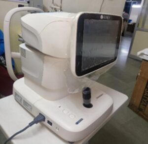
Figure 1: Goldmann applanation tonometer.
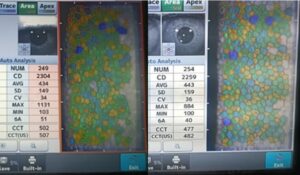
Figure 2: Specular microscopy.
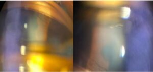
Figure 3: A: (Left) Open angle on gonioscopy; B: (Right) closed angle on gonioscopy.
|
Parameter |
Normal Value |
|
Endothelial cell density |
2000-3000 cells/mm220,21 |
|
Percentage of hexagonal cells (6A) |
46 ± 8%22 |
|
Central Corneal Thickness (CCT) |
554.78+/-32.6123 |
|
Coefficient of Variation (CV) |
0.22 – 0.3124-27 |
Table 1: Normal values in middle aged Indian population.
Statistical Analysis
Statistical analysis was performed using SPSS version 20.0 (SPSS, Chicago, IL, United States). Data are presented as mean ± SD. The unpaired Student’s t test was used to compare the means between normal subjects and patients with glaucoma. The Chi square test was applied to the comparison between untreated patients and patients receiving ophthalmic medication. Correlation between IOP and each characteristic of CECs was assessed by Pearson’s correlation coefficient. P-value less than 0.05 was considered statistically significant.
Results
In the present study majority of control group and glaucoma patients belonged to the elder age group i.e., 61-70 years (38%) as compared to only 17% in middle aged group of 40 to 50 years (Fig. 4).

Figure 4: a: Percentage of age distribution in glaucoma patients; b: Distribution of patients depending upon age in control group.
A slightly higher female preponderance was observed with 54% female and 46% male patients. Glaucoma patients were further divided into two subgroups, and it was observed that out of 50 glaucoma patients 36 patients (72%) were diagnosed as Primary Angle Closure Glaucoma (PACG) and 14 patients (28%) had Primary Open-Angle Glaucoma (POAG) (Fig. 5,6).
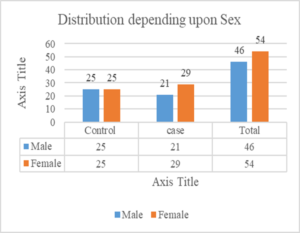
Figure 5: Distribution of patients depending upon sex in both groups.
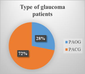
Figure 6: Distribution of patients depending upon type of glaucoma.
Endothelial Cell Characteristics of Healthy Eyes and PACG/POAG Eyes
Overall comparison of various parameters between diagnosed glaucoma patients on same antiglaucoma medications and age matched control group using specular examination was done. It was observed that glaucoma patients had a statistically significant(P<0.05) reduction in corneal ECD, Central Corneal Thickness (CCT) and hexagonality (6A) while an increase in average endothelial cell area and coefficient of variation as compared to age-matched healthy controls. The average endothelial cell density was significantly decreased in glaucoma patients (1610 ± 225 cells/mm2) compared with the control group (2210 ± 236 mm2) (Table 2).
|
ECD |
CCT |
CV |
6A |
|
|
Glaucoma patients |
(1610 ± 225cells/mm2) |
(515.5 ± 34.05 μm2) |
45.42 |
42.77 |
|
Control group |
(2210 ± 236cells/mm2) |
(520.50 ± 54.69 μ m2) |
42.20 |
49 |
Table 2: Comparison of various parameters between diagnosed glaucoma patients and age matched controls using specular examination.
The PACG patients had significantly lower ECD (1587 ± 233 cells/mm2), central corneal thickness and hexagonality as compared to the POAG subgroup (1617 ± 272 cells/mm2) whereas increased average endothelial cell area and coefficient of variation. However, the cell densities in both subgroups were less as compared to the controls (Fig. 7,8, Table 3).
|
|
ECD |
CCT |
CV |
6A |
|
PACG |
1587± 233 cells/mm2 |
513 |
46 |
42 |
|
POAG |
1617± 272 cells/mm2 |
518 |
44.42 |
43.7 |
Table 3: Comparison of various parameters between the two subgroups i.e., PACG and POAG.
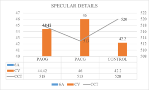
Figure 7: Comparison of various parameters between the two subgroups i.e., PACG and POAG and age and sex matched controls.
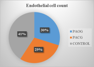
Figure 8: Comparison of Endothelial cell count between two subgroups with glaucoma and age sex matched controls.
Correlations Between Endothelial Cell Characteristics with IOP
The endothelial cell characteristics were also correlated with intraocular pressure in a patient on same antiglaucoma combination. The patients were categorized into 3 groups on the basis of IOP <20 mm of Hg, 21-25 mm of Hg and between 25-27 mm of Hg.
Correlation between ECD loss, hexagonality, endothelial cell area and intraocular pressure was studied between case and control groups and glaucoma subgroups as well. IOP showed positive correlation with Coefficient of Variation (CV) i.e. with increase in IOP value of CV increased. However, a negative correlation was found between IOP corneal ECD), hexagonality (6A) and CCT i.e., with increase in IOP the value of ECD, 6A and CCT decreased and vice versa (Fig. 9,10, Table 4,5).
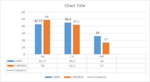
Figure 9: IOP showed positive correlation with Coefficient of Variation (CV) and negative correlation with (6A).
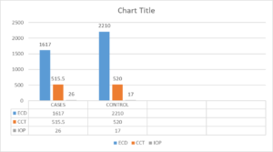
Figure 10: Patients showed negative correlation with IOP and corneal ECD.
|
IOP (mm of hg) |
PACG (CV) |
POAG CV |
PACG 6A |
PAOG 6A |
PACG CCT |
POAG CCT |
POAG ECD |
PACG ECD |
|
<20 |
37 |
35 |
46 |
43 |
520 |
524 |
2243 |
2176 |
|
21-24 |
43 |
42 |
41 |
39 |
515 |
517 |
2168 |
2053 |
|
25-27 |
49 |
46 |
38 |
33 |
505 |
515 |
1617 |
1587 |
Table 4: Correlations between endothelial cell characteristics with IOP in glaucoma subgroups.
|
Mean IOP |
ECD |
CCT |
CV |
6A |
|
|
Glaucoma Patients |
21 mm of hg |
(1610 ± 225 cells/mm2) |
(515.5 ± 34.05 μm2) |
45.42 |
42.77 |
|
Control group |
17 mm of hg |
(2210 ± 236 cells/mm2) |
(520.50 ± 54.69 μm2) |
42.20 |
49 |
Table 5: Correlations between endothelial cell characteristics with IOP in glaucoma patients and age matched healthy control group.
Discussion
The corneal endothelium is critical in preserving a healthy transparent cornea for proper functioning of corneal endothelium a definite cell density and morphology are necessary which are more sensitive indicators of functional alterations of the corneal endothelium, hence the maintenance of these cells is of paramount importance [1].
In current study we observed the corneal endothelium changes in glaucoma patients on same antiglaucoma combination and are compared with age matched healthy control group. Majority of patients in our study (38%) belonged to the elder age group of 61-70 years as compared to only 17% in younger age of 40 to 50 years. Similar results were reported by Yu ZY, et al., and Elagamy A, et al., who reported the mean age was 63 ± 13 years and 51.43 ± 0.02 years respectively [1,15]. A slightly higher female preponderance was observed with 54% female and 46% male patients. However, a slightly higher male prevalence was observed with. with 25 males (56.8%) and 19 females (43.2%) by Yu ZY, et al., [1].
Most of the patients were diagnosed to have Primary Angle Closure Glaucoma (PACG) i.e. 72% and only 28% had Primary Open Angle Glaucoma (PAOG), similar prevalence of PACG has been reported in a study performed by Palimkar A, et al., on the contrary the study performed by Nangia V, et al., reported POAG as the major type of glaucoma [16,17].
It was noticed that glaucoma patients had a reduction in corneal ECD (1610 ± 225 cells/mm2) 7.2% as compared to age-matched healthy controls (2210 ± 236 cells/mm2). The results were in concurrence with studies done by Elagamy A, et al., Yu ZY, et al., and Gagnon, et al., who reported a reduction of 5.47% and 5.8%, respectively in ECD in glaucoma patients as compared to healthy controls in which possible explanations include direct mechanical damage, elevated IOP, impaired endothelial metabolism due to disruptive aqueous humour circulation and long term use of antiglaucoma medications [1,7,12].
In PACG and POAG subgroups it was observed that PACG patients had significantly lower ECD (1587 ± 233 cells/mm2) as compared to the POAG subgroup (1617 ± 272 cells/mm2). However, the cell densities in both subgroups were less as compared to the controls (2210 ± 236 cells/mm2). Studies done by Malaise-Stals J, on 44 eyes of PACG patients on acute attack found that corneal endothelial cell density was significantly reduced in acute PACG eyes compared to normal eyes. On the other hand Studies done by Yu ZY, et al., Bhomaj, et al., Swamy Tasneem, et al., Knorr, et al., and Gagnon, et al., demonstrated a significant reduction in endothelial cell count in POAG patients compared to normal eyes, as the corneal endothelium has the capability to adapt to a gradual and modest increase in IOP, even if it continues for a prolonged period [7,11,18-20].
Additionally this study showed that hexagonality (6A) and Coefficient of Variation (CV) were significantly changed in glaucoma patients (42.77 in cases vs 49 in controls) and CV (45.42) in glaucoma cases as compared to controls(42.20).This was analogous to studies done by Gagnon, et al., Tham, et al., and Kang D, et al., who observed a significant decrease in the percentage of hexagonal cells (48.40% in cases vs 68.68% in controls) and a significantly high CV (35.7) in glaucoma cases as compared to controls (27.06), indicating considerable pleomorphism and polymegathism in glaucomatous eyes [7,21,22].
We also found the significant role of IOP and its correlation with corneal endothelial changes. It was reported that the mean IOP in the study group was higher (21 mm of Hg) as compared to healthy age-sex matched controls (17 mm of Hg),additionally all the patients on same combination (Brimonidine 0.2% and Timolol 0.5%) of antiglaucoma medication were categorized into 3 groups on the basis of IOP <20 mm of Hg, 21-25 mm of Hg and between 25-27 mm of Hg, suggesting that the IOP may be the contributing factor for reduction in ECD along with the use of antiglaucoma medications. Kang D, et al, and Gagnon, et al., in their study also reported an increase in IOP in glaucoma patients as compared to healthy controls [7,22]. They interpreted that a high IOP affected the cornea first by direct mechanical trauma to endothelial cells and later with a persistent high IOP injury to the active metabolic pump mechanism which keeps the cornea dehydrated. Swamy and Tasneem, et al., investigated the association between ECD and mean IOP in the newly detected POAG patients vs controls and found that the patients with 10-15 mm of Hg have ECD of 2500-3000 cells/mm2 compared to ECD of 2000-2500 cells/ mm2 (39%) with an increasing IOP beyond of 20-25 mm of Hg [19]. Thus, we observed that endothelial cell characteristics in primary glaucoma patients showed a statistically significant variations with rise in mean IOP as compared to healthy age-sex matched controls.
Moreover, we also noticed that IOP showed positive correlation with Coefficient of Variation (CV) and hexagonality (6A) whereas a statistically negative correlation was observed between mean IOP CCT and corneal ECD. Korey, et al., assumed that the corneal endothelium has the capability to adapt to a gradual and modest increase in IOP [23,24]. Similar studies were done by Elagamy A, et al., and Gagnon, et al., according to which high IOP directly damages the physical barrier function of CECs and affects the function of the endothelial pump [7,15].
Apart from ECD, CV and 6A Central Corneal Thickness (CCT) is also an important parameter in assessment of corneal endothelium health. Although no significant difference in CCT between glaucoma patients (515.23 ± 34.05 μm2) and the control groups (520.50 ± 54.69 μm2) were observed, studies by Kurna, et al., Beloway GW, et al., Sng C, et al., and Day AC, et al., which were Population-based studies, including groups from West Africa and East Asia, all these studies had also shown no difference in CCT between glaucomatous and non-glaucomatous eyes [23-26]. According to above studies in patients with established glaucoma the predictive role of CCT is not well proven in management or long-term prognosis its value in patients diagnosed with glaucoma is less certain. In contrast to the studies of Kniestedt C, et al., the Los Angeles Latino Eye Study which showed that patients with advanced damage have more likely to have a thin CCT [26-28]. They also reported significantly higher CCT in PACG eyes as compared to POAG and normal eyes and those with thin CCT had a significantly higher prevalence of POAG than those with normal or thick corneas [27,28]. There are several biological factors and genetic components that may influence glaucoma progression, which have been associated with thinner CCT [24].
Apart from ECD, CV and 6A Central Corneal Thickness (CCT) is also an important parameter in assessment of corneal endothelium health. Although no significant difference in CCT between glaucoma patients (515.23 ± 34.05 μm2) and the control groups (520.50 ± 54.69 μm2) were observed, studies by Kurna, et al., Beloway GW, et al., Sng C, et al., and Day AC, et al., which were Population-based studies, including groups from West Africa and East Asia, all these studies had also shown no difference in CCT between glaucomatous and non-glaucomatous eyes [23-26]. According to above studies in patients with established glaucoma the predictive role of CCT is not well proven in management or long-term prognosis its value in patients diagnosed with glaucoma is less certain. In contrast to the studies of Kniestedt C, et al., the Los Angeles Latino Eye Study which showed that patients with advanced damage have more likely to have a thin CCT [26-28]. They also reported significantly higher CCT in PACG eyes as compared to POAG and normal eyes and those with thin CCT had a significantly higher prevalence of POAG than those with normal or thick corneas [27,28]. There are several biological factors and genetic components that may influence glaucoma progression, which have been associated with thinner CCT [24].
Conclusion
The normal corneal endothelium is composed of uniform regular hexagonal cells. Once this structure is destroyed and the corneal endothelium is in an unstable state, the cells are easily damaged, which causes further deformation and loss. The present study suggests that use of antiglaucoma medications (Brimonidine 0.2% and Timolol 0.5%) affect the metabolism of the corneal endothelium and cause pathological changes. Hence for primary glaucoma patients, quantitative analysis of corneal endothelial cells morphology is necessary, despite the well-established benefits of antiglaucoma medications in reducing Intraocular Pressure (IOP), the potential impact of these drugs on the corneal endothelium remains a concern.
Limitations
As with the majority of studies, the design of the current study is subject to limitations which were observed during analysis of the study and are mentioned
- Effect of combination of topical antiglaucoma medications (Brimonidine (0.2%w/v) and Timolol (0.5%w/v)) without any preservative and also their individual drugs need to assess as well.
- Although this was the commonest combination used in our glaucoma clinics effect of other categories of topical antiglaucoma medications on the corneal endothelium needs to be studied.
- The current study was lacking in illustrating the complex relationship between antiglaucoma medications, IOP and disease itself to explain corneal endothelial damage in glaucoma patients.
- Although this study clarifies most of the concepts, however further continuation of this study with more sample size will be done to elucidate the findings.
Conflict of Interests
The authors declared no conflicts of interest with respect to the publication of this paper.
References
- YuZY, Wu L, Qu B. Changes in corneal endothelial cell density in patients with primary open- angle glaucoma. World J Clin Cases. 2019;7(15):1978-85.
- Bakunowicz-Łazarczyk A, Michalczuk M, Krętowska M. Evaluation of corneal endothelium in adolescents with juvenile glaucoma. J Ophthalmol. 2015;2015:895428.
- Cho SW, Kim JM, Choi CY, Park KH. Changes in corneal endothelial cell density in patients normal-tension glaucoma. Jpn J Ophthalmol. 2009;53:569-73.
- Buettner H, Bourne WM. Effect of trans pars plana surgery on the corneal endothelium. Dev Ophthalmol. 1981;2:28-34.
- Reznicek L, Muth D, Kampik A, Neubauer AS, Hirneiss C. Evaluation of a novel Scheimpflug-based non-contact tonometer in healthy subjects and patients with ocular hypertension and glaucoma. Br J Ophthalmol. 2013;(97):1410-4.
- Chen MJ, Liu CJ, Cheng CY, Lee SM. Corneal status in primary angle-closure glaucoma with a history of acute attack. J Glaucoma. 2012;21:12-6.
- Ates H, Palamar M, Yagci A, Egrilmez S. Trabeculectomy versus EX-PRESS shunt versus ahmed valve implant: short-term effects on corneal endothelial cells. Am J Ophthalmol. 2016;162:201-2.
- Gagnon MM, Boisjoly HM, Brunette I, Charest M, Amyot M. Corneal endothelial cell density in glaucoma. Cornea. 1997;16:314-8.
- Baratz KH, Nau CB, Winter EJ, McLaren JW, Hodge DO, Herman DC, et al. Effects of glaucoma medications on corneal endothelium, keratocytes, and subbasal nerves among participants in the ocular hypertension treatment study. Cornea. 2006;25:1046-52.
- Huber M, Kölzsch M, Stahlmann R, Hofmann W, Bolbrinker J, Dräger D, et al. Ophthalmic drugs as part of polypharmacy in nursing home residents with glaucoma. Drugs Aging. 2013;30(1):31-8.
- Yu L, Ding K, Luo L, Yu Z. Prescribing trends of glaucoma drugs in six major cities of China from 2013 to 2017. PLoS ONE. 2020;15(1):e0227595.
- Kandarakis SA, Togka KA, Doumazos L, Mylona I, Katsimpris A, Petrou P, et al. The multifarious effects of various glaucoma pharmacotherapy on corneal endothelium: A narrative review. Ophthalmology and Therapy. 2023;12(3):1457-78.
- Belamkar A, Harris A, Zukerman R, Siesky B, Oddone F, Verticchio Vercellin A, et al. Sustained release glaucoma therapies: novel modalities for overcoming key treatment barriers associated with topical medications. Ann Med. 2022;54(1):343-58.
- Conlon R, Saheb H, Ahmed II. Glaucoma treatment trends: a review. Can J Ophthalmol. 2017;52(1):114-24.
- Chaurasia S, Vanathi M. Specular microscopy in clinical practice. Indian J Ophthalmol. 2021;69(3):517-24.
- Elagamy A, Tawashi M, Berika M. Corneal endothelium and central corneal thickness changes in patients with primary open angle glaucoma. J Ophthalmol Res. 2022;5(4):141-6.
- Palimkar A, Khandekar R, Venkataraman V. Prevalence and distribution of glaucoma in central India (Glaucoma Survey-2001). Indian J Ophthalmol. 2008;56:57-62.
- Nangia V, Jonas JB, Matin A, Bhojwani K, Sinha A, Kulkarni M, et al. Prevalence and associated factors of glaucoma in rural central India. The central India eye and medical study. PLoS One. 2013;8:e76434 .
- Bhomaj P, Umarani S, Kudache J. Study of corneal endothelial morphology in primary open angle glaucoma and pseudo-exfoliative glaucoma patients compared with age related normal. J Clin Ophthalmol. 2020;8:95-9.
- Swamy NN, Tasneem AF. Evaluation of corneal morphology among newly detected patients with primary open angle glaucoma. J Clin Res Ophthalmol. 2020;7:008-014.
- Knorr HL, Handel A, Naumann GO. Morphometric and qualitative changes in corneal endothelium in primary chronic open angle glaucoma. Fortschritte der Ophthalmologie. 1991;88:118-20.
- Tham CCY, Kwong YYY, Lai JSM, Lam DSC. Effect of a previous acute angle closure attack on the corneal endothelial cell density in chronic angle closure glaucoma patients. J Glaucoma. 2006;15:482-5.
- Kang D, Kaur P, Singh K, Kumar D, Chopra R, Sehgal G. Evaluation and correlation of corneal endothelium parameters with the severity of primary glaucoma. Indian J Ophthalmol. 2022;70(10):3540-3.
- Kurna SA, Acikgoz S, Altun A, Ozbay N, Sengor T, Olcaysu OO. The effects of topical antiglaucoma drugs as monotherapy on the ocular surface: A prospective study. J Ophthalmol. 2014;2014:460-83.
- Belovay GW, Goldberg I. The thick and thin of the central corneal thickness in glaucoma. Eye (Lond). 2018;32(5):915-23.
- Sng C, Barton K, Kim H, Yuan S, Budenz DL. Central corneal thickness and its associations with ocular and systemic factors in an urban West African population. Am J Ophthalmol. 2016;169:268-75.
- Day AC, Machin D, Aung T, Gazzard G, Husain R, Chew PTK, et al. Central corneal thickness and glaucoma in East Asian people. Invest Ophthalmol Vis Sci. 2011;52:8407.
- Kniestedt C, Lin S, Choe J, Nee M, Bostrom A, Sturmer J, et al. Correlation between intraocular pressure, central corneal thickness, stage of glaucoma, and demographic patient data: prospective analysis of biophysical parameters in tertiary glaucoma practice populations. J Glaucoma. 2006;15:91-7.
- Francis BA, Varma R, Chopra V, Lai MY, Shtir C, Azen SP, et al. Intraocular pressure, central corneal thickness, and prevalence of open-angle glaucoma: the Los Angeles Latino Eye Study. Am J Ophthalmol. 2008;146:741-6.
- Wang HZ, Hong SJ, Wu KY. Change of calcium and cAMP concentration by adrenoceptor agents in cultured porcine corneal endothelial cells. J Ocul Pharmacol Ther. 2000;16(4):299-309.
- Grueb M, Mielke J, Rohrbach JM, Schlote T. Effect of brimonidine on corneal thickness. J Ocul Pharmacol Ther. 2011;27(5):503-9.
Article Type
Research Article
Publication History
Received Date: 05-07-2024
Accepted Date: 24-07-2024
Published Date: 31-07-2024
Copyright© 2024 by Thatte S, et al. All rights reserved. This is an open access article distributed under the terms of the Creative Commons Attribution License, which permits unrestricted use, distribution, and reproduction in any medium, provided the original author and source are credited.
Citation: Thatte S, et al. To Assess and Compare the Effect of Combination of Anti-Glaucoma Drug on Corneal Endothelium in Primary Glaucoma Patients with Age Matched Healthy Control. J Ophthalmol Adv Res. 2024;5(2):1-12.

Figure 1: Goldmann applanation tonometer.

Figure 2: Specular microscopy.

Figure 3: A: (Left) Open angle on gonioscopy; B: (Right) closed angle on gonioscopy.

Figure 4: a: Percentage of age distribution in glaucoma patients; b: Distribution of patients depending upon age in control group.

Figure 5: Distribution of patients depending upon sex in both groups.

Figure 6: Distribution of patients depending upon type of glaucoma.

Figure 7: Comparison of various parameters between the two subgroups i.e., PACG and POAG and age and sex matched controls.

Figure 8: Comparison of Endothelial cell count between two subgroups with glaucoma and age sex matched controls.

Figure 9: IOP showed positive correlation with Coefficient of Variation (CV) and negative correlation with (6A).

Figure 10: Patients showed negative correlation with IOP and corneal ECD.
Parameter | Normal Value |
Endothelial cell density | 2000-3000 cells/mm220,21 |
Percentage of hexagonal cells (6A) | 46 ± 8%22 |
Central Corneal Thickness (CCT) | 554.78+/-32.6123 |
Coefficient of Variation (CV) | 0.22 – 0.3124-27 |
Table 1: Normal values in middle aged Indian population.
ECD | CCT | CV | 6A | |
Glaucoma patients | (1610 ± 225cells/mm2) | (515.5 ± 34.05 μm2) | 45.42 | 42.77 |
Control group | (2210 ± 236cells/mm2) | (520.50 ± 54.69 μ m2) | 42.20 | 49 |
Table 2: Comparison of various parameters between diagnosed glaucoma patients and age matched controls using specular examination.
| ECD | CCT | CV | 6A |
PACG | 1587± 233 cells/mm2 | 513 | 46 | 42 |
POAG | 1617± 272 cells/mm2 | 518 | 44.42 | 43.7 |
Table 3: Comparison of various parameters between the two subgroups i.e., PACG and POAG.
IOP (mm of hg) | PACG (CV) | POAG CV | PACG 6A | PAOG 6A | PACG CCT | POAG CCT | POAG ECD | PACG ECD |
<20 | 37 | 35 | 46 | 43 | 520 | 524 | 2243 | 2176 |
21-24 | 43 | 42 | 41 | 39 | 515 | 517 | 2168 | 2053 |
25-27 | 49 | 46 | 38 | 33 | 505 | 515 | 1617 | 1587 |
Table 4: Correlations between endothelial cell characteristics with IOP in glaucoma subgroups.
Mean IOP | ECD | CCT | CV | 6A | |
Glaucoma Patients | 21 mm of hg | (1610 ± 225 cells/mm2) | (515.5 ± 34.05 μm2) | 45.42 | 42.77 |
Control group | 17 mm of hg | (2210 ± 236 cells/mm2) | (520.50 ± 54.69 μm2) | 42.20 | 49 |
Table 5: Correlations between endothelial cell characteristics with IOP in glaucoma patients and age matched healthy control group.


