Review Article | Vol. 6, Issue 1 | Journal of Ophthalmology and Advance Research | Open Access |
The Inappropriate Photoreceptor Depolarization May Give Rise to Age-Related Macular Degeneration: Clinical Evidence
Fabrizio Magonio1*
1Department of Ophthalmology, Igea Private Hospital, via Marcona, 69 20129 Milan, Italy
*Correspondence author: Fabrizio Magonio, Department of Ophthalmology, Igea Private Hospital, via Marcona, 69 20129 Milan, Italy; Email: [email protected]
Citation: Magonio F. The Inappropriate Photoreceptor Depolarization May Give Rise to Age-Related Macular Degeneration: Clinical Evidence. J Ophthalmol Adv Res. 2025;6(1):1-7.
Copyright© 2025 by Magonio F. All rights reserved. This is an open access article distributed under the terms of the Creative Commons Attribution License, which permits unrestricted use, distribution, and reproduction in any medium, provided the original author and source are credited.
| Received 15 January, 2025 | Accepted 07 February, 2025 | Published 15 February, 2025 |
Abstract
Age-related Macular Degeneration (AMD) is a complex, multifactorial, progressive retinal disease that affects millions of people worldwide. Alterations in phototransduction, renewal of the outer segments of photoreceptors, visual transduction and retinol metabolism have a major impact on retinal health.
Mutations within any of the molecules responsible for these visual processes cause different types of degenerative retinal diseases. Increase in membrane potential caused by accidental binding to a foreign molecule, increased temperature or ocular pressure could trigger an electrochemical overload in the photoreceptor which would be inappropriately depolarized. The signs of this process are clearly visible with Optical Coherence Tomography (OCT): hyper-reflective vertical lines that cross the choroid at the points of interruption of the ellipsoid zone resulting in atrophy and compromise of the outer hematoretinal barrier.
The occasional finding of these lines would represent the first clinical sign of a degenerative process which could subsequently give rise to those events characterizing AMD.
Keywords: Phototransduction; cGMP; CNG; Depolarization; Hyperpolarization; Poppers Maculopathy; Age-Related Macular Degeneration
Introduction
Age-related Macular Degeneration (AMD) is a complex, multifactorial and progressive retinal disease that affects millions of people worldwide and has become the leading cause of visual impairment in developed countries. Changes in the outer retina in the aging process include loss of photoreceptors, thickening of Bruch’s membrane and impairment of the choroidal microvasculature. This results in the accumulation of abnormal proteins and lipids that will form drusen often associated with pathological changes in the outer retina, including detachment of Retinal Pigment Epithelium (RPE) cells from their basement membrane and migration into the neurosensory retina [1]. Drusen trigger chronic inflammation of the subretinal space in which microglia, macrophages and the complement system are also involved [1]. The etiopathogenetic mechanisms of AMD involve defects in autophagy, proteolysis, lipid homeostasis, mitochondrial dysfunction and alterations in the Extracellular Matrix (ECM). The genetic component of the disease is attributed to alterations in the regulation of gene expression modulated by epigenetics and the participation of environmental factors such as pollution, smoking, hypertension, antioxidant-poor diet and obesity [1]. Mitochondrial dysfunction initiates a cascade of events that cause oxidative stress and the production of numerous intracellular reactive oxygen species consisting of hydrogen peroxide, superoxide ion (O2-) and hydroxyl radicals. Reactive nitrogen species such as Nitric Oxide (NO), peroxynitrite and the peroxyl radical also play a direct role in cell signaling, vasodilation and inflammation. In fact, reactive nitrogen species can cause damage to proteins, lipids, DNA and induce photoreceptor cell death [2]. Retina has a high demand for energy and oxygen, which it meets through a constant blood supply through the choroid. Photoreceptors, RP cells and the choriocapillaris act symbiotically with each other: RPE supplies glucose from the choroidal circulation to the photoreceptors, which in turn produce lactate to feed RPE [1]. RPE cells are polarized with the apical region expressing microvilli that interdigitate with the outer segment of photoreceptors and the basal region expressing specific transport enzymes. Ion transporters and tight junctions between RPE cells allow control of intercellular communication and the electrical potential difference between the two epithelial surfaces. In this way, RPE cells help regulate the composition of the interphotoreceptor ECM and create a selective barrier known as the outer Hematoretinal Barrier (HRB). Any failure in this sophisticated metabolic ecosystem leads to serious functional and structural consequences in the outer segments of photoreceptors [1]. The retina, like the brain, has traditionally been considered an immunologically privileged organ, but it is now known that it has an endogenous immune system consisting of immunocompetent cells including microglia, macrophages and RPE that play important roles in maintaining and restoring retinal homeostasis. We spend about 1/3 of our lives sleeping i.e., performing a fundamental function for our nervous system. No neurophysiological study, to my knowledge, has ever been conducted to understand what happens in the human retina while we sleep. However, it is likely that the retina, like the brain, also undergoes a process of synaptic plasticity and that a glymphatic system exists within it to enhance the elimination of metabolic waste [3].
Phototransduction
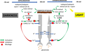
Each photoreceptor contains an outer segment, the photon capture site that initiates the visual process, an inner segment that houses the nucleus and cytoplasmic organelles and a synaptic terminal that contains neurotransmitter vesicles for signal transmission. The outer segments of photoreceptors are supported by an axonemal skeleton consisting of microtubules and from which originates the connecting cilium, an elongated and narrow structure through which all proteins and membrane components must pass, i.e., it functions as a gate that regulates access to the outer segment [4]. Each photoreceptor cell has a specific role depending on the light stimulus it receives. Rods are highly sensitive photoreceptors to light but have a slow response and are activated from the scotopic environment to the mesopic, the point at which they become saturated. In contrast, cones, which are less sensitive than rods, begin to respond rapidly under photopic conditions and do not saturate, thus being able to continue to respond to the light stimulus [4-6]. These characteristics, as will be described later, expose cones to an increased risk of experiencing electrochemical interference. The photoreceptor cells of invertebrate animals differ from those of vertebrates in morphology and physiology. The electrical signals of visual excitation have opposite character in vertebrates and invertebrates: the vertebrate photoreceptor cell is hyperpolarized because of a decrease in conductance and invertebrate photoreceptors are depolarized owing to an increase in conductance [7]. In humans, phototransduction begins in the outer segment of the photoreceptor where subthreshold electrotonic potentials ranging from -40 mV (at darkness) to -70 mV (at light) are generated and transmitted to the inner segment. However, these currents do not generate action potentials but are transmitted to other neurons (bipolar, horizontal, amacrine cells) for reprocessing. The action potential subsequently originates in the ganglion cells that go to make up the optic nerve. After light stimulation, the photosensitive protein rhodopsin/iodopsin undergoes a conformational rearrangement, which stimulates the G protein transducin. The latter activates 6-Phosphodiesterase (6-PDE), the enzyme that reduces the intracellular concentration of cyclic Guanosine Monophosphate (cGMP) leading to the closure of Cyclic Nucleotide-Regulated channels (CNGs), the reduction of intracellular Na+ and Ca++ influx and the subsequent activation of Guanylate Cyclase (GC), the enzyme that converts Guanosine Triphosphate (GTP) to cGMP. Levels of Na+ and Ca++ in the cytoplasm are then reduced, causing the photoreceptor to hyperpolarize and reduce Glutamate (GLU) release from its synaptic terminal in the outer plexiform layer, altering downstream signaling in bipolar and ganglion cells. Therefore, CNGs not only play a key role in phototransduction but also regulate cellular cGMP levels [8]. In the dark, CNG channels are instead kept open by cGMP binding, generating a constant Na+ and Ca++ input into the photoreceptor (the so- called “dark current”) that keeps the plasma membrane depolarized by promoting synaptic release of GLU. Elevated intracellular Ca++ concentration inhibits GC, which reduces cGMP synthesis by a feedback regulatory mechanism (Fig.1). Alterations in the activity of the phototransduction cascade, such as changes affecting the renewal and loss of photoreceptor outer segments, visual transduction and retinol metabolism have a great impact on retinal health. Mutations within any of the molecules responsible for these visual processes cause different types of retinal degenerative diseases. In fact, most retinal degeneration is caused by genetic defects that lead to alterations in protein synthesis [1].

Figure 1: Phototransduction. GC: Guanylate Cyclase; cGMP: cyclic Guanosine Monophosphate; 6-PDE: 6- Phosphodiesterase; CNG: Cyclic Nucleotide-Regulated channel; PK: Protein Kinase.
The Role of cGMP
In addition to acting as a second messenger, cGMP thus plays a key role in phototransduction by governing CNGs. Among the various cellular alterations that occur in photoreceptor degeneration, dysregulation of cGMP signaling represents a major pathogenic factor [8]. 6-PDE-bound cGMP constitutes about 90% of the total cGMP content in the photoreceptor; therefore, if cGMP increased, there would not be enough 6-PDE to hydrolyze it. In fact, 6-PDE is the only force capable of degrading cGMP. Dysregulation is mainly manifested by abnormally high cellular levels of cGMP that alter a wide range of cellular activities, resulting in impaired cellular homeostasis, oxidative stress and cell death [8]. Elevated cGMP signaling impairs Ca++ homeostasis in the Endoplasmic Reticulum (ER). The ER is a large cellular organelle dedicated to the biosynthesis, folding and assembly of membrane and secretory proteins and functions as a storehouse of free Ca++. ER homeostasis is critical for its function and its impairment will lead to ‘accumulation of abnormal proteins, cytotoxicity and cell death [8]. Normally, only a small fraction of the CNGs present is kept open by cGMP in dark-adapted photoreceptors, but an increase in cGMP would lead to the opening of too many channels, thereby abnormally increasing the influx of Na+ and Ca++. Defects and mutations in various molecules involved in the regulation of cGMP Production and Degradation, 6-PDE and CNG, are the main causes of dysregulation of cGMP signaling in photoreceptors [9,10].
The Inappropriate Depolarization of the Photoreceptor
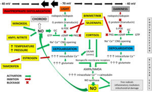
The electrical activity of neurons is characterized by a balance between excitation and inhibition, maintained by feedback mechanisms at the cellular, synaptic and neurohumoral levels. In the retina we have a vertical, excitatory electrochemical impulse pathway (photoreceptors, bipolar cells releasing GLU into the synaptic space) and a horizontal, inhibitory pathway (horizontal and amacrine cells releasing GABA). This electrochemical balance can be altered by numerous random factors, intrinsic and extrinsic, such as biogenic amines (histamine, catecholamines), peptides (glutamate, aspartate), hormones (cortisol, estrogen), drugs (cortisone, sildenafil, minoxidil, binimetinib, tamoxifen), gases (NO), temperature changes that can increase Na+ entry into the photoreceptor and depolarize it (Fig. 2) [11,12]. Furthermore, the ocular pressue and retinal stretch, as well as the pulse of arteries and the fluctuation of local pressure, shape and osmolarity, appear to be a suitable stimulus capable of opening Mechanosensitive Channels (MSC) [13]. Vertebrate retinal rods and the outer plexiform layer have been reported to express two groups of MSC, including the vanilloid transient receptor potential channel and K+ channels such as the big potassium channel (BK, also known as the calcium- and voltage-gated large conductance potassium channel). The K+ channels give rise to leak (also called background) K+ currents to stabilize the negative resting membrane potential and counterbalance membrane depolarization [13]. Unlike any other nerve cell, we said that the photoreceptor does not react to the (light) stimulus by depolarizing and generating the action potential but rather by hyperpolarizing i.e., reducing its own membrane potential. This condition makes it more exposed to potential depolarizing factors. Neuromodulatory disequilibrium toward greater depolarization (excitability) can occur transiently with subsequent compensation or in a cascade of events that will give rise to varying degrees of functional impairment [8,9]. Should this phenomenon occur under light conditions (hyperpolarization), the increase in membrane potential difference caused by interaction with a foreign molecule, increased temperature or ocular pressure could trigger an abrupt increase in potential in the photoreceptor, which would be inappropriately depolarized at light! (Fig. 2). The resulting increase in GLU in the extracellular space and intracellular Ca++ concentration can cause excitotoxicity, altering mitochondrial function, energy (ATP) production, ATP- dependent membrane pump activity and activating proteases (phospholipase A, NO synthase) [11,14]. NO synthase produces NO that can behave as a second messenger: it enters the photoreceptor, stimulates GC to synthesize cGMP that causes opening of the Na+ and Ca++ CNGs, depolarization and further release of GLU [14]. The NO → GLU circuit generates a vicious cycle that can be self-sustaining (Fig. 2). Excess NO can diffuse into the macular choroid where it will find numerous nitrergic receptors to bind to. The resulting choroidal vasodilation and increased hydrostatic pressure will promote the formation of epithelial lifts. This mechanism could also be involved in the pathogenesis of Central Serous Chorioretinopathy (CSC) [14,15]. The probability of unanticipated depolarization, excitotoxicity and increased NO will therefore be greater at the posterior pole, due to miosis, lens accommodation and macula morphology that promote hyperpolarization of photoreceptors, especially cones that do not saturate, are more exposed to light stimulus and temperature changes. A functional deficit in the choroidal microcirculation, especially in elderly subjects, may promote increased macular temperature and depolarization [12]. Cortisol may also influence phototransduction and glutamatergic transmission. This hormone controls neuronal activity either through a slow genomic action, regulating arrestin gene expression in the outer segments of photoreceptors or through a rapid nongenomic action, activating specific Glucocorticoid (GR) and Mineralcorticoid (MR) membrane receptors located at the presynaptic membranes of cones, causing Ca++ entry into the cell and modifying its neurotransmission [16,17]. Individual genetic predisposition may play an important role in these processes, being in a condition of higher or lower risk of developing the disease.

Figure 2: Conditions that could cause inappropriate photoreceptor depolarization at light.
Clinical Evidence
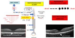
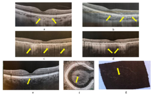
Early deterioration of the ellipsoidal zone in eyes with nonneovascular age-related macular degeneration has been observed by several authors [18,19]. Clinical evidence of this phenomenon can be explained by “Poppers Maculopathy” resulting from inhalation of drugs containing volatile alkyl nitrites [16]. Phototransduction creates an electrical circuit that allows free electrons to move continuously through the photoreceptor representing the conductor: this flow is called electric current or Intensity (I). The force that drives electrons to flow in an electrical circuit is called voltage (V). Voltage is a specific measure of potential energy that is always relative to two points (-40 mV and -70 mV in the photoreceptor). Free electrons tend to move through the conductor with a certain degree of friction i.e. opposition to movement. This friction is called resistance (R). These three factors are governed by Ohm’s 1st law: I=V/R [21]. Ohm’s 2nd law, on the other hand, states that “R” is directly proportional to the length of the conductor (L) and inversely proportional to the area of its cross- section (A): R=ρL/A; “ρ” represents the resistivity constant, which is the specific electrical resistance of each material and is affected by temperature [ρ=ρ0 (1+ αT), α= temperature coefficient] [21]. In fact, increased temperature causes greater difficulty in the movement of electrons due to collisions with atoms in the conductor. The movement of electric current along a conductor also requires energy, which, encountering resistance, produces heat (Joule effect) [22]. After inhalation, volatile alkyl nitrites release NO that can rapidly diffuse through cell membranes and reach the retina through the eyeballs or tear pathways. NO activates GC in the photoreceptor and increases cGMP levels. It has been previously explained that under a condition of excessive light exposure and thus extreme hyperpolarization, a high concentration of cGMP will promote, in the outer segment of the photoreceptor, a rapid increase in voltage and electric current (Ohm’s 1st law): the photoreceptor will then be found to be depolarized at light. The photoreceptor is a peculiar conductor because its cross-sectional area is not constant but is drastically reduced at the level of the cilium. Therefore, according to Ohm’s 2nd law (R=ρ. L/A), the greater flow of electric current moving through the outer segment will find a greater resistance to pass through the smaller cross-section of the cilium, which will burn out behaving like a fuse (Joule effect) and blocking transmission to the inner segment [20]. It is therefore likely that the function of the cilium in the photoreceptor is not only to regulate metabolic exchanges between the inner and outer segments but also to “sacrifice” itself to attenuate or stop high voltage currents that could trigger a chain of damaging events downstream. Disruption of the ellipsoid zone of the foveal cone (which will certainly be the one that will have undergone the greatest potential increase) is well evident with OCT and is pathognomonic of “Popper Maculopathy” (Fig.3). In the case of AMD, the continued occurrence of this phenomenon would over time result in the formation of more and more confluent points of atrophy until the function of the outer HRB is compromised. Clearly visible with OCT are the vertical hyperreflective lines crossing the macular choroid at the interruption of the ellipsoid zone or points of atrophy that might not be evident because of their small size. These would be clinical evidence of inappropriate photoreceptor depolarization and the resulting initial impairment of the outer HRB. Over time, these lines may become confluent and take on the appearance of a “bar code” at the areas of atrophy (Fig.4).

Figure 3: Popper Maculopathy: NO interacts with phototransduction of the foveal cones, which will undergo abrupt depolarization to light. OCT shows disruption of the ellipsoid zone and subsequent evolution into a point of atrophy from which a hyperreflective line starts and crosses the choroid.

Figure 4: In OCT, clinical signs of inappropriate depolarization of photoreceptors. a: the outer band of the retina appears normal although vertical lines that cross the choroid are already present at invisible points of atrophy; b: hyperreflective lines originate from points of interruption of the ellipsoid zone and atrophy; c, d: numerous vertical lines become confluent with “bar code” appearance; e: interruption of foveal ellipsoid zone; f: OCT en face of image “e” shows corresponding point of atrophy; g: 3D-OCT, tilted image “e” shows hyperreflective point at atrophy.
Conclusion
The occasional finding of hyperreflective vertical lines, which often escape the attention of the OCT operator, would thus represent the first clinical sign of a degenerative process that could subsequently give rise to the sequence of events that characterize AMD. Poppers Maculopathy, CSC and AMD could thus be considered the clinical manifestations of the same pathogenetic process that would occur, in predisposed individuals, with different intensity and timing: acute, sub-acute and chronic, respectively. A better understanding of cGMP signaling in photoreceptor degeneration may help the development of therapeutic interventions targeting this signaling pathway to protect photoreceptors and to counteract the degenerative process in the early stages.
Conflict of Interest
The authors declare no potential conflicts of interest with respect to the research, authorship and/or publication of this article.
Funding Details
No funding was received for this review. No other financial disclosures of all authors.
References
- Garcia-Garcia J, Usategui-Martin R, Sanabria MR, Fernandez-Perez E, Telleria JJ, Coco-Martin RM. Pathophysiology of age-related macular degeneration: implications for treatment. Ophthalmic Research. 2022;65(6):615-36.
- Toma C, De Cillà S, Palumbo A, Garhwal DP, Grossini E. Oxidative and nitrosative stress in age-related macular degeneration: A review of their role in different stages of disease. Antioxidants. 2021;10(5):653.
- Magonio F. REM phase: An ingenious mechanism to enhance clearance of metabolic waste from the retina. Experimental Eye Research. 2022;214:108860.
- Baehr W, Hanke-Gogokhia C, Sharif A, Reed M, Dahl T, Frederick JM, et al. Insights into photoreceptor ciliogenesis revealed by animal models. Prog Retinal Eye Res. 2019 1;71:26-56.
- Lamb TD. Photoreceptor physiology and evolution: cellular and molecular basis of rod and cone phototransduction. The Journal of Physiology. 2022;600(21):4585-601.
- Ingram NT, Sampath AP, Fain GL. Why are rods more sensitive than cones? J Physiol. 2016;594(19):5415-26.
- Rayer B, Naynert M, Stieve H. New trends in photobiology: phototransduction: different mechanisms in vertebrates and invertebrates. J Photochem and Photobiol B: Biology. 1990;7(2-4):107-48.
- Li S, Ma H, Yang F, Ding X. CGMP signaling in photoreceptor degeneration. Int Molecular Sci. 2023;24(13):11200.
- Das S, Chen Y, Yan J, Christensen G, Belhadj S, Tolone A, et al. The role of cGMP-signalling and calcium-signalling in photoreceptor cell death: Perspectives for therapy development. Pflügers Archiv-European Journal of Physiology. 2021;473:1411-21.
- Francis SH, Busch JL, Corbin JD, Sibley D. cGMP-dependent protein kinases and cGMP phosphodiesterases in nitric oxide and cGMP action. Pharmacological Revi. 2010;62(3):525-63.
- Magonio F. The role of excitotoxicity in the etiopathogenesis of central serous chorioretinopathy. Clin Ophthalmol J. 2022;3(1):1021.
- Coates TA, Woolnough O, Masters JM, Asadova G, Chandrakumar C, Baker MD. Acute temperature sensitivity in optic nerve axons explained by an electrogenic membrane potential. Pflügers Arch. 2015;467:2337-49.
- Ji-Jie Pang, Fan Gao, Samuel M Wu. Generators of pressure-evoked currents in vertebrate outer retinal neurons. Cells. 2021;10(6):1288.
- Ishikawa M. Abnormalities in glutamate metabolism and excitotoxicity in the retinal diseases. Scientifica (Cairo). 2013;2013: 528940.
- de Jong EK, Breukink MB, Schellevis RL, Bakker B, Mohr JK, Fauser S, et al. Chronic central serous chorioretinopathy is associated with genetic variants implicated in age-related macular degeneration. Ophthalmol. 2015;122(3):562-70.
- Oakley RH, Revollo J, Cidlowski JA. Glucocorticoids regulate arrest in gene expression and redirect the signalling profile of G protein-coupled receptors. Proceedings of the National Academy of Sciences. 2012;109(43):17591-6.
- Mirshahi M, Nicolas C, Mirshahi A, Hecquet C, d’Hermies F, Faure JP, et bal. The mineralocorticoid hormone receptor and action in the eye. Biochemical and biophysical Research Communications. 1996;219(1):150-6.
- Singh SR, Vaidya H, Borrelli E, Chhablani J. Foveal photoreceptor disruption in ocular diseases: An optical coherence tomography-based differential diagnosis. Surv Ophthalmol. 2023;68 (4):655-68.
- Borrelli E, Sacconi R, Zuccaro B, Cavalleri M, Bordato A, Zucchiatti I, et al. Alteration of photoreceptors in intermediate age-related macular degeneration. Sci Rep. 2020;10:21036.
- Magonio F. Popper maculopathy is an electrochemical overload. Arch Clin Exp Ophthalmol. 2022;4(1):1-3.
- Ohm GS. Die galvanische kette: Mathematisch. TH Riemann. 1827.
- Joule JP. On the mechanical equivalent of heat. Abstracts of the Papers Communicated to the Royal Society of London 1851. London: The Royal Society. 1851;5:839-9.
Author Info
Fabrizio Magonio1*
1Department of Ophthalmology, Igea Private Hospital, via Marcona, 69 20129 Milan, Italy
*Correspondence author: Fabrizio Magonio, Department of Ophthalmology, Igea Private Hospital, via Marcona, 69 20129 Milan, Italy; Email: [email protected]
Copyright
Fabrizio Magonio1*
1Department of Ophthalmology, Igea Private Hospital, via Marcona, 69 20129 Milan, Italy
*Correspondence author: Fabrizio Magonio, Department of Ophthalmology, Igea Private Hospital, via Marcona, 69 20129 Milan, Italy; Email: [email protected]
Copyright© 2025 by Magonio F. All rights reserved. This is an open access article distributed under the terms of the Creative Commons Attribution License, which permits unrestricted use, distribution, and reproduction in any medium, provided the original author and source are credited.
Citation
Citation: Magonio F. The Inappropriate Photoreceptor Depolarization May Give Rise to Age-Related Macular Degeneration: Clinical Evidence. J Ophthalmol Adv Res. 2025;6(1):1-7.


