Kwok Hei MOK1*
1Optometry Centre, Evangel Hospital, Hong Kong, China
*Corresponding Author: Kwok Hei MOK, BSc Optom (Hons), MPhil PhD (HKU) FAAO FACO, Optometry Centre, Evangel Hospital, Hong Kong, China; Email: [email protected]
Published Date: 01-10-2022
Copyright© 2022 by MOK KH. All rights reserved. This is an open access article distributed under the terms of the Creative Commons Attribution License, which permits unrestricted use, distribution, and reproduction in any medium, provided the original author and source are credited.
Letter to Editor
Spherical Refractive error (Rx) has been reported to be reliably estimated with Ocular Biometrics (OB) using intra-ocular lens calculation method such as SRK/T formula [1]. Further supplementary data was presented regarding the evaluation of the extent of Rx alterations with Axial Length (AL) elongation using this calculation method.
Rx were calculated using intraocular lens power (IOL) (22.5D A-Constant 118) and various AL (23.0 to 26.0 mm) and corneal curvature (K) readings (41.00 to 44.00 D) using SRK/T formula. IOL was estimated with Rx (Plano), AL (23.2 mm) and K reading (42.00 D).
RX | AL (mm) | 23.0 | 23.5 | 24.0 | 24.5 | 25.0 | 25.5 | 26.0 |
K (D) | 41.0 | +1.21 | +0.16 | -0.89 | -1.93 | -2.97 | -4.00 | -5.03 |
41.5 | +0.86 | -0.25 | -1.27 | -2.32 | -3.38 | -4.42 | -5.46 | |
42.0 | +0.51 | -0.57 | -1.65 | -2.72 | -3.79 | -4.85 | -5.99 | |
42.5 | +0.15 | -0.95 | -2.04 | -3.13 | -4.36 | -5.50 | -6.63 | |
43.0 | -0.25 | -1.33 | -2.44 | -3.54 | -4.75 | -5.89 | -7.02 | |
43.5 | -0.59 | -1.72 | -2.84 | -3.95 | -5.06 | -6.26 | -7.40 | |
44.0 | -0.97 | -2.11 | -3.25 | -4.37 | -5.50 | -6.63 | -7.76 |
Table 1: Rx was estimated by various AL and K readings with IOL (22.5 D A-constant 118).
RX Changes | AL (mm) Changes | 23.0-23.5 | 23.5-24.0 | 24.0-24.5 | 24.5-25.0 | 25.0-25.5 | 25.5-26.0 | MEAN (SD) |
K (D) | 41.0 | -1.05 | -1.05 | -1.04 | -1.04 | -1.03 | -1.03 | -1.04 (0.01) |
41.5 | -1.11 | -1.02 | -1.05 | -1.06 | -1.04 | -1.04 | -1.05 (0.03) | |
42.0 | -1.08 | -1.08 | -1.07 | -1.07 | -1.06 | -1.14 | -1.08 (0.03) | |
42.5 | -1.10 | -1.09 | -1.09 | -1.23 | -1.14 | -1.13 | -1.13 (0.05) | |
43.0 | -1.11 | -1.11 | -1.10 | -1.20 | -1.15 | -1.13 | -1.13 (0.04) | |
43.5 | -1.13 | -1.12 | -1.11 | -1.11 | -1.20 | -1.14 | -1.14 (0.03) | |
44.0 | -1.14 | -1.14 | -1.12 | -1.13 | -1.13 | -1.13 | -1.13 (0.01) | |
| Mean (SD) | -1.10 (0.03) | -1.09 (0.04) | -1.08 (0.03) | -1.12 (0.07) | -1.11 (0.06) | -1.11 (0.05) |
|
Table 2: Rx alterations with 0.5 mm AL increase at various K readings.
In Table 1, estimated Rx with various AL and K readings would only be regarded as clinical reference, not actual Rx, since only artificial lens power was used for calculation. Various human lens powers would likely result in different Rx outcomes. However the main objective of this letter was to estimate the extent of Rx alterations with AL elongation. In Table 2, interestedly it showed similar amount of Rx alterations, i.e. approximately -1.10 D Rx increase with 0.50 mm AL elongation between 23 to 26 mm AL. Comparable extent of Rx alteration was found among various K readings (41.00 to 44.00 D), but Rx tends to increase a bit more with higher K readings.
Conflict of Interest
The authors did not report any potential conflicts of interest in research, authorship and/or publication of this article.
References
- MOK K. Objective spherical refractive error estimation using ocular biometrics. J Ophthalmol Adv Res. 2022;3(1):1-4.
Article Type
Letter to Editor
Publication History
Received Date: 10-09-2022
Accepted Date: 23-09-2022
Published Date: 01-10-2022
Copyright© 2022 by MOK KH. All rights reserved. This is an open access article distributed under the terms of the Creative Commons Attribution License, which permits unrestricted use, distribution, and reproduction in any medium, provided the original author and source are credited.
Citation: MOK KH. Extent of Refractive Error Changes with Axial Length Elongation. J Ophthalmol Adv Res. 2022;3(3):1-2.


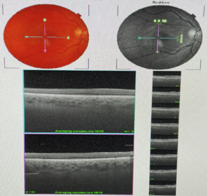
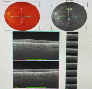
Figure 1: Spectral-domain optical coherence tomography showing grade 3 foveal hypoplasia in both eyes (a, Right eye; b, Left eye).

Figure 2: Multifocal ERG of the right eye showing signal depression in both foveal and parafoveal areas (90 nv/deg2 in the foveal area and 71 nv/deg2 respectively).

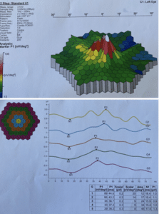
Figure 3: Multifocal ERG of the left eye showing signal depression in both foveal and parafoveal areas (89 nv/deg2 in the foveal area and 66 nv/deg2 respectively).


