Case Report | Vol. 6, Issue 1 | Journal of Ophthalmology and Advance Research | Open Access |
Modified Yamane Technique: A Case Series
Shu Yu Qian1, Annie Lam-Nguyen2, Marc Saab1,3*
1Faculty of Medicine, University of Sherbrooke, 3001 12 Ave N, Sherbrooke, J1H 5N4, QC, Canada
2Faculty of Medicine, University of Montreal, 2900 Edouard Montpetit Blvd, Montreal, H3T 1J4, QC, Canada
3Department of Ophthalmology, Charles LeMoyne Hospital, 3120 Taschereau Blvd, Greenfield Park, J4V 2H1, QC, Canada
*Correspondence author: Marc Saab, MD, Faculty of Medicine, University of Sherbrooke, 3001 12 Ave N, Sherbrooke, J1H 5N4, QC, Canada and Department of Ophthalmology, Charles LeMoyne Hospital, 3120 Taschereau Blvd, Greenfield Park, J4V 2H1, QC, Canada; Email: [email protected]
Citation: Qian SY, et al. Modified Yamane Technique: A Case Series. J Ophthalmol Adv Res. 2025;6(1):1-4.
Copyright© 2025 by Qian SY, et al. All rights reserved. This is an open access article distributed under the terms of the Creative Commons Attribution License, which permits unrestricted use, distribution, and reproduction in any medium, provided the original author and source are credited.
| Received 12 January, 2024 | Accepted 07 February, 2025 | Published 15 February, 2025 |
Abstract
Objective: Yamane, et al., introduced a sutureless Scleral Fixation of Intraocular Lens (SFIOL) technique using 30-gauge thin-wall needles to externalize both haptics of a three-piece Intraocular Lens (IOL) through transconjunctival sclerotomies. This technique has some limitations and challenges such as the creation of symmetrical sclerotomies and the externalization of haptics. Therefore, we propose a modified approach employing trocars and forceps as an alternative method to externalize the IOL haptics.
Methods: Six eyes of 6 patients underwent this modified technique consists of using two 27-gauce trocars to create symmetrical angled scleral tunnels and a three-piece IOL (MA50BM, Alcon Inc., Fort Worth, USA). A pair of 27-gauge microforceps is also used to externalize the IOL haptics instead of needles. Best Corrected Visual Acuity (BCVA) and occurrence of complications were the main outcomes measured.
Results: The mean BCVA was 1.18 ± 0.48 logMAR (range 0.7-2) and improved to 0.096 ± 0.123 logMAR (range 0-0.3) at their most recent postoperative consultation. There were no complications. Using forceps to directly grip and externalize the haptics not only avoids the complexities of threading the haptic through a thin needle, but also reduces surgical time. The increased robustness and larger optic diameter of the MA50BM IOL reduces the effects of a mild decentration compared to the CT Lucia 602 three-piece IOL (Carl Zeiss Meditec, Jena, Germany) that is commonly employed in the standard Yamane technique.
Conclusion: Our procedure achieved satisfactory results comparable to outcomes described by Yamane, et al., without occurrence of any complications.
Keywords: Yamane Technique; Cataract; Surgical Technique; Learning Curve; Intraocular Lens, Surgical Difficulty
Introduction
Phacoemulsification with Intraocular Lens (IOL) implantation remains the standard and first-line treatment for cataract patients. However, surgical complications, traumas or congenital lens anomalies may induce inadequate capsular support, in which case in-the-bag IOL placement is unfeasible [1]. Additionally, one report suggests that after 20 years post-cataract surgery, 1.2% of patients will need IOL dislocation surgery, with that risk being even greater among those with ocular comorbidities like pseudoexfoliations [2]. Longer lifespans combined with a greater numbers of phacoemulsification surgeries performed year after year contribute to this increase in IOL dislocation cases [3]. In these situations, Anterior Chamber IOLs (ACIOLs), iris-fixated Posterior Chamber IOLs (PCIOLS) and Scleral-Fixated IOLs (SFIOLs) become viable treatment options, each with its own pros and cons [4].
Notably, Yamane, et al., developed a sutureless SFIOL technique with 30-gauge thin-wall needles. That technique consisted of making angled incisions parallel to the limbus through which needles were used to externalize both haptics of a three-piece IOL. The ends of the haptics were then cauterized to create a flange, thereby holding it in place [5]. Despite the popularity of this approach, it is not without certain drawbacks and complexities. Therefore, we propose a modified approach employing trocars and forceps as a potentially safer, easier and less time-consuming alternative method to externalize the haptics.
Methods
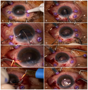
This is a retrospective interventional case series comprised of 6 eyes of 6 patients needing IOL placement who underwent a modified version of Yamane’s technique. All surgeries were performed by the same experienced surgeon (MS). Informed consent was obtained from all patients and our report adheres to the Declaration of Helsinki. The data and images used in the article contain no personal identifying information. Each patient’s preoperative workup included medical history assessments, slit-lamp exam, Best Corrected Visual Acuity (BCVA), Intraocular Pressure (IOP) and biometric measurements. Before IOL insertion, all patients underwent a standard 27-gauge Pars Plana Vitrectomy (PPV) with retrobulbar anesthesia to facilitate manipulations in the posterior chamber without disturbing the vitreous. Haptic placements were then marked at 2.5 mm from the limbus and 180° from each other. Two 27-gauge trocars were inserted at the marked points, creating two symmetrical angled scleral tunnels. Afterwards, the three-piece IOL (MA50BM, Alcon Inc., Fort Worth, USA) was injected through a limbal incision while maintaining the trailing haptic on the outside. A pair of 27-gauge micro forceps was used to grip and externalize the leading haptic through the port. The end of the haptic was melted with low-temperature cautery to form a flange. The trailing haptic was similarly gripped, externalized and its end cauterized. Lastly, the haptics are prolapsed through the conjunctiva and into the scleral tunnel to ensure proper positioning of the IOL. Fig. 1 summarizes the main steps of this procedure.

Figure 1: Main steps of our modified technique. (A): Create symmetrical angled sclerotomies 2.5 mm from the limbus using 27-gauge trocars; (B): Insert the 3-piece IOL and leave the trailing haptic hanging out through the main incision; (C): Grip the leading haptic using a pair of forceps with one going through the trocar. (D) Externalize the leading haptic while maintaining the trailing haptic outside the eye; (E): Cauterize the haptic to form a flange; (F and G): Externalize and cauterize the trailing haptic; (H): Push the haptics back into the scleral tunnel.
Postoperative follow-ups performed at 2 weeks, 1 month, 2 months and 4 months evaluated patient’s BCVA, IOP and IOL positioning. The BCVA values obtained from the Snellen chart were converted to logarithm of the Minimal Angle of Resolution (logMAR) values for statistical calculations. Light perception acuity was assigned a value of 20/2000 or 2.0 logMAR. These values are presented as means ± standard deviations.
Results
This case series included a total of 6 eyes of 6 patients, with 2 women and 4 men. Ocular comorbidities were documented in patient 1 (age-related macular degeneration) and patient 3 (glaucoma). The remaining patients had no additional known ocular problems. At the time of surgery, the mean age of the cohort was 75.6 ± 2.5 years (range 72-77 years). 3 patients were operated in the right eye and 3 in the left eye, with the main indications being IOL displacement (n=3), subluxated cataract (n=1), complicated cataract surgery (n=1) and traumatic aphakia (n=1). Preoperatively, the mean Best Corrected Visual Acuity (BCVA) was 1.18 ± 0.48 LogMAR (range 0.7-2), which is approximately 20/300 on the Snellen chart. This improved to 0.096 ± 0.123 LogMAR (range 0-0.3) or around 20/25 at their most recent postoperative consultation. All patients experienced BCVA improvements, with 3 of them achieving 20/20 vision. Table 1 details these patient demographics and clinical findings.
Patient ID | Age (years) | Sex | Diagnosis | Preoperative BCVA – Snellen (LogMAR) | Postoperative BCVA – Snellen (LogMAR) |
1 | 77 | F | Subluxated cataract | 20/100 (0.7) | 20/20 (0) |
2 | 75 | F | Complicated cataract surgery | 20/400 (1.3) | 20/25 (0.10) |
3 | 74 | M | IOL displacement | 20/2000 (2.0) | 20/40 (0.30) |
4 | 72 | M | Aphakia | 20/250 (1.1) | 20/30 (0.18) |
5 | 79 | M | IOL displacement | 20/400 (1.3) | 20/20 (0) |
6 | 77 | M | IOL displacement | 20/100 (0.7) | 20/20 (0) |
Table 1: Patient demographics and clinical characteristics.
Discussion
Since its devising in 2017, Yamane’s technique has been popularized as a highly effective procedure associated with rapid visual recovery. When performing this technique, surgeons may encounter numerous difficulties with the creation of symmetrical sclerotomies and the externalization of the haptics frequently being very challenging steps. Unequal tunnel lengths may cause IOL tilt or decentration that, in more significant cases, will need haptic trimming or redocking [6]. During the externalization process, lacing the trailing haptic into the needle lumen can be tricky to execute especially when using the non-dominant hand or if the haptic is bent at an awkward angle. If manipulated too vigorously, the haptic may break or kink, thus complicating the operation. To address these issues, the technique we proposed utilizes ports to more easily create reproducible scleral tunnel lengths and entries. Using forceps to directly grip and externalize the haptics not only avoids the complexities related to threading the haptic through a thin needle, but also reduces surgical time. Furthermore, doing so prevents having blind needles in the eye that risk accidentally damaging the retina. The standard Yamane technique frequently employs the CT Lucia 602 three-piece IOL (Carl Zeiss Meditec, Jena, Germany) since it possesses a good balance of strength and flexibility that facilitates haptic manipulation [7]. Despite those advantages and its popularity among surgeons, there have been numerous recent reports of optic-haptic junction distortions with that model due to weaknesses in that region [8]. Such occurrences would induce IOL tilt, with more severe situations may require re-operation. For our modified version, we prefer the MA50BM IOL due to its increased robustness because haptic malleability was deemed not as necessary for our approach. This model also has a larger optic diameter, which is more forgiving in cases of mild decentration compared to its smaller counterparts. In Yamane and colleagues’ cohort of 100 eyes of 97 patients, the mean BCVA improved from 0.25 logMAR preoperatively to 0.11 logMAR at 6 months and to 0.04 logMAR at their final follow-up 3 years after surgery [5]. In this report, our patients had worse BCVA at baseline, but nonetheless achieved similar VA outcomes at the 6-month mark. Jujo, et al., also described a series of 19 eyes treated with a comparable method using 27-gauge trocars. Their patients obtained a mean postoperative BCVA of 0.06 logMAR at 1 month [9]. In the 5 eyes treated by Gamal El-Din’s team (2022) with a 25-gauge trocar-assisted intrascleral IOL fixation technique, mean BCVA improved to 0.26 logMAR at 3 months postoperative without the occurrence of any complications [10]. Our study’s outcomes thus concurs with the findings reported in these similar investigations. Our report has certain limitations. First, the small number of patients prevents us from assessing the full-extent of our procedure’s safety and efficacy. Also, with a relatively short follow-up period of 6 months, we are unable to evaluate the long-term postoperative course and the potential development of late complications. Furthermore, we did not measure postoperative IOL tilt or decentration with anterior segment-optical coherence tomography imaging. We thus cannot more quantitatively analyze IOL positioning using our approach and compare with other studies. Moving forward, larger randomized controlled trials with longer follow-up periods would be necessary to provide more robust evidence and confirm these findings.
Conclusion
Despite these shortcomings, our investigation contributes to the current knowledge base on various secondary IOL implantation surgeries. Among the numerous existing procedures, the choice of technique should depend on patient characteristics, clinical data and the surgeon’s experience. We therefore encourage ophthalmologists to consider incorporating our approach to their armamentarium for its increased simplicity and short learning curve.
Conflict of Interest
The authors declare no potential conflicts of interest with respect to the research, authorship and/or publication of this article.
References
- Tsatsos M, Vartsakis G, Athanasiadis I, Moschos M, Jacob S. Intraocular lens implantation in the absence of capsular support: scleral fixation. Eye. 2022;36:1721-3.
- Mönestam E. Frequency of intraocular lens dislocation and pseudophacodonesis, 20 years after cataract surgery: A prospective study. Am J Ophthalmol. 2019;198:215-22.
- Fernández-Buenaga R, Alio JL, Pérez-Ardoy AL, Larrosa-Quesada A, Pinilla-Cortés L, Barraquer R, et al. Late in-the-bag intraocular lens dislocation requiring explantation: risk factors and outcomes. Eye. 2013;27:795-802.
- Holt DG, Stagg B, Young J, Ambati BK. ACIOL, sutured PCIOL or glued IOL: Where do we stand? Curr Opin Ophthalmol. 2012;23:62-7.
- Yamane S, Sato S, Maruyama-Inoue M, Kadonosono K. Flanged intrascleral intraocular lens fixation with double-needle technique. Ophthalmol. 2017;124:1136-42.
- Kurimori HY, Inoue M, Hirakata A. Adjustments of haptics length for tilted intraocular lens after intrascleral fixation. Am J Ophthalmol Case Reports. 2018;10:180-4.
- Randerson EL, Bogaard JD, Koenig LR, Hwang ES, Warren CC, Koenig SB. Clinical outcomes and lens constant optimization of the Zeiss CT lucia 602 lens using a modified Yamane technique. Clin Ophthalmol. 2020;14:3903-12.
- Kumar D, Tan J, Liu Y, Shah C, Stroh I, Shah N, et al. Axial instability of the zeiss ct lucia 602 intraocular lens with transconjunctival intrascleral haptic fixation. Invest Ophthalmol Visual Sci. 2023;64:5270.
- Jujo T, Kogo J, Sasaki H, Sekine R, Sato K, Ebisutani S, et al. 27-gauge trocar-assisted sutureless intraocular lens fixation. BMC Ophthalmol. 2021;21:8.
- GamalElDin SA, ElShazly MI, Salama MM. Trocar-assisted flanged transconjunctival intrascleral sutureless intraocular lens fixation. Euro J Ophthalmol. 2022;32:3699-702.
Author Info
Shu Yu Qian1, Annie Lam-Nguyen2, Marc Saab1,3*
1Faculty of Medicine, University of Sherbrooke, 3001 12 Ave N, Sherbrooke, J1H 5N4, QC, Canada
2Faculty of Medicine, University of Montreal, 2900 Edouard Montpetit Blvd, Montreal, H3T 1J4, QC, Canada
3Department of Ophthalmology, Charles LeMoyne Hospital, 3120 Taschereau Blvd, Greenfield Park, J4V 2H1, QC, Canada
*Correspondence author: Marc Saab, MD, Faculty of Medicine, University of Sherbrooke, 3001 12 Ave N, Sherbrooke, J1H 5N4, QC, Canada and Department of Ophthalmology, Charles LeMoyne Hospital, 3120 Taschereau Blvd, Greenfield Park, J4V 2H1, QC, Canada; Email: [email protected]
Copyright
Shu Yu Qian1, Annie Lam-Nguyen2, Marc Saab1,3*
1Faculty of Medicine, University of Sherbrooke, 3001 12 Ave N, Sherbrooke, J1H 5N4, QC, Canada
2Faculty of Medicine, University of Montreal, 2900 Edouard Montpetit Blvd, Montreal, H3T 1J4, QC, Canada
3Department of Ophthalmology, Charles LeMoyne Hospital, 3120 Taschereau Blvd, Greenfield Park, J4V 2H1, QC, Canada
*Correspondence author: Marc Saab, MD, Faculty of Medicine, University of Sherbrooke, 3001 12 Ave N, Sherbrooke, J1H 5N4, QC, Canada and Department of Ophthalmology, Charles LeMoyne Hospital, 3120 Taschereau Blvd, Greenfield Park, J4V 2H1, QC, Canada; Email: [email protected]
Copyright© 2025 by Qian SY, et al. All rights reserved. This is an open access article distributed under the terms of the Creative Commons Attribution License, which permits unrestricted use, distribution, and reproduction in any medium, provided the original author and source are credited.
Citation
Citation: Qian SY, et al. Modified Yamane Technique: A Case Series. J Ophthalmol Adv Res. 2025;6(1):1-4.


