Research Article | Vol. 6, Issue 1 | Journal of Ophthalmology and Advance Research | Open Access |
The “Eye Movement Test” in the Clinical Management of Chronic Open-Angle Glaucoma
Fabrizio Magonio1*
1Department of Ophthalmology, Igea Private Hospital, Milan, Italy
*Correspondence author: Fabrizio Magonio, Department of Ophthalmology Igea Private Hospital, via Marcona, 69 20129 Milan, Italy; Email: [email protected]
Citation: Magonio F. The “Eye Movement Test” in the Clinical Management of Chronic Open-Angle Glaucoma. J Ophthalmol Adv Res. 2025;6(1):1-5.
Copyright© 2025 by Magonio F, et al. All rights reserved. This is an open access article distributed under the terms of the Creative Commons Attribution License, which permits unrestricted use, distribution, and reproduction in any medium, provided the original author and source are credited.
| Received 30 March, 2025 | Accepted 21 April, 2025 | Published 28 April, 2025 |
Abstract
Glaucoma is a neurodegenerative disease characterized by increased Intraocular Pressure (IOP), progressive apoptosis of retinal ganglion cells, increased optic nerve head excavation, reduced visual field and visual acuity. The most common form of glaucoma is Chronic Open-Angle Glaucoma (COAG) and the main cause of ocular hypertension is increased resistance to aqueous humor outflow at the level of the iuxtacanalicular tissue of the trabecular meshwork. The aim of this study is to investigate the usefulness of a new diagnostic technique called “EYE MOVEMENT TEST” in the clinical management of COAG. Two hundred subjects (400 eyes) were enrolled and divided into two groups. Group A consisted of 100 patients with COAG and group B consisted of 100 patients without a diagnosis of ocular hypertension. Group B was divided into two subgroups: B1 consisting of 80 patients (160 eyes) with IOP ≤ 18 mmhg and B2 consisting of 20 patients (40 eyes) with IOP ≥ 19 mmhg classified as “subjects at risk of glaucoma.” After performing Goldmann applanation tonometry, subjects were asked to move their eyes in all gaze positions by fixing an aim for 1 minute and immediately afterwards, their IOP was measured again. In 172 eyes of group A (subjects with COAG) and in 16 eyes of group B2 (subjects at risk of glaucoma) there was a non-significant decrease in mean IOP (< 1 mmHg), while in group B1 the decrease in mean IOP was ≥ 1 mmHg (range 1 – 3 mmHg). This technique, through the changes in IOP induced by eyeball movement, aims to investigate the degree of elasticity and efficiency of the trabecular meshwork, verify the effectiveness of therapies that act on trabecular outflow and most importantly help identify subjects with borderline IOP, at risk of developing the disease.
Keywords: Eye Movement Test; Chronic Open-Angle Glaucoma; Trabecular Outflow; Intraocular Pressure; Goldmann Applanation Tonometry; Extracellular Matrix; Ocular Biomechanics
Objective
The purpose of this study is to investigate the usefulness of a new diagnostic technique called “EYE MOVEMENT TEST” (EMT) in the clinical management of Chronic Open-Angle Glaucoma (COAG).
Materials and Methods
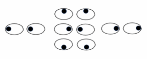
Two hundred patients (400 eyes) were enrolled and divided into two groups. The first group (A) consisted of 100 patients (200 eyes), 48 males and 52 females, mean age 70.3 years (range 53-88 years), with COAG in treatment with hypotonizing eyedrops (β-blockers, carbonic anhydrase inhibitors, prostaglandins, α2- agonists, RHO-kinase inhibitors) or undergoing Selective Lasertrabeculoplasty (SLT) for at least 3 months. In contrast, the second group (B) consisted of 100 patients (200 eyes), 42 males and 58 females, mean age 62.5 years (range 34-87 years) with no diagnosis of ocular hypertension. After performing basal tonometry by Goldmann applanation tonometry, group B was divided into two subgroups: B1 consisting of 80 patients (160 eyes) with IOP ≤ 18 mmHg and B2 consisting of 20 patients (40 eyes) with IOP ≥ 19 mmHg classified as “subjects at risk of glaucoma.” Immediately after basal tonometry the patients’ eyes, while fixing an aim, were induced to look up, down, right, left and converge for 1 minute, alternating between rapid, saccadic and slow, tracking movements i.e., simulating the eye movements that each person performs daily (Fig. 1). The EMT did not require Ethics Committee approval and signing of informed consent by the enrolled subjects.

Figure 1: The “Eye Movement Test” asseses the efficiency of trabecular outflow by measuring the IOP before and after moving the patient’s eyes for 1 minute.
Results
An IOP value ≥ 1 mmHg was considered “EMT+”. In 172 eyes in group A, the baseline mean IOP was 14.5 mmHg (range 8-20 mmHg), 16.4 mmHg in the other 28 eyes undergoing SLT or being treated with RHO- kinase inhibitor (Netarsudil) and 14.4 mmHg (range 10-18 mmHg) in the 160 eyes of group B1. In group B2, 8 eyes with IOP > 22 mmHg were underwent therapy and were excluded from the study. In the other 32 eyes, the mean baseline IOP was 21.5 mmHg (range 19-22 mmHg). Immediately after EMT, applanation tonometry gave the following results. In 172 eyes of group A the mean IOP was 14.1 mmHg (range 8-19 mmHg) with a mean reduction of 0.4 mmHg compared to baseline (pressure drop < 1 mmHg, EMT-). In addition, only in 20 eyes undergoing SLT and 8 treated with Netarsudil the mean IOP was 14.6 mmHg with a mean IOP reduction of 1,8 mmHg (≥ 1 mmHg, EMT+). In 44 pseudophakic eyes and 16 eyes with pseudoexfoliation glaucoma, there was no significant drop in IOP (EMT-). The sensitivity of the test was 81% and 95% when excluding subjects on Netarsudil therapy or SLT treatment. In group B1, the mean IOP was 12.4 mmHg (range 10-18 mmhg) with a reduction of 2 mmHg compared to baseline (pressure drop > 1 mmHg, EMT+) reaching, in some cases, pressure drops of 3 mmhg. This result was also obtained in 28 pseudophakic eyes. In group B2 the mean IOP was 21 mmHg in 16 eyes with a mean reduction of 0.5 mmHg (EMT-) and in others 16, 20 mmHg with a mean drop of 1.5 mmHg (EMT+) (Table 1). Finally, we report that in 14 eyes, 10 with pseudoexfoliation glaucoma in group A and 4 in group B2, there was a rise in IOP after the test. After routine examinations, 8 patients of group B2 with baseline IOP ≥ 19 ≤ 22 mmHg/ EMT-, classified as “subjects at risk of glaucoma” were underwent therapy. In contrast, 8 subjects with IOP ≥ 19 ≤ 22 mmHg/EMT+ were not prescribed any therapy. No subjects reported discomfort when performing the test.
400 EYES (200 subjects) | Mean basaline IOP (mmHg) | Mean IOP after EMT (mmHg) | Mean IOP drop after EMT (mmHg) | EMT (+) ≥ 1 mmHg (-) < 1 mmHg |
Group A 172 eyes with COAG 28 eyes with COAG (rho-Kinase inhibitors or SLT) | 14,5 16,4 | 14,1 14,6 | 0,4 1,8 | (-) (+) |
Group B1 160 healthy eyes (IOP ≤ 18 mmHg) | 14,4 | 12,4 | 2 | (+) |
Group B2 32 eyes at risk of glaucoma (IOP ≥ 19 ≤ 22 mmHg) | 21.5 | 16 eyes 21 16 eyes 20 | 0.5 1.5 | (-) (+) |
Table 1: Mean IOP.
Discussion
Glaucoma is a neurodegenerative disease characterised by increased IOP, progressive apoptosis of retinal ganglion cells, increased optic nerve head excavation, reduced visual field and visual acuity. COAG represents the most common form of glaucoma and has a multifactorial aetiology. Several risk factors have been highlighted as contributing to the damage and progression of the disease, such as elevated IOP, ageing, gender, familiarity, oxidative stress, mitochondrial damage, excitotoxicity, sleep disturbances, vascular and glymphatic system alterations [1,10,11]. In COAG, increased IOP is currently the only modifiable risk factor. The eyeball is filled with two fluids, the vitreous Humour and Aqueous humour (HA) which, among other functions, also regulate IOP. HA is secreted by the ciliary bodies and through the pupil, passes from the posterior chamber to the anterior chamber where it encounters two pathways for its outflow. The first pathway (conventional) consists of the Trabecular meshwork (TR), a highly contractile tissue similar to smooth muscle, Schlemm’s Canal (SC), collector canals and the episcleral venous system until it reaches the systemic circulation [1,2,5]. The filtering portion of the TR is composed of three different structures: the inner uveal reticulum, the deep sclerocorneal reticulum and the iuxtacanalicular tissue consisting of a fenestrated endothelial layer in contact with the inner wall of the SC [1,2,4]. HA passes through the two reticular layers passively along a pressure gradient, whereas the endothelial layer also appears to utilise an active transport system [4]. In the second pathway (unconventional), HA passes through the interstitium of the ciliary muscle adjacent to the TR, then reaches the suprachoroidal space, the episcleral vessels (uveo-scleral pathway), the vortical veins (uveo- vortex pathway) or the lymphatic vessels (uveo-lymphatic pathway) and finally flows into the systemic circulation [3]. In humans, approximately 90% of HA outflow occurs via the trabecular pathway [1]. Probably, during the REM phase of sleep, the uveo-scleral outflow increases due to the combined action of the glymphatic system and rapid eye movements, which may be at the origin of nocturnal IOP fluctuations [10]. The mechanical stress of these rapid eye movements, which is useful for the homeostasis of a healthy eye, interacting with an altered trabecular network, might result in morphogenic changes in the structure of the TR, affecting its elasticity, ECM activity, cytoskeleton rearrangement, increased outflow resistance and IOP, ultimately giving rise to proinflammatory and neurodegenerative factors [10,11]. There is growing evidence that external mechanical factors can influence the configuration of biomacromolecules, cell fate and tissue structure [6]. Mechanistic evidence indicates that experimental ocular hypertension can increase cell contractility by activating Rho GTPases, which cause increased resistance to AH drainage [7,8]. Within the eye, the balance between HA production and outflow contributes to maintaining an IOP of about 12-20 mmHg. In COAG, the main cause of ocular hypertension is the increased resistance to trabecular outflow at the level of the iuxtacanalicular tissue due to remodelling of the Extracellular Matrix (ECM), which would lead to its stiffening and reduced porosity as is usually the case in atrophic or aged tissues [1]. The same changes also appear to occur in the endothelial cells of the SC [1]. The ECM is a crucial component of the ocular microenvironment, playing an essential role in every part of the eye, both by maintaining the transparency and hydration of the cornea and vitreous and by modulating angiogenesis, IOP maintenance and vascular signalling [6]. The ECM is a three-dimensional non-cellular macromolecular network composed of water, glycosaminoglycans, proteoglycans, hyaluronic acid, collagen, fibrin, elastin, fibronectin and several other glycoproteins. It continuously undergoes remodelling mediated by various matrix-degrading enzymes such as metalloproteinases and glycosidases. Environmental stimulation, such as mechanical stretching or a variety of growth factors, cytokines and drugs (such as dexamethasone), can alter the expression of ECM components and its mechanical properties, triggering the continuous increase in trabecular outflow resistance, leading to an increase in IOP [6,7]. In conclusion, in COAG, the pump that allows HA drainage fails due to the loss of elasticity of the trabecular tissue and ciliary muscle fibres [4,5]. Due to its elasticity, the TR stretches and contracts in response to physiological changes in IOP caused by ocular and extraocular factors. The vitreous moves within the eyeballs through head movements, the voluntary action of extraocular muscles and the accommodation of the lens, directly or indirectly exerting positive and negative pressure on the structures contained within the eyeball [9,10]. This “vitreal pump” contributes to the maintenance of tissue elasticity and the movement of fluids entering and leaving the eyeballs [10]; it also increases blood flow, shear stress, nitric oxide release and indirectly stimulates the contraction of the trabecular meshwork [10,11]. COAG is more frequent in elderly people in whom, in addition to reduced tissue elasticity, atherosclerosis, cardiovascular disease and diabetes may contribute to impaired fluid circulation and elimination from the eyeball. The “heart pump” also acts on the trabecular meshwork. Indeed, during systole the left ventricle contracts giving rise to a wave that causes the choroid to expand, thus increasing IOP [4]. The increase in IOP causes the trabecular meshwork to move into the lumen of the SC, constricting it and increasing its internal pressure, which will favour the outflow of HA into the collecting ducts, aqueous and episcleral veins [4]. However, during diastole the choroidal volume and IOP decrease. The recoil of the TR creates a negative pressure in the SC while the valves of the collector canals and veins prevent the retrograde flow of HA and blood [4]. In daily life, the eyes are never at rest, even while we sleep and can generate voluntary and involuntary movements [10]. Saccadic (rapid) eye movements are the most common, produced by both eyes simultaneously and in the same direction when we stare at an object. Tracking (slow) eye movements, on the other hand, are used to follow the object. Finally, involuntary movements are due to the contraction of the ciliary muscle (accommodation), which favours near vision. The blinking of the eyelids, which helps keep the ocular surface hydrated and pumps tears into the tear ducts, also indirectly exerts a mechanical action on the trabecular meshwork. In addition to contributing to image capture, eye movements therefore perform biomechanical actions that are useful to the physiology of the eye [10,12]. Many of the drugs that lower IOP act by reducing HA production (β-blockers, carbonic anhydrase inhibitors, alpha2-agonists) or by increasing its outflow through the uveo-scleral pathway (prostaglandins, prostanoids, alpha2-agonists). On the other hand, drugs that act on the conventional pathway are the muscarinic cholinergic agonists (pilocarpine) which, by contracting the ciliary muscle, increase HA outflow by widening the trabecular meshwork and the SC [1,2]. Indeed, contraction (pilocarpine) or relaxation of the ciliary muscle (atropine) respectively abolish or increase the presence of muscle fissures to increase or reduce uveoscleral outflow [1,2]. However, these drugs have seen a reduction in clinical use in recent years, mainly due to the side effects (headache, myopia, blurred vision especially at night, frequency of instillation) resulting in poor patient compliance. A new class of drugs (RHO- kinase inhibitors) has recently been introduced into clinical practice. By acting on the cytoskeleton of trabecular cells, they weaken their intercellular bonds and promote the formation of new vacuolar spaces and the dilation of episcleral veins, facilitating HA outflow [8]. EMT, through IOP changes induced by eye movement, investigates the degree of elasticity of the TR and thus the efficiency of conventional outflow. It is known that cataract surgery can lower IOP. This study shows that in pseudophakic eyes with COAG, EMT detected pressure drops < 1 mmHg: the reduction in IOP would therefore be attributable not so much to the increase in trabecular outflow as to the reduction in intraocular mass. In healthy, even pseudophakic eyes, EMT showed a mean IOP drop of 2 mmHg with peaks of 3 mmHg while in glaucomatous eyes the mean drop was < 1 mmhg. Finally, in 16 eyes (10 with pseudoexfoliation glaucoma in group A and 6 in group B2), we had a rise in IOP of 2-3 mmHg after EMT, a phenomenon that can be explained by the recall of fluid at the level of the TR, which is not compensated by an efficient outflow. This result would demonstrate a severe impairment of trabecular outflow and, in the case of pseudoexfoliative glaucoma, the presence of frequent and unpredictable increases in IOP caused by transient obstruction of the trabecular network.
Conclusion
The clinical management of COAG includes numerous diagnostic examinations such as biomicroscopy of the optic papilla, tonometry, corneal pachymetry, gonioscopy, coherent light optical tomography and perimetry. It was explained how the regulation of trabecular HA outflow is a biomechanical action supported by a series of pumps (cardiac, palpebral, muscular and vitreal) that act on the contractility of the TR. EMT is a new diagnostic technique that, by exploiting ocular motility and thus the muscular-vitreal-palpebral pump, has proved useful in assessing the efficiency of trabecular outflow, the effectiveness of therapies acting on the TR and above all in identifying subjects with borderline IOP, at risk of developing the disease. Finally, perhaps we are not realising that nowadays our lifestyle forces us to use our eyes, much of the time, for near vision due to the excessive use of electronic devices that could represent a further risk factor. In fact, in addition to the damage we already know about (photo-oxidation, dry eye, conjunctival hyperemia, asthenopia, headache, sleep disturbances), these devices restrict the movement of our eyes in a narrow range of action and consequently could reduce the elasticity of ocular tissues including the trabecular meshwork. If this statement were correct, we would see the incidence of glaucoma increase in the coming years.
Conflict of Interest
The author declares no potential conflicts of interest with respect to the research, authorship and/or publication of this article.
Funding Details
No funding was received for this review.
References
- Tamm ER, Braunger BM, Fuchshofer R. Intraocular pressure and the mechanisms involved in resistance of the aqueous humor flow in the trabecular meshwork outflow pathways. Prog Mol Biol Transl Sci. 2015;134:301-14.
- Sunderland DK, Sapra A. Physiology, aqueous humor circulation. 2023. StatPearls Publishing. 2025.
- Johnson M, McLaren JW, Overby DR. Unconventional aqueous humor outflow: A review. Exp Eye Res. 2017;158:94-111.
- Johnstone M, Xin C, Tan J, Martin E, Wen J, Wang RK. Aqueous outflow regulation – 21st century concepts. Prog Retin Eye Res. 2021;83:100917.
- Lee JY, Akiyama G, Saraswathy S, Xie X, Pan X, Hong YK, et al. Aqueous humour outflow imaging: Seeing is believing. Eye (Lond). 2021;35(1):202-15.
- Zhang R, Li B, Li H. Extracellular-matrix mechanics regulate the ocular physiological and pathological activities. J Ophthalmol. 2023;2023:7626920.
- Soundararajan A, Wang T, Sundararajan R. Multiomics analysis reveals the mechanical stress-dependent changes in trabecular meshwork cytoskeletal-extracellular matrix interactions. Front Cell Dev Biol. 2022;10:874828.
- Ellis DZ. Guanylate cyclase activators, cell volume changes and IOP reduction. Cell Physiol Biochem. 2011;28(6):1145-54.
- Silva AF, Pimenta F, Alves MA, Oliveira MSN. Flow dynamics of vitreous humour during saccadic eye movements. J Mech Behav Biomed Mater. 2020;110:103860.
- Magonio F. REM phase: An ingenious mechanism to enhance clearance of metabolic waste from the retina. Exp Eye Res. 2022;214:108860.
- Vernazza S, Tirendi S, Bassi AM, Traverso CE, Saccà SC. Neuroinflammation in primary open-angle glaucoma. J Clin Med. 2020;9(10):3172.
- Aloy MÁ, Adsuara JE, Cerdá-Durán P, Obergaulinger M, Esteve-Taboada JJ, Ferrer-Blasco T, et al. Estimation of the mechanical properties of the eye through the study of its vibrational modes. PLoS One. 2017;12(9): e0183892.
Author Info
Fabrizio Magonio1*
1Department of Ophthalmology, Igea Private Hospital, Milan, Italy
*Correspondence author: Fabrizio Magonio, Department of Ophthalmology Igea Private Hospital, via Marcona, 69 20129 Milan, Italy; Email: [email protected]
Copyright
Fabrizio Magonio1*
1Department of Ophthalmology, Igea Private Hospital, Milan, Italy
*Correspondence author: Fabrizio Magonio, Department of Ophthalmology Igea Private Hospital, via Marcona, 69 20129 Milan, Italy; Email: [email protected]
Copyright© 2025 by Magonio F, et al. All rights reserved. This is an open access article distributed under the terms of the Creative Commons Attribution License, which permits unrestricted use, distribution, and reproduction in any medium, provided the original author and source are credited.
Citation
Citation: Magonio F. The “Eye Movement Test” in the Clinical Management of Chronic Open-Angle Glaucoma. J Ophthalmol Adv Res. 2025;6(1):1-5.


