ISSN (Online): 2582-6751

Review Article | Vol. 3, Issue 1 | Journal of Clinical Medical Research

The Periodontal Ligament-A Link between the Cementum and Alveolar Bone

Michel Goldberg1*

1Department of Oral Biology, Faculty of Fundamental and Biomedical Sciences, INSERM UMR-S 1124 Paris Cite University, France

*Corresponding Author: Michel Goldberg, Department of Oral Biology, Faculty of Fundamental and Biomedical Sciences, INSERM UMR-S 1124 Paris Cite University, France; Email: mgoldod@gmail.com

Published Date: 05-03-2022

Copyright© 2022 by Goldberg M. All rights reserved. This is an open access article distributed under the terms of the Creative Commons Attribution License, which permits unrestricted use, distribution, and reproduction in any medium, provided the original author and source are credited.

Received
24 Jan, 2021
Accepted
26 Feb, 2022
Published
05 Mar, 2022

Abstract

The Periodontal Ligament (PDL) provides support to the tooth in the bonny socket, in connecting the cementum and alveolar bone.  The PDL width vary around 25 mm, including collagens and oxytalan fibers (Sharpey’s fibers). Metalloproteinases play role in the turnover of type I collagen and other matrix proteins.  The PDL is implicated in bone remodeling, cells nutrition and sensory events, namely as periodontal mechanoreceptors. The PDL generates and controls tooth eruption and ensures homeostasis between fibroblasts and bone cells.  The PDL form, maintain and repair this interface. After dissociation of the cells of the Hertwig’s Epithelial Root Sheath, epithelial rests of Malassez represent a unique source of stem cells population within the periodontal ligament.  Extracellular matrix proteins (collagen, fibronectin and Secreted Proteins Acidic and Rich in Cysteine (SPARC), Glycosaminoglycans (GAGs) and Proteoglycans (PGs), Metalloproteinases (MMPs) and Tissue Inhibitors of Metalloproteinases (TIMP- 1 to – 4) are structural components of the PDL. This review explore:

  1. The attachment function of the PDL (eruption, formative and supportive functions)
  2. Summarize the implication of stem cells in healing, in the regulation of bone volume, PDL repair and regeneration
  3. Focus on the contribution of fibroblast-like and pericytes to periodontal inflammation through cytokines and chemokines and the release of molecules implicated in periodontal inflammation
  4. Sensory events requiring nerve corpuscles (Ruffini-like mechanoreceptors)
  5. Provide vascular supply and nutrients to the cementum, alveolar bone and to the PDL itself
  6. Because the cells of the PDL may differentiate into cementoblasts and osteoblasts, this phenotypic availability may contribute to heal the tooth supporting tissues

Keywords

Periodontal Ligament; Anchorage; Radicular Tooth; Alveolar Bone; Cementum; Collagen Fibers; Oxytalan; Matrix Metalloproteinases; Mechanoreceptors; Epithelial Rest of Malassez
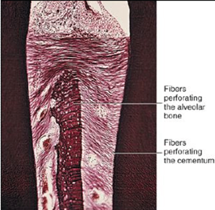
Figure 1: Fibers of the periodontal ligament.

Structure of the periodontium

Figure 2: Structure of the periodontium. A: alveolar bone, C: cementum, D: dentin; G: gingiva, P: periodontal ligament, SF: Sharpey’s fibers.

Supramolecular assemblies

Figure 3: Supramolecular assemblies formed by collagens.

Specific Domains of MMP, ADAM and ADAMTS.

Figure 4: Specific Domains of MMP, ADAM and ADAMTS.

Epithelial rests of Malassez.

Figure 5: Epithelial rests of Malassez.

dental pulp

Figure 6: Vascularization of the dental pulp. Branches rum horizontally and penetrate the alveolar bone to enter the periodontal ligament.

Vascularization of the periodontal ligament

Figure 7: Vascularization of the periodontal ligament.

Gene

Name

Aliases

Location

Description

MMP1

Interstitial collagenase

CLG, CLGN

secreted

Substrates include Col I, II, III, VII, VIII, X, gelatin

MMP2

Gelatinase-A, 72 kDa gelatinase


secreted

Substrates include Gelatin, Col I, II, III, IV, Vii, X

MMP3

Stromelysin 1

CHDS6, MMP-3, SL-1, STMY, STMY1, STR1

secreted

Substrates include Col II, IV, IX, X, XI, gelatin

MMP7

Matrilysin, PUMP 1

MMP-7, MPSL1, PUMP-1

secreted

membrane associated through binding to cholesterol sulfate in cell membranes, substrates include: fibronectin, laminin, Col IV, gelatin

MMP8

Neutrophil collagenase

CLG1, HNC, MMP-8, PMNL-CL

secreted

Substrates include Col I, II, III, VII, VIII, X, aggrecan, gelatin

MMP9

Gelatinase-B, 92 kDa gelatinase

CLG4B, GELB, MANDP2, MMP-9

secreted

Substrates include Gelatin, Col IV, V

MMP10

Stromelysin 2

SL-2, STMY2

secreted

Substrates include Col IV, laminin, fibronectin, elastin

MMP11

Stromelysin 3

SL-3, ST3, STMY3

secreted

MMP-11 shows more similarity to the MT-MMPs, is convertase-activatable and is secreted therefore usually associated to convertase-activatable MMPs. Substrates include Col IV, fibronectin, laminin, aggrecan

MMP12

Macrophage metalloelastase

HME, ME, MME, MMP-12

secreted

Substrates include elastin, fibronectin, Col IV

MMP13

Collagenase 3

CLG3, MANDP1, MMP-13

secreted

Substrates include Col I, II, III, IV, IX, X, XIV, gelatin

MMP14

MT1-MMP

MMP-14, MMP-X1, MT-MMP, MT-MMP 1, MT1-MMP, MT1MMP, MTMMP1, WNCHRS

membrane-associated

type-I transmembrane MMP; substrates include gelatin, fibronectin, laminin

MMP15

MT2-MMP

MT2-MMP, MTMMP2, SMCP-2, MMP-15, MT2MMP

membrane-associated

type-I transmembrane MMP; substrates include gelatin, fibronectin, laminin

MMP16

MT3-MMP

C8orf57, MMP-X2, MT-MMP2, MT-MMP3, MT3-MMP

membrane-associated

type-I transmembrane MMP; substrates include gelatin, fibronectin, laminin

MMP17

MT4-MMP

MT4-MMP, MMP-17, MT4MMP, MTMMP4

membrane-associated

glycosyl phosphatidylinositol-attached; substrates include fibrinogen, fibrin

MMP18

Collagenase 4, xcol4, xenopus collagenase


No known human orthologue

MMP19

RASI-1, occasionally referred to as stromelysin-4

MMP18, RASI-1, CODA


MMP20

Enamelysin

AI2A2, MMP-20

secreted

MMP21

X-MMP

MMP-21, HTX7

secreted

MMP23A

CA-MMP

membrane-associated

type-II transmembrane cysteine array

MMP23B

MIFR, MIFR-1, MMP22, MMP23A

membrane-associated

type-II transmembrane cysteine array

MMP24

MT5-MMP

MMP-24, MMP25, MT-MMP 5, MT-MMP5, MT5-MMP, MT5MMP, MTMMP5

membrane-associated

type-I transmembrane MMP

MMP25

MT6-MMP

MMP-25, MMP20, MMP20A, MMPL1, MT-MMP 6, MT-MMP6, MT6-MMP, MT6MMP, MTMMP6

membrane-associated

glycosyl phosphatidylinositol-attached

MMP26

Matrilysin-2, endometase

MMP27

MMP-22, C-MMP

MMP-27

MMP28

Epilysin

EPILYSIN, MM28, MMP-25, MMP-28, MMP25

secreted

Discovered in 2001 and given its name due to have been discovered in human keratinocytes. Unlike other MMPs this enzyme is constitutivley expressed in many tissues (Highly expressed in testis and at lower levels in lung, heart, brain, colon, intestine, placenta, salivary glands, uterus, skin). A threonine replaces proline in its cysteine switch (PRCGVTD) [14].

Table 1: Classification of MMPs and their functions.