
Research Article | Vol. 7, Issue 2 | Journal of Ophthalmology and Advance Research | Open Access |
Rafael Bella-Gala1,2, Jesús Carballo-Alvarez2* , Ricardo Cuiña Sardiña2
1Clinico San Carlos Hospital, Madrid, Spain
2Faculty of Optics and Optometry, Complutense University of Madrid, Madrid, Spain
*Correspondence author: Jesús Carballo-Alvarez, OD, PhD, Faculty of Optics and Optometry, Complutense University of Madrid, Madrid, Spain;
Email: jcarballo@ucm.es
Citation: Bella-Gala R, et al. Topographic, Aberrometric and Visual Changes After Symmetrical and Asymmetrical Intrastromal Corneal Ring Implantation in Patients with Keratoconus. J Ophthalmol Adv Res. 2026;7(2):1-12.
Copyright: © 2026 The Authors. Published by Athenaeum Scientific Publishers.
This is an open access article distributed under the terms of the Creative Commons Attribution 4.0 International License (CC BY 4.0), which permits unrestricted use, distribution, and reproduction in any medium, provided the original work is properly cited.
License URL: https://creativecommons.org/licenses/by/4.0/
| Received 10 July, 2026 | Accepted 27 July, 2026 | Published 03 August, 2026 |
Purpose of the study: To evaluate the short-term effects of two 6.0-mm Intrastromal Corneal Ring Segment (ICRS) designs, Symmetrical (S-ICRS) and Asymmetrical (A-ICRS), on visual performance, corneal morphology and optical quality in patients with keratoconus.
Methodology: This prospective study included 48 eyes of 48 patients with keratoconus implanted with either S-ICRS (n = 30) or A-ICRS (n = 18). Comprehensive ophthalmic examinations were performed before surgery and 6 months postoperatively, including manifest refraction, Pentacam tomography, corneal topometric indices, Higher-Order Aberrations (HOAs) and best-corrected High- and Low-Contrast Visual Acuity (HCVA and LCVA) under photopic and mesopic conditions, together with Contrast Sensitivity (CS). Associations between optical aberrations and visual function were also analyzed.
Results: Both ICRS designs produced significant improvements in refractive error, corneal regularity and topographic parameters. Anterior corneal flattening, reduced astigmatism, improved asphericity and significant enhancement of corneal asymmetry indices (ISV, IVA, KI, CKI, IHA and IHD) were observed. Corneal HOAs decreased, particularly after S-ICRS implantation. Visual function improved under both photopic and mesopic conditions, with greater gains in mesopic LCVA and CS. Significant correlations were found between reductions in total and anterior corneal HOA root mean square values and improvements in visual acuity and contrast sensitivity, indicating that enhanced optical quality was associated with better functional outcomes.
Key findings: Implantation of 6.0-mm Intrastromal Corneal Ring Segments (ICRS) significantly improved refractive status, corneal regularity and visual performance in patients with keratoconus after 6 months. Both symmetrical and asymmetrical ICRS designs produced significant anterior corneal flattening, reduced corneal astigmatism and improved corneal topographic asymmetry indices. Corneal higher-order aberrations were significantly reduced after surgery, particularly following symmetrical ICRS implantation, indicating improved optical quality. Best-corrected visual acuity and contrast sensitivity improved under both photopic and mesopic conditions, with the greatest functional gains observed under mesopic illumination.
Conclusion: Implantation of 6.0 mm ICRS significantly improves corneal morphology, optical quality and visual performance in keratoconus. Both symmetrical and asymmetrical designs were effective, although their effects differed according to disease severity and ring geometry.
Keywords: Cyclodiode; Transscleral Diode Cyclophotocoagulation; Refractory Glaucoma
Keratoconus is an ectatic corneal disorder characterized by progressive corneal thinning that result in corneal protrusion, regular and irregular astigmatism, increased Higher-Order Aberrations (HOAs) and decreased vision [1,2]. Early keratoconus is detectable first on the back surface but is masked by the epithelium on the anterior surface and is consistent with progressive biomechanical weakening. Implantation of Intrastromal Corneal Ring Segments (ICRS) has been shown to flatten the cone and reinforce the cornea. This flattening of the stroma produces an epithelial remodeling that seeks to reduce astigmatism and HOAs [3-5].
Keraring ICRS (Mediphacos, Belo Horizonte, Brasil) have a triangular cross-section and flat posterior surfaces that induce a prismatic effect on the cornea to reduce glare; however, patients frequently report the presence of nighttime visual disturbances after implantation. The effects of implantation are greater on the central cornea. However, as described by Hamdi, it is possible that the changes in the paracentral cornea could be responsible for residual night-vision disturbances [6,7]. The Keraring ICRS was implanted in the past at 5.00 mm from the visual axis, results in more central overlap of the mesopic pupil and theoretically has a greater influence on mesopic visual function [8]. The ICRS that are currently implanted can be placed at a greater distance, usually 6.00 mm, to reduce night-vision disturbances. However, this increased distance can result in reduced central corneal function, as shown in a study by Kubaloglu, et al., that compared the results of Keraring ICRS implantation with those of peripheral implantation of Intacs ICRS® (Addition Technology Inc. USA) [9]. It should be considered that the shorter the arc length, the greater the effect on the refractive cylinder, whereas a 210° arc length provides corneal flattening and reduces the myopia [5,10-12]. Despite significant advances in corneal biomechanics and structure, the prediction of visual outcomes remains unsatisfactory in some cases. ICRS implantation, in addition to improving corneal regularity and being effective in the treatment of astigmatism, should improve higher-order aberrometric values to enhance visual function after implantation [13,14].
The ICRS implantation with the customization of parameters with different arc lengths, cross-sectional shape and thicknesses, dimensions and diameters, to the different types of keratoconus try to get a better prediction in the results [15,16]. It should be considered that the shorter the arc length, the greater the effect on the refractive cylinder, whereas a 210° arc length provides corneal flattening and reduces the myopia [5,10-12].
The purpose of this study was to analyze the short-term changes in distant visual acuity with high and low contrast sensitivity in photopic and mesopic conditions in keratoconus before and after 6 months of implantation with 2 types of ICRS, one Symmetric (S-ICRS) and the other Asymmetric (A-ICRS) with the same geometry and the same (6.00 mm) treatment area. Aberrometric and topographic changes were also analyzed.
This prospective study adhered to the tenets of the Declaration of Helsinki as well as Spanish legislation. The Ethics Committee, Clínico San Carlos Hospital, Madrid, Spain, approved the study protocol (code 16/564-E) and all patients provided written informed consent before the start of the study.
The sample includes forty-eight (48) participants in this study, aged between 14 and 59 years, all of them previously successfully implanted with two different types of rings (30 S-ICRS (symmetrical) and 18 A-ICRS (asymmetrical). Inclusion criteria were a diagnosis of keratoconus with a clear central cornea (no scars), a CDVA no worse than 20/200 (1.00 logMAR), a maximum Keratometry reading (K) of less than 65.0 Diopters (D) and minimal corneal thickness. Greater than 350 mm and at least 400 mm at the site where the ICRS would be placed. Exclusion criteria included previous intraocular or corneal surgery, a history of herpetic keratitis or ocular inflammation, endothelial cell density less than 2000 cells/mm2, cataract, glaucoma, retinal detachment and other retinopathies and neuropathies and any diagnosed autoimmune disease and systemic connective tissue disease. For this, we have used two types of rings that act on the same pupillary area. The S-ICRS (Symmetric ICRS) is a ring of similar design and that follows the same philosophy as those manufactured for 5.00 mm in diameter but designed for a 6.00 mm area around the center of the pupillary corneal area. The A-ICRS (Asymmetric ICRS) are available in different arc lengths, cross-sectional shapes, thickness and diameters.
The patients were classified according to the ABCD grades of Belin-Ambrosio. The Belin ABCD classification system was developed based on the results obtained by the Scheimpflug imaging system, the Oculus Pentacam® (Oculus GmbH, Wetzlar, Germany) in response to the shortcomings of the AK system and, in part, in response to the needs outlined in the Global Consensus on Keratoconus and Ectatic Disease [17].
The Belin ABCD keratoconus staging system incorporates anterior and posterior curvature centered on the thinnest point of the cornea, thinnest pachymetry values and distance visual acuity in grades from 0-4. By including posterior curvature and thickness measurements based on the thinnest point, as opposed to apical. The advantages of the ABCD classification are that it describes each corneal layer independently. In keratoconic eyes, differences between apical and thinnest point pachymetry readings can often exceed 100 microns. The parameters used for the staging system ABCD were found to be among the most reliable and repeatable and can be recommended to accurately portray the individual’s disease [18].
Surgical Technique
All Keraring ICRS were implanted 6.00 mm from the visual axis using a manual system according to the manufacturer’s standards for each nomogram and after assessing the position of the cone, the flattest main axis and the comatic axis. With this protocol, one or two rings were implanted. All surgeries were performed by a single experienced surgeon (RCS) using the mechanical canal dissection procedure. With this manual technique, precise depth, tunnel resections and entry incisions may not be as reproducible as in femtosecond laser-assisted surgery. One week before surgery and 6 months after surgery, all patients had an ophthalmic examination that included UDVA and refraction with a trial frame and trial lens set.
The same optometrist (RBG) performed all manifest refractions. Keratometry, asphericity and pachymetry were measured using (Pentacam, Oculus, Inc.). Keratometry values were recorded in diopters for the central 4.00 mm zone and the central 6.00 mm zone. Also, asphericity was calculated at a 4.00 mm diameter corneal area and a 6.00 mm diameter corneal area. Pachymetry was measured at the thinnest point.
Corneal topometric indices were evaluated with Scheimpflug Pentacam Camera: Index of Surface Variance [ISV], Index of Vertical Asymmetry [IVA], Keratoconus Index [KI], Central Keratoconus Index [CKI], Index of Height Asymmetry [IHA], Index of Height Decentration [IHD] and corneal wavefront parameters from 3rd to 6th Zernike order evaluated at 6.00 mm.
Visual performance was measured with best spectacle correction and after ICRS implantation under photopic (85 cd/m2) and mesopic (≤3 cd/m2) conditions. High-(96%) and Low-(10%) Contrast Visual Acuity (HCVA and LCVA respectively) were assessed using an ETDRS illumination cabinet (Precision Vision, USA) at 4 m. For the mesopic luminance level required, illumination was reduced by using a large filter designed for use in the ETDRS cabinet with the room lighting turned off. For photopic conditions, the normal room lighting was left on. The cylinder axis values affected by enantiomorphism were recalculated in left eyes (180 minus axis value) to allow comparison between right and left eyes. Next, the relative power vector values of M, J0 and J45 were obtained. The method uses 3 fundamental vectors, including M = S + C/2, J0 = (− C/2) cos2α and J45 = (− C/2) sin2α, where S is the sphere power, C is the cylinder power, α is the cylinder axis and J is the Jackson astigmatic vector. M is the spherical lens equal to the spherical equivalent of the refractive error. J0 value is the cylinder power set at 90 and 180 degrees and J45 value refers to a cross-cylinder set at 45 and 135 degrees. Contrast Sensitivity (CS) was assessed using the Pelli-Robson chart (Clement Clarke International, UK) placed at 1 m distance. The test was externally illuminated with a halogen lamp behind a screen and connected to a potentiometer to adjust the exact voltage needed to reach an adequate mesopic luminance level with the room lighting turned off. This setup provided uniform luminance over the entire chart. For photopic conditions, the normal room lighting was left on. Luminance measurements for the tests were obtained using a Mavo-Spot 2 USB luminance meter (Gossen, Germany).
Statistical Analysis
All statistical tests were performed using Statgraphics Centurion-XIX software®. Descriptive statistics, including the mean and standard deviations of the quantitative data, were calculated. Descriptive statistics for quantitative data included mean, range and standard deviation. The normality of the data was verified with the Shapiro-Wilk test. The student’s t test for paired groups with normal distribution was used to compare the values before and after ICRS implantation within each KC classification and in the entire sample and to compare the values of visual and refractive function. The analysis of the relationship between the variation of the aberrometric morphological variables and the visual function in the follow-up period was carried out using Pearson’s correlation in normal distributions or Spearman’s correlation with non-normal distributions. Statistical significance was established at a level of 0.05. Marking the values p <0.05 with an asterisk (*), p <0.01 with two asterisks (**) and p <0.001 with three asterisks (***).
The initial sample comprised 51 patients. Two patients were excluded after extrusion of the segments and one after visual discomfort and corneal hypersensitivity, which required removal of the segment. Thus, 48 eyes of 48 patients were evaluated. The mean age of the 17 women and 31 men was 32.4 ± 15.3 years (range 14 to 60 years). Patients were classified according to the Belin-Ambrosio ABCD criteria keratoconus stage and no scarring. The descriptive results of mean and standard deviation, for each sorting criteria, for each type of ICRS and for the whole sample. Statistically significant differences were found between both samples in A (p=0.002), B (p=0.001) and C (p=0.003) (Table 1).
S-ICRS = A4B5C2D2
A-ICRS = A3B3C2D1
TOTAL = A3B5C2D2

Table 1: Refractive outcomes pre and 6-Months after Implantation of Symmetric Intrastromal Rings (S-ICRS), asymmetric (A-ISCR) and for the Whole Sample in 49 keratoconic eyes. Mean ± SD (min; max).
The refractive error (Table 2) shows the values obtained in the refractive measurements before and after 6 months. In the M values, a statistically significant mean improvement of 2.53 D in the A-ICRS values was observed and in the total sample, a statistically significant improvement of 1.48 D in the A-ICRS values was also observed. A statistically significant mean improvement in the total sample was also observed in the J45 values of 0.36 D.
In the Blur values, statistically significant enhancement was obtained in the A-ICRS values of 1.01 D, statistically significant improvements were also found in the S-ICRS values of 1.53 D. In addition, for the total sample, statistically significant improvements of 1.23 D were found.
Decomposition of SE in the horizontal meridian (RxH) showed a statistically significant enhancement for both types of rings, thus in the S-ICRS an improvement of 1.08 D, in the A-ICRS of 1.96 D and for the total sample of 1.45 D was obtained. For the decompositions of SE in the vertical meridian (RxV), statistically significant improvements were found in the A-ICRS values of 3.09 D, as well as in the total sample of 1.51 D.
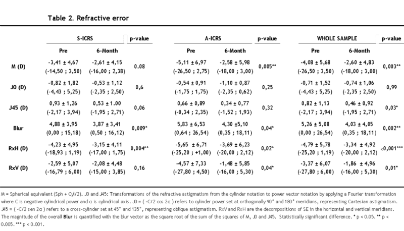
Table 2: Refractive error.
Analyzing the results of topographic and tomographic data measured for a pupillary area of 6 mm (Table 3). For the anterior corneal surface (Front): Results show a statistically significant mean enhancement with respect to the simulated K reading in the flattest meridian (K1) in the S-ICRS of 0.97 D, in the S-ICRS, they also present a statistically significant improvement of 0.80 D and for the whole sample, a statistically significant improvement of 0.90 D is also obtained.
About simulated K reading in the steepest meridian (K2), a statistically significant mean enhancement of 2.14 D is found for the S-ICRS, a statistically significant improvement of 1.34 D is obtained for the A-ICRS and a statistically significant improvement of 1.80 D is obtained for the whole sample. In the horizontal meridian curvature values (Rh), there is a statistically significant improvement in the S-ICRS of 0.20 mm on average. In turn, the whole sample there is a statistically significant improvement of 0.19 mm on average in that meridian.
In the vertical meridian curvature values (RxV), there is a statistically significant mean enhancement in the S-ICRS of 0.19 mm on average. In the A-ICRS, there is also a statistically significant improvement of 0.36 mm on average. In the whole sample, there is a statistically significant improvement of 0.26 mm on average. Regarding the astigmatism calculated as the difference between K1 and K2; the results show a statistically significant improvement in the S-ICRS of 1.40 D. Also, for the whole sample, a statistically significant improvement of 1.19 D is produced. In relation to asphericity (Q), a statistically significant enhancement there is in the S-ICRS of 0.20 points. Also in the whole sample, there is a statistically significant improvement of 0.18 points.
For the posterior corneal surface (Back): Results show a statistically significant mean improvement regarding simulated K reading in the steepest meridian (K2) in S-ICRS of 0.18 mm. There is also a statistically significant enhanced for the whole sample of 0.20 mm.
In the horizontal meridian curvature values (Rh), there is a statistically significant mean improvement in the A-ICRS of 0.17 mm on average. Also, in the vertical meridian curvature values (RxV), there is a statistically significant main improvement in the A-ICRS of 1.74 D. In the A-ICRS, there is also a statistically significant improvement of 0.36 mm on average. In the whole sample, there is a statistically significant improvement of 0.14 mm.

Table 3: Topographic and tomographic outcomes at 6 mm.
Analyzing the results of topographic and tomographic data measured for a pupillary area of 4 mm (Table 4). The flatter axis (K1) showed a statistically significant mean reduction in corneal refractive power in each type of rings, being 1.87 D in the S-ICRS and 2.13 D in the A-ICRS. There was also a statistically significant mean reduction in the whole sample of 1.95D.
The axis more curved (K2) also present a statistically significant mean reduction in corneal refractive power in each type of rings, being 2.63 D in the S-ICRS and 2.83 D in the A-ICRS. There was moreover a statistically significant mean reduction in the whole sample of 2.68 D.
The asphericity of the anterior surface of the cornea (Q), showed a statistically significant mean flattening of 0.15 microns in the S-ICRS. PminX shows a statistically significant mean improvement in nasal centering of the corneal apex in A-ICRS of 0.13 mm.
The point of minimum corneal thickness shows a statistically significant mean thickness increase in both types of rings. Being 10.33 mm in the S-ICRS and 16.04 mm in the A-ICRS. There was furthermore a statistically significant mean increase for the whole sample of 12.21 mm.
The point of least thickness at the corneal apex also shows a statistically significant mean increase in thickness at this more curved point in both types of rings, being 6.68 microns in the S-ICRS and 16.04 microns in the A-ICRS. A statistically significant increase in thickness of 12.02 microns was also produced for the whole sample.
In reference to corneal asymmetry indices: ISV: Index of surface variance, shows the standard deviation of corneal sagittal radii from the mean curvature. A statistically significant mean improvement in the S-ICRS of 16.90 points was observed. Further on average enhancement of 12.33 points was obtained in the whole sample. IVA: Index of vertical asymmetry, average difference between superior and inferior corneal curvature. This presents a statistically significant mean difference in the two types of rings analyzed, being 0.20 mm in the S-ICRS and 0.13 mm in the A-ICRS. Moreover, in the whole sample analyzed, a statistically significant mean improvement of 0.16 mm was obtained. KI: Keratoconus Index, relationship between the mean values of radius in the upper and lower segments of the cornea. A statistically significant mean enhancement was found in both types analyzed. Being 0.09 points in the S-ICRS and 0.07 points in the A-ICRS. Moreover, in the whole sample we found a statistically significant mean improvement of 0.08 points. CKI: Central Keratoconus Index. A statistically significant mean improvement was found in the A-ICRS of 0.02 points. In the whole sample, mean differences of 0.02 points were also found. IHA: Index Height Asymmetry. Statistically significant mean differences of 8.86 microns were found in the S-ICRS. Further, in the whole sample it was obtained a mean difference of 10.69 microns. IHD: Index of height decentration. In all cases there was a statistically significant mean improvement, being 0.05 microns in the S-ICRS, 0.02 microns in the A-ICRS and 0.03 microns in the whole sample.

Table 4: Topographic outcomes at 4 mm.
Table 5 shows the aberrometric outcomes assessed both in front and back corneal surfaces. A statistically significant mean difference of 0.41 microns in total corneal HOA RMS was found between pre- and 6 months after S-ICRS implantation. It was found a statistically significant difference between the effect of both ring designs of 1.29 + -0.62 microns (p=0.045) and in the S-ICRS HOA of the anterior corneal side of the cornea of 0.24 microns. Statistically significant differences in the Zernike Coefficient for the horizontal secondary coma (Z5. 1) were also found in the anterior corneal surface for the total sample of rings with a difference of 0.14 microns.
On the posterior corneal side, statistically significant mean differences were obtained in the Zernike Coefficient for the vertical primary coma (Z3. -1) in the S-ICRS of 0.81 microns. Statistically significant differences were moreover found in the Zernike Coefficient for vertical primary coma (Z3. -1) in the A-ICRS of 0.72 microns and in the total sample statistically significant differences were also observed in the Zernike Coefficient for vertical primary coma (Z3. -1) of 0.77 microns.
On the posterior corneal side, statistically significant differences were also found for the A-ICRS in the Zernike Coefficient for secondary astigmatism (Z4. 2) with an improvement of 0.09 microns. On the posterior corneal side, statistically significant differences were also found for the total sample in the Zernike Coefficient for 4th order spherical aberration (C4. 0) by 0.04 microns.

Table 5: Aberrometric outcomes cornea front and back.
Visual Function values obtained before and after 6-Months of surgery under photopic and mesopic conditions are shown in Table 6. Regarding the Photopic Visual Function, a statistically significant mean difference of 1 line and 1 letter was found in HCVA after S-ICRS implantation and a difference of 1 logMAR line in the whole sample. Respect to logMAR LCVA, an improvement of 1 line and 4 letters in the S-ICRS and 1 line and 2 letters was obtained in S-ICRS and A-ICRS, respectively. Moreover, an improvement of 1 line and 4 letters was found for the whole sample. Regard to CS a statistically significant enhancement of 14 logarithmic units and of 12. When the Mesopic Visual Function was analyzed, a statistically significant improvement of one line was obtained in logMAR HCVA after S-ICRS implantation, whereas a not clinically significant difference of 4 letters was found after A-ICRS implantation. Furthermore, an improvement of 1 line was found in the whole sample. Concerning logMAR LCVA, there was a statistically significant improvement of 2 lines and 3 letters after S-ICRS implantation whereas an improvement of two lines was found in the whole sample. With reference to CS, a statistically significant difference of 14 log. Units and 10 log. Units were found in S-ICRS and the whole sample, respectively.
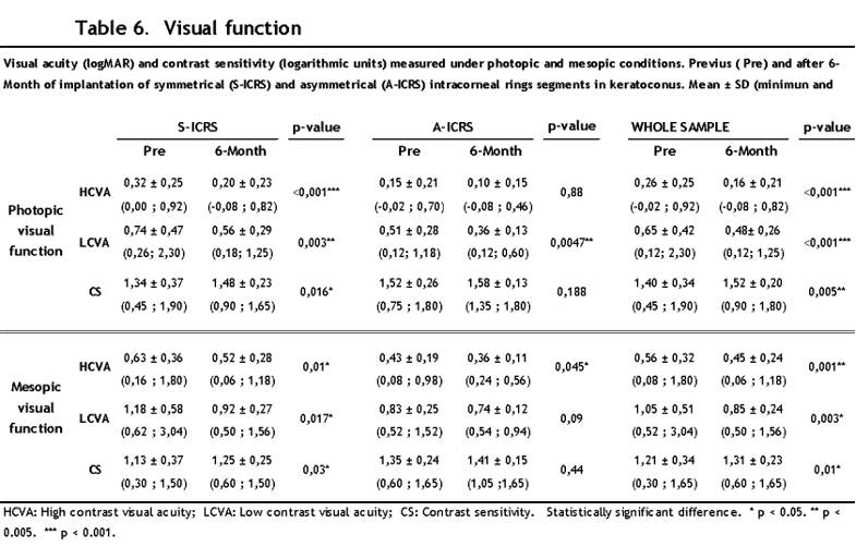
Table 6: Visual function.
Regarding the photopic visual function, for the HCVA in the case of S-ICRS, a high and statistically significant correlation was obtained between this and the total RMS of the cornea (r = 0.6089, p = 0.036). In the A-ICRS, statistically significant high correlations were obtained between this and the total RMS HOA of the cornea (r = 0.7112, p = 0.006) and the RMS HOA Front (r = 0.7211, p = 0.005). For the whole of ICRS, there was a statistically significant moderate correlation in the correlations between the HCVA and all the RMS analyzed, for RMS Cornea (r = 0.4944, p = 0.036), for RMS HOA Cornea (r = 0, 4969, p = 0.012), for RMS Front (r = 0.4764, p = 0.016) and for RMS HOA Front (r = 0.5443, p = 0.004).
For the LCVA in the S-ICRS, a statistically significant high correlation was found in all the RMS analyzed. For RMS Cornea (r = 0.7531, p = 0.003), for RMS HOA Cornea (r = 0.6528, p = 0.016), for RMS Front (r = 0.7171, p = 0.006) and for RMS HOA Front (r = 0.6750, p = 0.008). In the A-ICRS, a statistically significant high correlation was also obtained in all the RMS analyzed, for RMS Cornea (r = 0.5789, p = 0.038), for RMS HOA Cornea (r = 0.7751, p = 0.002), for RMS Front (r = 0.6017, p = 0.029) and for RMS HOA Front (r = 0.7941, p = 0.001). For the whole sample, a statistically significant high correlation was also obtained in all the RMS analyzed, for RMS Cornea (r = 0.6444, p ˂0.001), for RMS HOA Cornea (r = 0.6445, p ˂0.001), for RMS Front (r = 0.6363, p ˂0.001) and for RMS HOA Front (r = 0.6748, p ˂0.001).
Respect to CS under photopic conditions, a high inverse correlation was obtained for the S-ICRS with statistical significance in all the RMS analyzed. For RMS Cornea (r = -0.6733, p = 0.008), for RMS HOA Cornea (r = -0.6438, p = 0.013), for RMS Front (r = -0.6238, p = 0.017) and for RMS HOA Front (r = -0.6857, p = 0.005). The same results were obtained for the A-ICRS, the inverse correlations being moderate in the two total RMS, for RMS Cornea (r = -0.5305, p = 0.062) and for RMS Front (r = -0.5740, p = 0.040) and high in both RMS HOA, for RMS HOA Cornea (r = -0.7497, p = 0.003) and for RMS HOA Front (r = -0.7860, p = 0.001). For all implants, the correlations were moderate in all the RMS with high statistical significance. for RMS Cornea (r = -0.5285, p = 0.005), for RMS HOA Cornea (r = -0.5514, p = 0.003), for RMS Front (r = -0.5340, p = 0.004) and for RMS HOA Front (r = -0.6307, p ˂0.001).
In relation to mesopic visual function, for HCVA in the case of S-ICRS, a high correlation with statistical significance was obtained in all the RMS analyzed, for RMS Cornea (r = 0.6823, p = 0, 01), for RMS HOA Cornea (r = 0.5722, p = 0.041), for RMS Front (r = 0.6736, p = 0.012) and for RMS HOA Front (r = 0.6236, p = 0.017). For the A-ICRS, statistically significant correlations were obtained in all cases, moderate in the Cornea RMS (r = 0.5739, p = 0.04) and Front RMS (r = 0.5947, p = 0.032) and high in the RMS HOA Cornea (r = 0.7832, p = 0.002) and RMS HOA Front (r = 0.7979, p = 0.002). In the whole sample analyzed, moderate correlations were obtained in all the RMS with high statistical significance, for RMS Cornea (r = 0.5641, p = 0.003), for RMS HOA Cornea (r = 0.5448, p = 0.004), for RMS Front (r = 0.5718, p = 0.002) and for RMS HOA Front (r = 0.5998, p ˂0.001).
For the LCVA, in the A-ICRS, high correlations were obtained in the Cornea RMS (r = 0.7036, p = 0.007) and Front RMS (r = 0.7220, p = 0.005) and very high in the RMS HOA Cornea (r = 0.8471, p ˂0.001) and RMS HOA Front (r = 0.8774, p ˂0.001) with high statistical significance in all cases. Regarding the whole sample, statistically significant moderate correlations were obtained in all cases. For RMS Cornea (r = 0.5407, p = 0.004), for RMS HOA Cornea (r = 0.5135, p = 0.007), for RMS Front (r = 0.5075, p = 0.008) and for RMS HOA Front (r = 0.5105, p = 0.007).
Regarding SC in the case of S-ICRS, statistically significant high inverse correlations were obtained for RMS Cornea (r = -0.7813, p ˂0.001), for RMS HOA Cornea (r = -0.7950, p ˂0.001) and RMS Front (r = -0.7556, p ˂0.001) and very high inverse correlation for RMS HOA Front (r = -0.8120, p ˂0.001). For the A-ICRS, a moderate inverse correlation was obtained for the Cornea RMS (r = -0.5961, p = 0.032) and high inverse correlations with high statistical significance for the Cornea HOA RMS (r = -0.8471, p ˂ 0.001), the RMS Front (r = -0.6382, p = 0.019) and the RMS HOA Front (r = -0.8748, p ˂0.001). For whole sample, we obtained a high correlation with high statistical significance in all the RMS analyzed. For RMS Cornea (r = -0.6261, p ˂0.001), for RMS HOA Cornea (r = -0.6806, p ˂0.001), for RMS Front (r = -0.6381, p ˂0.001) and for the RMS HOA Front (r = -0.7456, p ˂0.001).
The result of our sample was different according to the type of ICRS implanted when comparing both designs, statistically significant differences were found in factors A, B, C, but not in factor D. If we compare the values obtained for the first 3 factors, the degree of keratoconus was higher for the S-ICRS for the bending in the central 3.00 mm of the anterior surface, the posterior surface and for the pachymetry, but not for the VA measurements.
According to the criteria used by Sedaghat, (19) our sample would be classified as follows:
S-ICRS = A4B5C2D2
A-ICRS = A3B3C2D1
TOTAL = A3B5C2D2
Taken the numerical values rounded to whole number values. This difference found in the initial state of KC between both implants is a relevant fact that must be assessed and considered in the comparative analysis of the two samples
Regarding refractive changes in our study, a statistically significant improvement was obtained for the spherical equivalent (M) of A-ICRS of 2.53 D (p = 0.005) on average and 1.48 D (p = 0.003) for the total sample. These results are therefore in line with those provided by other authors and this is consistent with the findings reported with other types of asymmetric and symmetric ring segments in keratoconus cases [4,14]. In comparative studies between types of ICRS. In relation to the blur magnitude, we obtained superior results than those obtained by Cuiña, et al., for the A-ISCR and lower for the S-ICRS, but the same as those obtained for the whole sample [13,20]. Obtaining an improvement in both types of implants.
In the present work, an improvement was obtained in the results of refractive decomposition on the horizontal axis (RxH) for the A-ICRS of 4.34 D and for the total sample of 1.94 D. Also, for the vertical axis (RxV) of 1.99 D (Table 2). Although we have not found authors who analyze this decomposition, this general improvement and its even greater difference on the horizontal axis may be related to the improvements obtained in the tomographic and aberration components, perhaps confirmed by the improvement in vertical asymmetry indices that we will discuss later.
Corneal topography has been found to be sensitive for detecting subtle changes on the anterior corneal surface due to corneal ectatic disorders before the appearance of clinical signs. The automatically calculated decentration IHD index, is not only an index to detect corneal asymmetry and consequently important in KC detection, but also it was reported as one of the most sensitive and specific criteria for follow up of KC [21-23].
Studies have demonstrated the short-term and long-term efficacy of ICRS implantation in patients with keratoconus.18 In most of the studies, ring segments implantation contributed to a mean reduction of the K readings between 3 and 5 D, with anastigmatic reduction between 1.5 to 2.88 (although Zare, et al., in a prospective study with intacs presented a mean reduction of 0.75 D) and these have proven effective in both types of ICRS and with different types of ICRS, mainly on the anterior surface [19,20]. Although the effectiveness of ICRS in controlling the progression of keratoconus has been questioned to date, it appears that results in significant changes in corneal densitometry specifically in the central corneal layer and in the 2-6 mm annulus, with an inverse correlation with keratometric values of the front corneal surface. Besides, ICRS leads to substantial improvements of refractive status, visual acuity and corneal keratometry and asphericity [24]. However, these changes have not been strongly correlated by corneal resistance and hysteresis parameters [25]. It could be that in the future, due also to the improvement of surgical techniques mainly using Femtosecond Laser, they will help to better adjust the results [26].
In conclusion, although the result in some parameters is not statistically significant, there is an improvement after its implantation in corneal regularization, corrected VA under photopic conditions and mainly in corrected VA under mesopic conditions.
The authors declared no potential conflicts of interest with respect to the research, authorship and/or publication of this article.
This research did not receive any specific grant from funding agencies in the public, commercial or non-profit sectors.
The authors have no acknowledgments to declare.
The data supporting the findings of this study are available from the corresponding author upon reasonable request.
The study protocol was approved by the Ethics Committee of the Hospital Clínico San Carlos, Madrid, Spain.
Informed consent was obtained from all participants included in the study.
All authors contributed equally to this paper.
Rafael Bella-Gala1,2, Jesús Carballo-Alvarez2* , Ricardo Cuiña Sardiña2
1Clinico San Carlos Hospital, Madrid, Spain
2Faculty of Optics and Optometry, Complutense University of Madrid, Madrid, Spain
*Correspondence author: Jesús Carballo-Alvarez, OD, PhD, Faculty of Optics and Optometry, Complutense University of Madrid, Madrid, Spain;
Email: jcarballo@ucm.es

